Diffuse Large B-Cell Lymphoma with Synchronous Breast and Ovarian Localization: A Rare and Misleading Presentation ()
1. Introduction
Primary extranodal lymphomas are defined as lymphomas developing in tissues outside the lymphatic system, with or without regional lymph node involvement [1]. Primary non-Hodgkin breast lymphomas are rare, accounting for less than 0.5% of all breast cancers [2]. A primary breast lymphoma (PBL) is defined when the breast is the main organ involved or, in most cases, the sole site affected by lymphomatous proliferation [3]. Non-Hodgkin lymphoma (NHL) localized in the female genital tract is rare. The ovary represents the most frequent site, which may be involved either as a primary tumor or in the context of secondary involvement during systemic NHL [4]. Distinguishing primary breast lymphoma from systemic lymphoma with secondary breast involvement is important because these entities differ in terms of staging, disease classification, and prognosis. This distinction relies on the extent of disease at diagnosis and the criteria proposed for primary breast lymphoma.
2. Case Report
This is a 32-year-old woman with no significant past medical history, who consulted for self-palpation of a right breast nodule and non-cyclic pelvic pain described as heaviness, evolving over 4 months. Clinical examination revealed a 5 cm mass in the upper outer quadrant (UOQ) of the right breast, along with three additional contiguous lesions in the same quadrant, associated with a 3 cm homolateral axillary lymphadenopathy, mobile in both planes, and a left latero-uterine mass on gynecological examination.
Mammography completed by breast ultrasound, as showed in Figures 1-2, revealed in the right breast multiple lesions, including three well-defined hypoechoic oval masses in the UOQ measuring 30.6 × 47.6 mm, 30 × 28 mm, and 20 × 12 mm, a similar lesion of 23.5 × 21 mm at the junction of the upper quadrants, a well-defined solid-cystic oval lesion of 39 × 21.6 mm in the subareolar region, scattered cystic formations, and two axillary lymph nodes; one inflammatory-looking measuring 23 × 9 mm and the other hypoechoic oval measuring 11.4 × 17 mm.
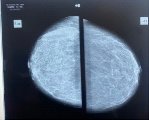
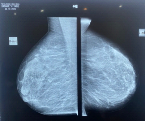
Figure 1. Bilateral mammography demonstrating type D breast density with no suspicious mammographic findings.
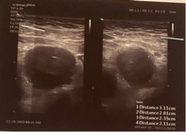
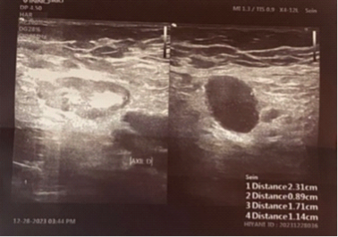
Figure 2. Right breast ultrasound demonstrating multiple hypoechoic masses and axillary lymphadenopathy.
Breast MRI demonstrated, as showed in Figure 3, multiple nodular lesions in the right breast distributed as follows: two adjacent retro-mammary lesions of irregular shape with microlobulated margins measuring 44 × 37 mm and 19 × 15 mm, at least three nodules in the axillary extension ranging from 12 to 6 mm in largest axis, and multiple suspicious axillary lymph nodes, the largest measuring 40 × 36 mm. In the left breast, a medial subareolar nodule measuring 10 × 7 mm was observed. The examination was classified as BI-RADS 5 on the right and BI-RADS 4 on the left.
Figure 3. Distribution of bilateral breast lesions with suspicious right axillary lymphadenopathy. Lesion dimensions are indicated.
A Tru-Cut biopsy of the right UOQ lesion showed a diffuse, undifferentiated, infiltrative cellular proliferation. Immunohistochemistry (IHC) concluded to diffuse large B-cell non-Hodgkin lymphoma phenotype.
Pelvic ultrasound showed in Figure 4 a left latero-uterine solid mass measuring 10 × 8 cm, whose ovarian or uterine origin could not be determined.
Pelvic MRI demonstrated a left latero- and supra-uterine tumor mass with predominantly solid components measuring 91 × 87 × 90 mm, with a central necrotic area, an anteverted uterus of normal appearance and size, a small peritoneal effusion, and multiple suspicious lymph nodes involving internal iliac and lower mesenteric regions, the largest measuring 34 × 24 mm (Figure 5).
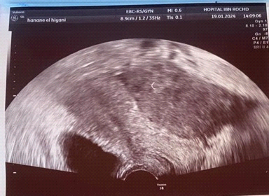
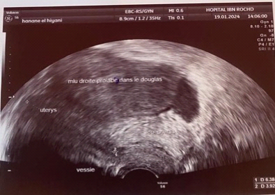
Figure 4. Pelvic mass lesion (10 × 8 cm) of indeterminate uterine or ovarian origin, without associated effusion.
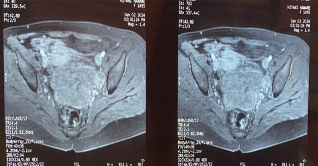
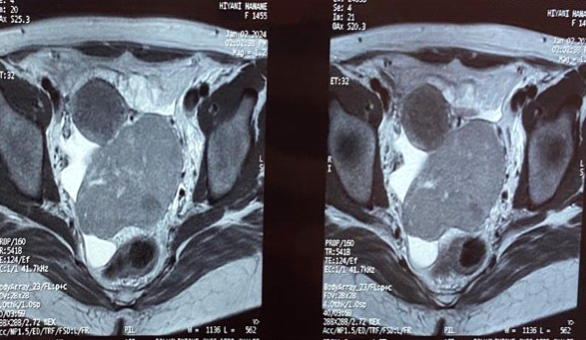
Figure 5. Pelvic MRI demonstrating a large left latero-uterine mass with central necrosis and associated suspicious lymphadenopathy.
CA125 was negative at 24.1.
After multidisciplinary tumor board discussion, the decision was to perform excision of the biopsy-proven breast lesion and lymph node, and not to intervene on the latero-uterine mass, considered a probable localization of non-Hodgkin lymphoma, followed by chemotherapy.
A right lumpectomy was performed, confirming diffuse large B-cell non-Hodgkin lymphoma, phenotype B.
The patient was then started on chemotherapy, with evolution marked by complete regression of the latero-uterine mass.
3. Discussion
The breast is an uncommon site for malignant lymphomas due to its low content of lymphoid tissue [5]. Their frequency is estimated at 0.04% to 0.53% of all breast cancers and 2.2% of extranodal NHLs [6] [7]. This disease mainly affects women; however, cases in men have been reported. Regarding age, two incidence peaks have been described: an initial peak in young women of reproductive age, often during pregnancy, and a second more prominent peak between 50 and 60 years, associated with a more favorable prognosis [3]. In our case, the patient was 32 years old.
Breast involvement is usually unilateral [8] [9]. However, bilaterality is possible, either synchronous in 13% of cases or metachronous in 9% [7] [8].
Clinical presentation is usually limited to a breast nodule, as in our case. It is most often a single, large, well-circumscribed mass without inflammatory signs [6] [10] [11], although cases of gigantomastia or inflammatory mastitis may occur [12]. Axillary lymphadenopathy is found in 20% - 40% of cases [13].
Imaging features are non-specific. Mammography typically shows a well-circumscribed homogeneous mass with benign appearance, sometimes mimicking cysts, fibroadenomas, or phyllodes tumors. Less frequently, diffuse mastitis-like changes, ill-defined masses, or spiculated lesions may be observed [14]. Ultrasound usually shows a hypoechoic, homogeneous lesion with regular and well-defined margins. MRI descriptions are less common. Yang et al. reported lesions with regular margins, hyperintense on T2-weighted images, iso-intense on T1-weighted images, with homogeneous and rapid enhancement, most often with type II (plateau) kinetic curves [15].
Diagnosis is confirmed by histological examination obtained by surgical or image-guided biopsy [1] [11]. The most common histological subtype is diffuse large B-cell lymphoma, while MALT lymphoma represents the second most frequent entity [2]. In our case, it was diffuse large B-cell non-Hodgkin lymphoma, phenotype B.
First described by Selye in 1946 [9], ovarian non-Hodgkin lymphoma is rare and most often represents secondary involvement of systemic lymphomatous disease. Primary ovarian lymphoma is extremely rare, accounting for only 0.5% of NHLs and 1.5% of ovarian tumors [16].
Most patients are between 20 and 50 years old [17], which was the case in our observation. The most common clinical manifestations are abdominal or pelvic pain, pelvic mass, and ascites, often mimicking epithelial ovarian carcinoma [16] [18]. B symptoms occur in 10% - 33% of patients [16] [19]. In our case, non-cyclic pelvic heaviness was the main symptom.
Although CA-125 is widely used in the follow-up of epithelial ovarian cancer, it may also serve as a prognostic marker in non-Hodgkin lymphoma. Zidan et al. [20] reported elevated CA-125 levels in 45% of lymphoma patients, with higher levels associated with advanced disease, poorer treatment response, and reduced survival. In our case, CA-125 was negative.
Radiological features are non-specific. Ovarian masses are usually large, homogeneous, moderately enhancing lesions, sometimes associated with central necrosis and the “touching ovaries” sign [21]. In our case, pelvic MRI showed a large left latero- and supra-uterine solid mass with central necrosis, strongly suggestive of lymphoma in the context of associated breast involvement.
The classification of this case deserves particular consideration. Although breast lymphoma was the initial histologically confirmed manifestation, the simultaneous presence of breast lesions, axillary lymphadenopathy, pelvic lymphadenopathy, and a large pelvic mass at diagnosis favors the diagnosis of systemic diffuse large B-cell lymphoma with extranodal breast and probable ovarian involvement rather than primary breast lymphoma according to the established diagnostic criteria. Histological confirmation of the ovarian lesion was not obtained; however, its complete regression following R-CHOP chemotherapy strongly supported lymphomatous involvement.
Cytoreductive surgery is not recommended in ovarian lymphoma due to the absence of demonstrated benefit. Treatment is based on R-CHOP chemotherapy (rituximab, cyclophosphamide, doxorubicin, vincristine, and prednisone) [22].
In our observation, the patient received chemotherapy with a favorable outcome marked by complete regression of both breast lesions and the latero-uterine mass.
4. Conclusion
Primary or secondary non-Hodgkin lymphomas of the breast and ovary are rare entities that represent a major diagnostic challenge due to their non-specific clinical and radiological presentation, often mimicking epithelial tumors. Histological confirmation is essential for diagnosis. Treatment relies mainly on systemic chemotherapy, particularly the R-CHOP regimen, while surgery is limited to diagnostic purposes. A multidisciplinary approach is crucial to avoid unnecessary surgical procedures and optimize therapeutic management, as illustrated in our case with a favorable outcome under chemotherapy.
Patient Consent
Written informed consent was obtained from the patient for publication of this case report and the accompanying images. All identifying information has been removed to protect patient confidentiality.