The purpose of this study was to observe the histopathological lesions and oxidative damage induced by dietary nickel chloride (NiCl
2) in the thymus. A total of 280 one-day-old avian broilers were divided into four groups and fed on a corn-soybean basal diet as the control diet or the same basal diet supplemented with 300, 600, and 900 mg/kg of NiCl
2 for 42 days. In the NiCl
2-treated groups, the broiler weight and thymic relative weight were significantly (
P < 0.05 or
P < 0.01) decreased. Histopathologically, thymic corpuscles were increased and enlarged; the reticular cells were degenerate and necrotic, and lymphocytes were slightly decreased and loosely arranged in the medulla of thymus in the 600 mg/kg and 900 mg/kg groups. The activities of superoxide dismutase (SOD), catalase (CAT) and glutathione peroxidase (GSH-Px), and the ability to inhibit hydroxy radical and glutathione (GSH) content were significantly (
P < 0.05 or
P < 0.01) lower in the NiCl
2-treated groups than those in the control group, while MDA content was higher. The above-mentioned results demonstrated that dietary NiCl
2 in excess of 300 mg/kg could reduce the broiler weight and thymic relative weight, and cause histopathological lesions and oxidative damage in the thymus, which finally impaired the thymic function.
Nickel (Ni) is the 24th most abundant element in the earth’s crust, comprising about 3% of the composition of the earth [1] , and used in a wide variety of industrial and consumer applications [2] . At the same time, it is known to be an essential element for animals [3] [4] . Anke et al. [5] reported an absolute requirement for nickel in the ruminants (sheep, goat, and so on). Ni deficiency inhibits growth, reduces reproductive rate, and alters glucose and lipid metabolism, which are associated with anemia, hemoglobin reduction, alternations of metal ion contents, and reduced activity of several enzymes in animals [6] . However, Ni or Ni compounds can enter the food chain and may be toxic to living organisms. Ni gets into human and animals mainly through inhalation, drinking water and food, and food is the most important among these pathways [7] . The previous studies have shown that long-term exposure to Ni can also be toxic to the upper respiratory tract, skin, kidney, immune system, embryo, and breeding system [8] [9] . Even short-term exposure to Ni can significantly influence the cardiovascular system [10] [11] . Ni exposure causes formation of free radicals in various tissues in both human and animals, and enhances lipid peroxidation [12] . Thus, the toxic effects of Ni and Ni compounds have been major environmental health problems.
It had been reported that Ni can enhance lipid peroxidation (LPO) and cause cellular damage and reduced glutathione (GSH) contents, and catalase (CAT) and glutathione peroxidase (GSH-Px) activities in the liver and kidney of rats [13] [14] . NiCl2-induced human lymphocyte toxicity may be mediated by oxygen radical intermediates [15] . Our previous studies have also shown that dietary NiCl2 causes intestinal and splenic oxidative damage [12] [16] . However, very limited data are available on the oxidative damage induced by dietary NiCl2 in the thymus in human beings and animals. Also, there are no reports about effects of NiCl2 on thymic morphology in human and animals at present. In the present research, the experiment was conducted to examine the effects of dietary NiCl2 on thymus, induced clinical observation, histological lesions, relative weight and oxidative stress parameters (the activities of SOD, CAT, GSH-Px, and ability to inhibit hydroxyl radical, and GSH and MDA contents), which provided helpful materials for the same or similar studies in both human and other animals in the future.
2. Materials and Methods
Chickens and diets
Two hundred eighty one-day-old healthy broilers were randomly divided into four equal groups of 70. The broilers were housed in separate cages with electrically heated units and were provided with water and the control or experimental diets ad libitum for 42 days.
Our experiments involving the use of broilers and all experimental procedures involving animals were approved by Sichuan Agricultural University Animal Care and Use Committee.
A corn-soybean basal diet formulated by the National Research Council (1994) [17] was the control diet. NiCl2∙6H2O (Cheng Du Kelong Chemical Co., Ltd., Chengdu, China) was mixed into the corn-soybean basal diet to produce experimental diets with 300, 600, and 900 mg/kg of NiCl2, respectively.
Clinical signs and relative weight of thymus
Clinical signs were observed and recorded everyday. At 7, 14, 21, 28, 35 and 42 days of age during the experiment, five broilers in each group were euthanized and necropsied after their body weight were weighed. Thymus was dissected from each broiler and weighed after dissecting connective tissue around the organ, and the macroscopic changes of thymus were observed and recorded. Related weight of thymus was calculated by the following formula:
Related weight = organ weight (g)/body weight (kg)
Pathological observation
After weighed, thymuses were fixed in 4% paraformaldehyde and routinely processed in paraffin. Thin sections (5 μm) of each tissue were sliced from each block and mounted on glass. Slices were stained with hematoxylin and eosin (H&E) and were examined on an Olympus light microscope.
Detection of oxidative damage parameters in the thymus
At 14, 28 and 42 days of age, after five broilers in each group were humanely killed, thymuses were immediately removed and chilled to 0˚C in 0.85% NaCl, and then dried, weighed and homogenized in 9 vol of ice- cold 0.85% NaCl in a chilled homogenizer and centrifuged at 3500 × g at 4˚C for 10 min. Then the supernatant were collected. After determining the amount of total protein in the supernatant by the method of Bradford [18] , the CAT, SOD and GSH-Px activities, and ability to inhibit hydroxyl radical, and MDA and GSH contents in the supernatant were detected by biochemical methods following the instruction of the reagent kits (CAT, A007; SOD, A001-1; GSH-Px, A005; abilities to inhibit hydroxy radical, A018; GSH, A006-1; MDA, A003-1; total protein, A045-2, purchased from Nanjing Jiancheng Bioengineering Institute of China, Nanjing, China). The absorbance was measured at 240, 550, 412, 550, 532, 420 and 590 nm, respectively using a microtiter plate reader (Thermo, Varioskan Flash , USA ).
Statistical analysis
The significance of difference between the control groups and the NiCl2-treated groups was analyzed by use of variance analysis, and the results are presented as means ± standard deviation (± SD). The analysis was performed with the one-way analysis of variance (ANOVA) test of SPSS 16.0 for windows. A value of P < 0.05 was considered significant.
3. Results
Clinical observation
From 14 to 42 days of age during the experiment, the feed intake of broilers in the 300 mg/kg, 600 mg/kg, and 900 mg/kg groups began to decline when compared with that in the control group, except the 300 mg/kg group at 14 days of age. From 21 to 42 days of age during the experiment, broilers in the 300 mg/kg, 600 mg/kg, and 900 mg/kg groups showed poor appetite, poor growth and depression. A few broilers showed polypnea. No death was found during the experiment. Change of broiler weight was shown in Figure 1.
Change of thymic relative weight
From 21 to 42 days of age during the experiment, the thymic relative weight of broilers were significantly decreased (P < 0.05 or P < 0.01) in the 300 mg/kg, 600 mg/kg, and 900 mg/kg groups in comparison with those in the control group. The results were shown in Figure 2.
Pathological lesions
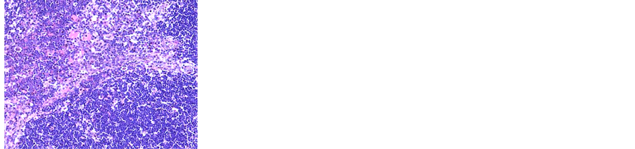
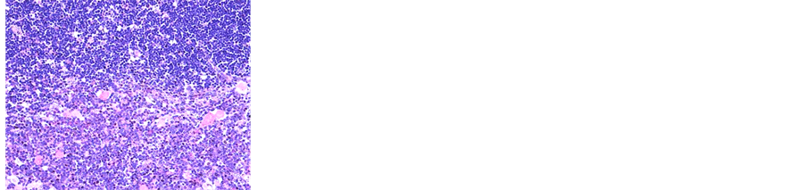
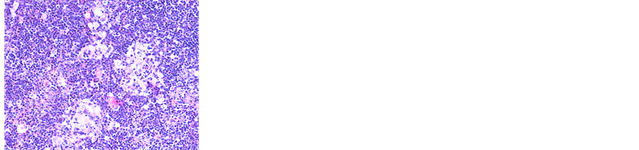
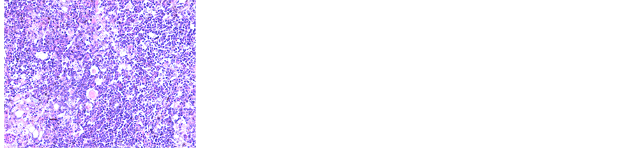
There were no obvious lesions among NiCl2-treated groups at 7, 14, and 21 days of age when compared with the control group. From 21 to 42 days of age, the thymic corpuscles were increased and enlarged, the cells were degenerate and necrotic reticular, and lymphocytes were slightly decreased and loosely arranged in the medulla of thymus in the 600 mg/kg and 900 mg/kg groups (Figure 3).
Changes of oxidative damage parameters in the thymus
The CAT, SOD, and GSH-Px activities were significantly lower (P < 0.05 or P < 0.01) in the 900 mg/kg group at 14 days of age, and in the 300 mg/kg, 600 mg/kg, and 900 mg/kg groups from 28 to 42 days of age than those in the control group, except the SOD and GSH-Px activities in the 300 mg/kg group at 28 days of age. The ability to inhibit hydroxyl radical were significantly decreased (P < 0.05 or P < 0.01) in the 600 mg/kg and 900 mg/kg groups at 14 to 42 days of age when compared with those of the control group, except that in the 600 mg/kg group at 14 days of age. The GSH contents were significantly lower (P < 0.05 or P < 0.01) in the 900 mg/kg group at 14 days of age, and in the 300 mg/kg, 600 mg/kg, and 900 mg/kg groups at 28 to 42 days of age than those in the control group, except the GSH contents in the 300 mg/kg group at 28 days of age. The MDA contents were significantly higher (P < 0.05 or P < 0.01) in the 300 mg/kg, 600 mg/kg, and 900 mg/kg groups at 28 to 42 days of age than those in the control group, except the MDA contents in the 300 mg/kg group at 28 days of age. The results were shown in Figure 4.
4. Discussion
In the present study, the growth of the NiCl2-treated broilers was inhibited, and the above-mentioned clinical signs showed that dietary NiCl2 in excess of 300 mg/kg was toxic to broilers from 3-week-old, which was consistent with those described in broilers by Ling and Leach [19] .
Thymus is the central immune organs. The situation of thymus development is usually judged by the thymic relative weight [20] . The relative weight of thymus in NiCl2-treated groups was lower than that in control group, which implied dietary NiCl2 in excess of 300 mg/kg inhibited the thymus growth, and could finally impair the function of thymus. Histopathologically, thymic corpuscles were increased and enlarged, the reticular cells were degenerate and necrotic, and lymphocytes were decreased and loosely arranged in the medulla of thymus in the 600 mg/kg and 900 mg/kg groups, implying that dietary NiCl2 in excess of 300 mg/kg could influence or/and impair thymic function and growth.
It is well known that there are close relationships between lesions and oxidative damage caused by NiCl2.
Living organisms possess several antioxidative agents (including enzymes such as SOD, GSH-Px, and CAT,
Changes of broiler weight (g).
Changes of relative weight in the thymus.
(a) (b)
(c) (d)
(a) Thymus in the control group; (b) Thymus in the 300 mg/kg group; (c) Thymus in the 600 mg/kg group. The thymic corpuscles are increased and enlarged, the reticular cells are degenerate and necrotic, and lymphocytes are slightly decreased and loosely arranged in the medulla; (d) Thymus in the 900 mg/kg group. The thymic corpuscles are increased and enlarged, the reticular cells are degenerate and necrotic, and lymphocytes are decreased and loosely arranged in the medulla. H.E × 400.
Figure 3. Histopathological changes in the thymus at 42 days of age.
Changes of oxidative stress parameters in the thymus.
GSH, and the ability to inhibit hydroxyl radical, and nonenzymatic antioxidants such as extra calcium and vitamin E) and protect them against the harmful effect of oxidative stress [16] [21] [22] . These antioxidants inhibit overproduced free radicals (including ROS and .OH), which induces lipid peroxidation (LPO) [23] . It has been reported that Ni ion accumulation may be responsible for the generation of ROS and the enhancement of LPO [24] . LPO may be a contributing factor in Ni-induced tissue oxidative stress [25] . As the first line of cellular defence against oxidative damage, SOD and CAT play an important role in these enzymatic mechanisms [26] . SOD can break up ROS and repair cellular damage caused by ROS [16] . CAT is responsible for the breakdown of hydrogen peroxide (H2O2) [27] . Our results showed that SOD and CAT were decreased in NiCl2-treated groups when compared with those in the control group, which demonstrated that free radicals species are accumulation in the thymus of NiCl2-treated groups, and reduction of the SOD and CAT activities enhances ROS production and H2O2 generation [28] .
GSH is considered to be an important bio-marker in LPO and is important in maintaining the cellular redox status [29] , and depletion of intracellular free radical scavengers such as GSH is one of Ni-caused oxidative stress mechanisms [30] [31] . GSH-Px activity depends on the balance between the levels of GSH and glutathione disulfide [32] , and GSH-Px works in tandem with CAT to scavenge excess hydrogen peroxide and lipid peroxides in response to oxidative stress [33] . In the present study, we found the significantly reduced of GSH- Px and CAT activities, and GSH contents in the NiCl2-treated groups in comparison with those in the control group, which was agree with previous studies [13] [14] . The hydroxyl radical is one of the major oxygen radicals, and causes oxidative stress and damages virtually all types of macromolecules: carbohydrates, nucleic acids, lipids and amino acids, and the scavenging peroxyl radicals for the protection of cellular structures includes endogenous antioxidants such as melatonin and glutathione [34] . In this study, the ability to inhibit hydroxyl radical was reduced, suggesting that more hydroxyl radicals were accumulating in the thymus.
The MDA levels elevated by free radicals are recognized as a good marker of increased LPO in tissues [35] . Besides, MDA inhibits various enzyme reactions and exerts mutagenicity and carcinogenicity by forming DNA adducts [36] . The MDA contents were increased in NiCl2-treated groups when compared with that in the control group in our study, which were similar to the findings by Rao et al. [37] and Lou et al. [38] , implying that the thymus was undergoing the oxidative stress.
In our study, the activities of CAT, SOD, GSH-Px, and ability to inhibit hydroxyl radical and the GSH contents were decreased, while MDA contents were increased in the NiCl2-treated groups, demonstrating dietary NiCl2 in excess of 300 mg/kg in broiler chicks induced oxidative damage in the thymus.
5. Conclusion
Based on the results observed in the present study and the aforementioned discussion, it is concluded that dietary NiCl2 in excess of 300 mg/kg in broiler chicks is toxic to thymus by inducing oxidative damage, then causing lesions and reducing relative weight. Toxicity caused by NiCl2 likely has important effects on the thymic function. This study provided new evidences and an animal model to further understand the mechanism of the effects of NiCl2 on the thymic function.
Acknowledgements
The study was supported by the program for Changjiang scholars and innovative research team in university (IRT 0848) and the Shuangzhi Project of Sichuan Agricultural University (03570327).
Conflict of Interest
The authors declare no conflict of interest.
NOTESReferencesCempel, M. and Nikel, G. (2006) Nickel: A Review of Its Sources and Environmental Toxicology. Polish Journal of Environmental Studies, 15, 375-382.Grandjean P. ,et al. (1983)Human Exposure to Nickel IARC Scientific Publications 5, 469-485.Marzec, Z. (2004) Alimentary Chromium, Nickel, and Selenium Intake of Adults in Poland Estimated by Analysis and Calculations Using the Duplicate Portion Technique. Die Nahrung, 48, 47-52. http://dx.doi.org/10.1002/food.200300355Spears J. ,et al. (1984)Nickel as a “Newer Trace Element” in the Nutrition of Domestic Animals Journal of Animal Science 59, 823-835.Anke, M., Partschefeld, M., Grün, M. and Groppel, B. (1978) Nickel—Ein essentielles Spurenelement. Archives of Animal Nutrition, 28, 83-90.Samal, L. and Mishra, C. (2011) Significance of Nickel in Livestock Health and Production. International Journal for Agro Veterinary and Medical Sciences, 5, 349-361. http://dx.doi.org/10.5455/ijavms.20110331111304Haber, L., Erdreicht, L., Diamond, G., Maier, A., Ratney, R., Zhao, Q. and Dourson, M. (2000) Hazard Identification and Dose Response of Inhaled Nickel-Soluble Salts. Regulatory Toxicology and Pharmacology, 31, 210-230.http://dx.doi.org/10.1006/rtph.2000.1377Kasprzak, K.S., Sunderman Jr, F.W. and Salnikow, K. (2003) Nickel Carcinogenesis. Mutation Research: Fundamental and Molecular Mechanisms, 533, 67-97. http://dx.doi.org/10.1016/j.mrfmmm.2003.08.021Haley, P.J., Bice, D.E., Muggenburg, B.A., Hann, F.F. and Benjamin, S.A. (1987) Immunopathologic Effects of Nickel Subsulfide on the Primate Pulmonary Immune System. Toxicology and Applied Pharmacology, 88, 1-12.http://dx.doi.org/10.1016/0041-008X(87)90264-XDonskoy, E., Donskoy, M., Forouhar, F., Gillies, C., Marzouk, A., Reid, M., Zaharia, O. and Sunderman, F. (1986) Hepatic Toxicity of Nickel Chloride in Rats. Annals of Clinical & Laboratory Science, 16, 108-117.Doreswamy, K., Shrilatha, B. and Rajeshkumar, T. (2004) Nickel-Induced Oxidative Stress in Testis of Mice: Evidence of DNA Damage and Genotoxic Effects. Journal of Andrology, 25, 996-1003.Wu, B., Cui, H., Peng, X., Fang, J., Zuo, Z., Deng, J. and Huang, J. (2013) Dietary Nickel Chloride Induces Oxidative Intestinal Damage in Broilers. International Journal of Environmental Research and Public Health, 10, 2109-2119. http://dx.doi.org/10.3390/ijerph10062109Chakrabarti, S.K. and Bai, C. (1999) Role of Oxidative Stress in Nickel Chloride-Induced Cell Injury in Rat Renal Cortical Slices. Biochemical Pharmacology, 58, 1501-1510. http://dx.doi.org/10.1016/S0006-2952(99)00232-4Misra, M., Rodriguez, R.E. and Kasprzak, K.S. (1990) Nickel Induced Lipid Peroxidation in the Rat: Correlation with Nickel Effect on Antioxidant Defense Systems. Toxicology, 64, 1-17. http://dx.doi.org/10.1016/0300-483X(90)90095-XChen, C.-Y., Wang, Y.-F., Lin, Y.-H. and Yen, S.-F. (2003) Nickel-Induced Oxidative Stress and Effect of Antioxidants in Human Lymphocytes. Archives of Toxicology, 77, 123-130.Huang, J., Cui, H., Peng, X., Fang, J., Zuo, Z., Deng, J. and Wu, B. (2013) The Association between Splenocyte Apoptosis and Alterations of Bax, Bcl-2 and Caspase-3 mRNA Expression, and Oxidative Stress Induced by Dietary Nickel Chloride in Broilers. International Journal of Environmental Research and Public Health, 10, 7310-7326. http://dx.doi.org/10.3390/ijerph10127310National Research Council (1994) Nutrient Requirements of Poultry. National Academy Press, Washington DC.Bradford, M.M. (1976) A Rapid and Sensitive Method for the Quantitation of Microgram Quantities of Protein Utilizing the Principle of Protein-Dye Binding. Analytical Biochemistry, 72, 248-254.http://dx.doi.org/10.1016/0003-2697(76)90527-3Ling, J. and Leach, R. (1979) Studies on Nickel Metabolism: Interaction with Other Mineral Elements. Poultry Science, 58, 591-596. http://dx.doi.org/10.3382/ps.0580591Chen, K., Shu, G., Peng, X., Fang, J., Cui, H., Chen, J., Wang, F., Chen, Z., Zuo, Z. and Deng, J. (2013) Protective Role of Sodium Selenite on Histopathological Lesions, Decreased T-Cell Subsets and Increased Apoptosis of Thymus in Broilers Intoxicated with Aflatoxin B1. Food and Chemical Toxicology, 59, 446-454. http://dx.doi.org/10.1016/j.fct.2013.06.032Chlubeka D. ,et al. (2003)Fluoride and Oxidative Stress Fluoride 36, 217-228.Maiti, R. and Ghosh, D. (2006) Management of Fluoride Induced Testicular Disorders by Calcium and Vitamin-E Co-Administration in the Albino Rat. Reproductive Toxicology, 22, 606-612.http://dx.doi.org/10.1016/j.reprotox.2006.05.001Paller, M.S., Hoidal, J. and Ferris, T.F. (1984) Oxygen Free Radicals in Ischemic Acute Renal Failure in the Rat. Journal of Clinical Investigation, 74, 1156-1164. http://dx.doi.org/10.1172/JCI111524Stohs, S. and Bagchi, D. (1995) Oxidative Mechanisms in the Toxicity of Metal Ions. Free Radical Biology and Medicine, 18, 321-336. http://dx.doi.org/10.1016/0891-5849(94)00159-HChen, C.-Y., Sheu, J.-Y. and Lin, T.-H. (1999) Oxidative Effects of Nickel on Bone Marrow and Blood of Rats. Journal of Toxicology and Environmental Health Part A, 58, 475-483. http://dx.doi.org/10.1080/009841099157106Ferreccio, C., Psych, C.G., Stat, V.M., Gredis, G.M. and Sancha, A.M. (1998) Lung Cancer and Arsenic Exposure in Drinking Water: A Case-Control Study in Northern Chile. Cadernos de Saúde Pública, 14, S193-S198.http://dx.doi.org/10.1590/S0102-311X1998000700021Altintas, L., Essiz, D., Eraslan, G., Ince, S. and Arslanbas, E. (2010) Prophylactic Effect of N-Acetylcysteine against Sodium Fluoride-Induced Blood Oxidative Stress in Mice. Food and Chemical Toxicology, 48, 2838-2841.http://dx.doi.org/10.1016/j.fct.2010.07.015Kubrak, O.I., Husak, V.V., Rovenko, B.M., Poigner, H., Mazepa, M.A., Kriews, M., Abele, D. and Lushchak, V.I. (2012) Tissue Specificity in Nickel Uptake and Induction of Oxidative Stress in Kidney and Spleen of Goldfish Carassius auratus, Exposed to Waterborne Nickel. Aquatic Toxicology, 118, 88-96.http://dx.doi.org/10.1016/j.aquatox.2012.03.016Streit, W.J. and Kincaid-Colton, C.A. (1995) The Brain’s Immune System. Scientific American, 273, 54-61.http://dx.doi.org/10.1038/scientificamerican1195-54Salnikow, K., Gao, M., Voitkun, V., Huang, X. and Costa, M. (1994) Altered Oxidative Stress Responses in Nickel-Resistant Mammalian Cells. Cancer Research, 54, 6407-6412.Boadi, W.Y., Harris, S., Anderson, J.B. and Adunyah, S.E. (2013) Lipid Peroxides and Glutathione Status in Human Progenitor Mononuclear (U937) Cells Following Exposure to Low Doses of Nickel and Copper. Drug and Chemical Toxicology, 36, 155-162. http://dx.doi.org/10.3109/01480545.2012.660947Luczaj, W. and Skrzydlewska, E. (2004) Antioxidant Properties of Black Tea in Alcohol Intoxication. Food and Chemical Toxicology, 42, 2045-2051. http://dx.doi.org/10.1016/j.fct.2004.08.009Chen, L., Xi, S. and Cohen, D. (1995) Liver Antioxidant Defenses in Mice Fed Ethanol and the AIN-76A Diet. Alcohol, 12, 453-457. http://dx.doi.org/10.1016/0741-8329(95)00030-UReiter, R.J., Melchiorri, D., Sewerynek, E., Poeggeler, B., Barlow-Walden, L., Chuang, J., Ortiz, G.G. and Acuna-Castroviejo, D. (1995) A Review of the Evidence Supporting Melatonin’s Role as an Antioxidant. Journal of Pineal Research, 18, 1-11. http://dx.doi.org/10.1111/j.1600-079X.1995.tb00133.xGawel, S., Wardas, M., Niedworok, E. and Wardas, P. (2003) Malondialdehyde (MDA) as a Lipid Peroxidation Marker. Wiadomosci Lekarskie, 57, 453-455.Marnett, L.J. (1999) Lipid Peroxidation—DNA Damage by Malondialdehyde. Mutation Research-Fundamental and Molecular Mechanisms, 424, 83-95. http://dx.doi.org/10.1016/S0027-5107(99)00010-XRao, M.V., Chawla, S.L. and Sharma, S.R. (2009) Protective Role of Vitamin E on Nickel and/or Chromium Induced Oxidative Stress in the Mouse Ovary. Food and Chemical Toxicology, 47, 1368-1371. http://dx.doi.org/10.1016/j.fct.2009.03.018Lou, J., Jin, L., Wu, N., Tan, Y., Song, Y., Gao, M., Liu, K., Zhang, X. and He, J. (2013) DNA Damage and Oxidative Stress in Human B Lymphoblastoid Cells after Combined Exposure to Hexavalent Chromium and Nickel Compounds. Food and Chemical Toxicology, 55, 533-540. http://dx.doi.org/10.1016/j.fct.2013.01.053