A significant increase in dental diseases in adolescents who spend a long time at the computer, according to modern concepts, is mediated by the electromagnetic field of the computer, the impact of which has not been fully studied. The paper presents data from a comparative study of the dental status of the oral cavity and the state of periodontal tissues in adolescents aged 18 to 15 who spend more than 5 (target group of 55 adolescents) and less than 5 hours (control group of 20 adolescents) at the computer. The selection of the contingent was carried out randomly. According to the dental characteristics of the oral cavity, the number of dental plaque was assessed according to the simplified Turkish OHI-S hygiene index, in the modification Hi Fedorov-Volotkina, the severity of inflammatory and destructive changes in periodontal tissues according to the Pi index (Russell), and their prevalence according to the Papillary-Marginal-Alveolar index PMA in Parma modification. The conducted comparative study showed that in adolescents who worked 8 - 12 hours at the computer, periodontal changes occur much more often and are more pronounced than in adolescents who worked less than 5 hours. In addition, the neurohormonal parameters of the saliva of adolescents who are at the computer for less than 5 hours practically do not change, but in adults who work longer than 8 hours the saliva data in the mouth changes while.
Some studies suggest that EMF is another Fenton reaction mechanism, suggesting that it promotes free radical activity in cells [1]. Although some researchers report that ROS (reactive oxygen species) perform a beneficial function, high levels of ROS production can cause cell damage, leading to a variety of diseases. These radicals react with various biomolecules, including DNA (Figure 1). Namely, the energy of free radicals is not enough, and for this reason, they behave like robbers who take energy from other cells and rob a person for their own satisfaction [2] [3]. Many studies have shown that EMF can induce the formation of reactive oxygen species in exposed cells in vitro [4] and in vivo [5]. The initial stage of ROS production in the presence of RF is controlled by the NADPH oxidase enzyme located in the plasma membrane. Consequently, ROS activate matrix metalloproteases, thereby initiating intracellular signaling cascades to alert the nucleus to the presence of external stimulation. These changes in transcription and protein expression are observed after RF exposure [6] [7]. The effect of 900 MHz EMF on oxidative stress induction and intracellular ROS levels was studied in human mononuclear cells. An excessive increase in ROS levels is an important cause of oxidative damage to lipids, proteins, and nucleic acids. Therefore, it causes changes in enzyme activity and gene expression, which ultimately leads to various diseases, including sleep disorders, arthrosis, loss of appetite, diabetes mellitus, dizziness, rheumatoid arthritis, cardiovascular disease, nausea, and stroke [8] [9]. In addition, the degradation of the pro-oxidant-antioxidant balance due to an uncontrolled increase in ROS can also lead to lipid peroxidation. Lipid peroxidation is a process in which cell membranes are rapidly destroyed due to the oxidation of phospholipid components containing unsaturated fatty acids. Continuing this reaction, lipid peroxides (-CO, H) accumulate in the membrane and transform polyunsaturated fatty acids into biologically active substances [10] [11]. Consequently, lipid peroxidation leads to significant damage in cells, such as impaired membrane transport, structural changes, cell membrane fluidity, damage to protein receptors in membrane structures, and altered activity of cell membrane enzymes [12]. Significant changes in lipid peroxidation parameters were obtained after exposure to EMF on SH-groups of mouse cells. Non-ionizing radiation also causes changes in HSP in various tissues, including the brain [13] [14], myocardium [15] [16], testis [17] [18] and skin [19] [20], which causes oxidative stress (OS), therefore, based on the above, we decided to study d-ROMs, PAT, OBRI, OSI REDOX in the blood [21].
With rapid technological progress and the rapid development of computerization, the flow of information is constantly increasing. A person working at a computer inevitably encounters the action of such a factor as electromagnetic radiation, which, according to various authors [22], depending on the dose, time of exposure and condition of the person, can have an adverse impact. The impact of these factors occurs on the personal-psychological, psychophysiological,
morphological, biochemical levels of the systemic response of the human body. The aim of the study was to study the dental morbidity of adolescents (18 - 25 years old) who spend a lot of time at the computer and mobile phone (more than 5 hours). At the same time, it should be noted that the users themselves rated their own condition as “healthy”. Such a level of psychological stress, as noted earlier, inevitably leads to disruption of the body’s self-regulation mechanisms and the development of somatic pathology [11].
Because adolescents sometimes use a cell phone and sometimes a leptop computer for both gaming and learning, we used both devices in the study with the power of a cell phone and a computer being almost the same (SAR = 1.6 Wat). The longer the syndrome of neuroemotional stress persists, the slower the recovery processes in the body proceed. The following stages of dysfunction during prolonged psycho-emotional stress are distinguished: physiological, adaptive and pathological state of stress. Due to a long-term chronic stress reaction, in response to the impact of a complex of factors of the industrial environment, multisystem disorders of the type of polypathies are formed in the body [23] [24]. Dysmetabolic computer syndrome—a disruption of lipid, carbohydrate, mineral, hormonal metabolism (OS), which is characterized by more than 5 hours of use, is determined by computer syndrome and is manifested by a change in humoral immunity and the phagocytosis system against the background of previously occurring disorders. It indicates the formation of a complete computer syndrome that develops after 8 - 12 hour use of mobile phones [12] [23] [24]. It should be noted that the main feature of the factors affecting PC users is their possible cumulative effect—which does not have immediate or instant consequences. All described pathological syndromes and conditions develop as a result of a long-term and systematic impact of these factors on the user’s body [25].
It is logical to assume that such a variety of factors could not but affect the state of tissues and organs of the oral cavity of users of mobile phones and computers. A team of authors led by Tamuna Bakradze described a new nosological form among non-carious lesions—“computer necrosis”. This phenomenon is a specific subspecies of necrosis of hard tissues of the tooth, which is characterized by systemicity, multiplicity and extensiveness of the lesion. Foci of necrosis cover a significant part of the crowns of the teeth, especially the caries-immune surface. These lesions are pigmented, filled with a softened mass of tooth tissues, painless and easily removed by an excavator. Undamaged areas are cloudy white, without lively luster. Electroodontometry indicates a weak reaction of the pulp to electrical stimulation (25 - 30 μA). All patients have hyposalivation, sometimes turning into xerostomia. Based on the analysis of world and domestic literature, it can be concluded that the effect of long-term work with a PC on the hard tissues of teeth and other organs of the oral cavity is currently a little-studied area of dentistry. Therefore, we decided to study (OS) and the physicochemical properties of oral fluid.
2. Methods
We measured the concentration of free radicals in adolescents who were at the laptop computer or mobile phone for more than 3 hours (d-ROM)—active oxygen metabolites in blood plasma, using a photometric test, we measured the concentration of hydroperoxides (ROOH). Hydroperoxides, also called reactive oxygen metabolites (ROM), are formed during oxidative attack when reactive oxygen species (ROS) react with various organic substrates (e.g. carbohydrates, lipids, amino acids, proteins, nucleotides, etc.).
To assess the antioxidant capacity of plasma, we used PAT (antioxidant concentration test) by measuring the ability to restore iron, and to assess the effectiveness of antioxidants, we determined OSI (oxidative stress index) and OBRI (oxidative balance status). All these measurements were performed using the FRAS5 photometric analytical system (H&D, Parma, Italy).
Data processing algorithms and statistical procedures. Modern multidimensional and multiparametric statistical methods such as dispersion analysis (MANOVA) were used for the complex processing-generalization of the obtained experimental material. STATISTICA-7.
We have also studied the mineralization of saliva. Mineralization of saliva is of great importance in maintaining the mineralization of the oral cavity. Saliva is a solution saturated with calcium and phosphorus, which is the basis of its mineralization. Mineralization of saliva with calcium and phosphorus causes their diffusion from the oral cavity into tooth enamel, thereby strengthening and increasing the structure of the tooth, as there is a constant saturation of the enamel and an increase in tooth strength with age. Determination of calcium: The principle of calcium determination is based on the formation of a complex between a calcium ion and an EDTA ion that is stable even during a strong alkaline reaction. Magnesium ions are broken down in this area of the pH-12 - 13-ion complex, it is released in the form of hydroxide. The absence of free calcium is confirmed by titration with Trilon B in the presence of the indicator murexide. Test procedure: Dilute 0.5 - 1.0 mL of saliva in distilled solution to 50 mL. Add 1 mL of 1% hydroxylamine hydrochloride, and 2 mL of 2 normal sodium hydroxide, a few crystals of murexide, and titrate with 0.05 normal trillon B until discolored. The lower limit for calcium detection in 0.5 mL of saliva is 8.0 mg/L. Determination of phosphorus. This method is based on the reaction of orthophosphates in an acidic area with ammonium molybdate to form a yellow-colored heteropolyacid, which is regenerated by ascorbic acid and converted to a blue-colored compound. Test procedure: 1 mL of saliva was added to 2.4 mL of 7% TXY saliva to make a protein precipitate, centrifuged, using a supernatant of the solution (0.1 - 2.0 mL) for analysis. Color intensity is determined on a spectrophotometer. The lower limit for phosphorus detection is 1 mg/L. Carbohydrate metabolism, glucose determination: Glucose was determined by the glucose oxidant method—photometrically at a wavelength of 500 (480 - 520) nm. The amount of glucose in the oral cavity was determined in mmol/L units. Determination of total protein: by biuret method in the presence of lactotoxidase and peroxidase. The color intensity of the obtained complex is proportional to the concentration of lactate in the solution. The obtained result was determined in mmol/L units.
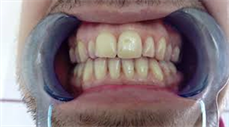
The principle of determining the total protein: The violet-blue hue of the resulting complex is proportional to the amount of total protein in this sample [11]. The experiment involved 55 adolescents aged 18 - 25 years who spent at the computer from 8 to 12 hours (target group of 55 adolescents) and from 3 to 5 hours (control group of 20 adolescents). The assessment of the oral cavity was carried out according to the plaque index (photo 1) in the Tureski modification [20]. The index is similar to the Quigley Hayne index, according to which each facial and lingual unrestored surface of all teeth (except third molars) is assigned a score from 0 to 5 on the following scale:
0 points: no plaque
1) Point—individual spots of plaque in the cervical region of the tooth.
2) Points—a thin continuous strip of plaques (up to 1 mm) in the cervical region of the tooth.
3) Points—a strip of plaques more than 1 mm wide, but covers less than 1/3 of the tooth crown.
4) Points—plaque covers more than 1/3, but less than 2/3 of the tooth crown.
5) Points—plaque covers 2/3 of the tooth crown and more.
The results were assessed as follows: Tureski Index − PI = total plaque score/2 × 2 = n, where n is the number of teeth (Picture 1).
To assess the dental status, the level of oral hygiene and the condition of periodontal tissues were determined. Oral hygiene was assessed according to the Fedorov-Volodkina hygiene index (HI), and the hygienic condition was assessed based on the Simplified Green-Vermillion index [8] [19], and the OHI-S index. The prevalence of inflammatory changes in periodontal tissues was assessed by the Parma modification of the papillary-marginal-alveolar index (PMA) [22] and the periodontal community index CPI (MMCI 1987).
The CPI index is used to assess the degree of damage to periodontal tissues. Examination of the oral cavity was carried out using a dental mirror, a dental probe and a periodontal probe (to measure periodontal pockets). According to the method of Fedorov-Volodkina [9], the hygienic state of the oral cavity was determined by staining the vestibular surface of teeth 43, 42, 41, 31, 32, 33 with Schiller-Pisarev, Lugol or other dyes.
The simplified hygiene index determined the level of oral hygiene by assessing the amount of plaque. Erythrosin tablets or iodine solution was used as a dye. 6 teeth were examined: 16, 11, 26, 31—from the vestibular surface, 36, 46—from the lingual surface.
CPI. The index was calculated using the following Formula (1):
I R = c i n (1)
where ci is the sum of the scores; n is the number of teeth.
All adolescents underwent a clinical examination. The condition of periodontal tissues was assessed according to the dental index: PMA.
In adolescents of the target and control groups, the levels of histamine 11-OCS, serotonin, norepinephrine, and dopamine in saliva were also determined, which was collected 1 hour after eating. The ELISA system test (“Immun Diagnostik”) was applied.
Oxidative stress measurement with photometric analytical system FRAS-5, dedicated solely to the global assessment of oxidative stress in biological systems
Picture 1. The Oral Index is determined by dividing the total score by the number of surfaces examined (maximum 2 × 2 × 14 = 56 surfaces).
by enabling the measurement of pro-oxidant status in plasma samples by means of d-ROMs (reactive oxygen metabolites), fast test and of anti-oxidant status in plasma by means of PAF (Plasma Antioxidant fragment) test, as well as measurement of OBRI (Oxidation balance risk index) and OSI (oxidation index).
The participation of adolescents in the study was agreed with the parents and ethical norms were observed.
3. Results and Discussion
According to Table 1, Figure 1, when assessing the condition of the periodontium, the dependence of changes associated with the time spent by adolescents at the computer was revealed. When questioning the dental status, the most common complaints were: the presence of an unpleasant taste and smell, the presence of tartar, partial absence of teeth, and bleeding. Hygienic condition deteriorated with age, before treatment, on visual examination, most patients showed gingival swelling, hyperemia, hypertrophy, retraction, cyanosis, bleeding.
Oxidative stress:
As is known from the literature [5] [15] [16] [17] [24], it is of great importance to study the dynamics of the metabolism of biogenic amines under stress, so we decided to study biogenic amines in the saliva of adolescents.
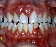
It is known that stress has a significant and not always positive effect on the activity of all organs and systems [2] [7] [17]. The study of the effect of stress induced by electromagnetic fields and their consequences on the body is an important prerequisite for determining the effect of EMF on periodontium in adolescents (Picture 2).
A 23-year-old patient complains of discomfort in the mouth, bleeding from a mechanical stimulus, mainly when brushing teeth (Gingival thickness ≥ 2.0 mm, height 5 - 6 mm).
Picture 2. Clinical picture.
Oxidative stress
Adolescents
D-ROMs Fast
PAT
OBRI
OSI REDOX
Control Experiment
258 ± 2.95
2081 ± 3.95
1.23 ± 0.2
111 ± 2.3
Experiment Control
403 ± 3.67 Is average (p < 0.05).
2763 ± 5.85 Average level
1.76 ± 0. High
122 ± 2.3 The body is on alert to protect itself
Adolescents who were at the computer for 12 hours with a simultaneous combination of several complaints were higher in the main group (p < 0.001) (Table 2, Figure 2). Bleed gums when brushing their teeth bothered 31% of the surveyed adolescents who were at the computer for more than 8 hours on the day of the examination. In the control group, they did not exceed 10% of those examined (p < 0.001). The presence of an unpleasant taste and smell from the mouth in the main group was noted in 27%, and in the control group −10% (p < 0.001) (Picture 3).
A 20-year-old patient has gingival hyperemia with a bluish tinge, a large number of subgingival and supragingival stones and plaques, the depth of the periodontal pocket is 4 - 5 mm. The gum is white, loosely adjacent to the tooth (Picture 4).
The most common complaints of the studied adolescents who were at the computer (percentage)
Complaints
Target group n = 55
Control group n = 20
Abs
%
Abs.
%
Unpleasant taste and smell
15
27
2
10
Bleeding
17
31
2
10
Tartar
12
22
-
-
Partial absence of teeth
11
20
1
5
Picture 3. Moderately severe chronic periodontitis with exacerbation.
Picture 4. A 25-year old patient complains of swollen gums, itching and buring, bad breath, bleeding from a slight mechanical stimulus, absence of teeth.
According to the given data, the percentage of adolescents with a mild degree of periodontitis who spent 8 and 10 hours at the computer is almost the same (p < 0.05), and significantly increases among adolescents with a 12-hour duration of being at the computer. In contrast to the studied group of adolescents with a mild degree of periodontitis, in the group of adolescents with an average degree of periodontitis, the time spent at the computer significantly affected the quantitative indicators. The percentage of adolescents studied by us with an average degree of periodontitis according to the time spent at the computer (8, 10 and 12 hours) increases significantly (Table 3, Figure 3, Picture 3).
According to Table 4, the severity of gingivitis according to the PMA index, (average values) in adolescents who were at the computer for more than 8 hours, is much higher than those of the control group. According to Table 4, the initial changes in periodontal tissues, according to the PMA index, are more aligned in the target group than in the control group and are directly dependent on the time spent at the computer. The dental status of adolescents of the target group according to the Russell index (PI) according to the time spent at computers is much higher in adolescents of the main group. It should be noted that the degree of gingival inflammation, assessed by the Russell indices, clearly confirms the changes assessed by the Fedorova-Volodkina index (Table 4).
We also studied saliva mineralization.
The study of salivary mineralization showed that there was a change in mineralization during both mild and moderate periodontitis, which indicates the development of an inflammatory process and tissue damage.
Clinical studies have shown that in patients with periodontitis there are some changes in oral fluid, which were manifested in the mineral metabolism Ca and P, as shown in (Table 5 and Table 6) as well as patients with moderate
The state of the oral cavity of adolescents spending time at the Laptop computer (% ratio)
Time
Number of people examined
Periodontitis of mild severity, %
Number of people examined
Periodontitis of moderate severity, %
8 h
15
27
11
20
10 h
16
29
18
33
12 h
24
44
26
47
n = 55
CPI, and PMA, Russell (PI), and Fedorov-Volodkina indices for adolescents who were at the computer
CPI index
CPI index
Target group n = 55
Control group n = 20
8 h
10 h
12 h
3 h
4 h
5 h
4.8 ± 0.2
6.9 ± 0.2
10.1 ± 1.02
2.0 ± 0.2
3.1 ± 0.2
3.9 ± 0.2
n = 15
n = 19
n = 21
n = 4
n = 7
n = 9
PMA index
PMA index
8 h
10 h
12 h
3 h
4 h
5 h
5 ± 0.1
7 ± 1.3
12.8 ± 2.1
1.1 ± 0.2
2.8 ± 0.2
3.9 ± 1.2
n = 15
n = 19
n = 21
n = 4
n = 7
n = 9
Russell (РI) index
Russell (РI) index
8 h
10 h
12 h
3 h
4 h
5 h
1.1 ± 0.1
1.2 ± 1.3
1.6 ± 2.1
0.1 ± 0.2
0.2 ± 0.2
0.4 ± 1.2
n = 15
n = 19
n = 21
n = 4
n = 7
n = 9
Fedorov-Volodkina index
Fedorov-Volodkina index
8 h
10 h
12 h
3 h
4 h
5 h
2.2 ± 0.1
2.5 ± 0.3
3.6 ± 0.1
1.1 ± 0.2
1.2 ± 0.2
1.4 ± 0.2
n = 15
n = 19
n = 21
n = 4
n = 7
n = 9
Shift in oral cavity mineralization at acidic pH and saliva mineralization (Mild severity)
Mild severity
Mild severity
Target group n = 55 Ca shift
Control group n = 20 Ca shift
8 h
10 h
12 h
3 h
4 h
5 h
2.30 ± 0.05
2.38 ± 0.2
2.45 ± 0.05
1.95 ± 0.02
2.10 ± 0.2
2.15 ± 0.02
n = 15
n = 19
n = 21
n = 4
n = 7
n = 9
P shift
P shift
8 h
10 h
12 h
3 h
4 h
5 h
0.18 ± 0.04
0.17 ± 0.03
0.16 ± 0.06
0.23 ± 0.02
0.21 ± 0.02
0.20 ± 0.02
n = 15
n = 19
n = 21
n = 4
n = 7
n = 9
pH acidic alkalinity
pH acidic alkalinity
8 h
10 h
12 h
3 h
4 h
5 h
pH-7.09
pH-7.1
pH-7.25
pH-7.0
pH-7.01
pH-7.02
n = 15
n = 19
n = 21
n = 4
n = 7
n = 9
Shift in oral cavity mineralization at acidic pH and saliva mineralization (Moderate severity)
Moderate severity
Moderate severity
Target group n = 55 Ca shift
Control group n = 20 Ca shift
8 h
10 h
12 h
3 h
4 h
5 h
2.25 ± 0.2
2.38 ± 0.2
2.49 ± 0.17
1.95 ± 0.02
2.10 ± 0.2
2.15 ± 0.02
n = 15
n = 19
n = 21
n = 4
n = 7
n = 9
P shift
P shift
8 h
10 h
12 h
3 h
4 h
5 h
0.16 ± 0.04
0.15 ± 0.03
0.14 ± 0.06
0.23 ± 0.02
0.19 ± 0.02
0.18 ± 0.02
n = 15
n = 19
n = 21
n = 4
n = 7
n = 9
pH acidic alkalinity
pH acidic alkalinity
8 h
10 h
12 h
3 h
4 h
5 h
pH-7.2
pH-7.3
pH-7.35
pH-7.08
pH-7.09
pH-7.1
n = 15
n = 19
n = 21
n = 4
n = 7
n = 9
periodontitis have some changes in Ca metabolism, namely calcium is elevated. If it was in control −1.95 ± 0.65, at light weight it increases −2.45 ± 0.05, while at medium weight it increases even more and becomes 2.49 ± 0.17.
A similar expression is observed for the P shift. In control, it is 0.23 ± 0.03, while in the mild form it is 0.16 ± 0.06, in the medium form 0.14 ± 0.01, which indicates the development of an inflammatory process.
So, the pH index (Table 6) at the initial level of 7.08 (7.09 - 7.1) increased to 7.2 (7.3 - 7.35). Such dynamics is explained by the natural process of alkalization of the oral fluid due to the release of CO2 [20].
Emphasizing the hygienic significance of the state of the oral cavity, we determined (Table 7) in the saliva of adolescents of the target and control groups 11-OCS histamine, serotonin, dopamine and norepinephrine, which play an important role not only in the functional state of the periodontium, but also in the processes of general adaptation of the whole organism [21] and neuroendocrine system to the electromagnetic effects of the computer. The content of these physiologically active substances in saliva and blood is interrelated [9] [11] [16].
About 0.05 to 3.0 mL of saliva is usually secreted per minute, which amounts to approximately 150.0 mL of saliva per day, it was collected (0.5 mL) in special polypropion vessels and stored at −200˚C. ELISA analysis was carried out according to the ELISA test system (Immun Diagnostik).
It appears in (Table 7) the levels of serotonin, histamine, norepinephrine and 11-OKS, powerful vasoconstrictors, remained consistently high throughout the experiment in adolescents of the target group, while dopamine levels remained consistently low and did not reach control after twelve hours of computer work, which indicates a deficiency of vasodilating factors in the systemic and organ circulation.
The above data can be predetermined [3] [12] [14] [15] [23] by the protective reaction of the body of adolescents (release of hormones of the adrenal cortex into the blood) to electromagnetic exposure during work at the computer for no
Hormonal content of saliva in adolescents of the target and control groups
Target group
Control group
n = 55
n = 20
Time spent at the computer
3 h
4 h
5 h
8 h
10 h
12 h
11-OCS histamine, Mcg/mL
0.095 ± 0.001
0.091 ± 0.002
0.094 ± 0.003
0.129 ± 0.002
0.078 ± 0.004
0.145 ± 0.004
Histamine, Mcg/mL
0.076 ± 0.001
0.074 ± 0.001
0.077 ± 0.001
0.126 ± 0.002
0.099 ± 0.013
0.125 ± 0.004
Serotonin, Mcg/mL
0.107 ± 0.001
0.109 ± 0.001
0.105 ± 0.001
0.157 ± 0.002
0.198 ± 0.002
0.165 ± 0.002
Norepinephrine, Mcg/mL
0.123 ± 0.001
0.121 ± 0.001
0.122 ± 0.001
0.137 ± 0.002
0.168 ± 0.002
0.195 ± 0.002
Dopamine, Mcg/mL
0.137 ± 0.001
0.139 ± 0.001
0.136 ± 0.001
0.077 ± 0.002
0.042 ± 0.002
0.065 ± 0.002
longer than 8 hours, while in adolescents, working much longer (from 10 to 12 hours). A tendency to an excess of vasoconstrictive amines and a deficiency of vasodilators is apparently formed, which clearly indicates a decrease in the body’s defense processes caused by prolonged exposure (more than 10 hours) to the electromagnetic field [21] [22] [25]. It leads to the development of oxidative stress in the body of a teenager; oxidative stress then affects the oral cavity and causes a number of problems that were discussed in the article.
4. Conclusions
1) Adolescents who spend more than 5 hours in an electromagnetic field cause a change in the OHI hygiene index with a Turecki modification.
2) Causes the severity of inflammatory and destructive changes in periodontal tissues according to Pi and their distribution Papillary-Marginal-Alveolar index PMA in Parma modification.
3) The conducted comparative study showed that in adolescents who worked 8-12 hours at the computer, periodontal changes occur much more often and are more pronounced than in adolescents who worked less than 5 hours.
4) In addition, the neurohormonal parameters of the saliva of adolescents who are at the computer for less than 5 hours practically do not change, but in adults who work longer than 8 hours the saliva data in the mouth changes while.
5) We cannot avoid the need to use the electromagnetic field, but the need for its judicious use in adults should be determined and this time should not exceed 5 - 6 hours a day.
Conflicts of Interest
The authors declare no conflicts of interest regarding the publication of this paper.
Cite this paper
Bakradze, T., Nikolaishvili, M., Gogiberidze, M., Galogre, A., Pkhaladze, M., Khimshiashvili, N. and Sakvarelidze, N. (2022) The State of the Oral Cavity of Adolescents Who Spend More than 3 Hours at the Computer. Journal of Biosciences and Medicines, 10, 99-113. https://doi.org/10.4236/jbm.2022.104010
ReferencesCalcabrini, C., Mancini, U., De Bellis, R., Diaz, A.R., Martinelli, M., Cucchiarini, L., et al. (2017) Effect of Extremely Low-Frequency Electromagnetic Fields on Antioxidant Activity in the Human Keratinocyte Cell Line NCTC 2544. Biotechnology and Applied Biochemistry, 63, 415-422.Deepa, T. and Thirrunavukkarasu, N. (2010) Saliva as a Potential Diagnostic Tool. Indian Journal of Medical Sciences, 64, 293-306.Dondoladze, K., Nikolaishvili, M., Museliani, T., Jikia, G. and Zurabashvili, D. (2018) The Impact of Electromagnetic Field on Conditioned Reflex Memory. NeuroQuantology, 16, 93-99. https://doi.org/10.14704/nq.2018.16.11.1777Dondoladze, K., Nikolaishvili, M., Museliani, T. and Jikia, G. (2018) Effect of Electromagnetic Field on Aggressive and Non-Aggressive Rats’ Memory. Systemic, Cellular and Molecular Mechanisms of Physiological Functions and Their Disorders Series, 43, 71-79.Gherardini, L., Ciuti, G., Tognarelli, S. and Cinti, C. (2014) Searching for the Perfect Wave: The Effect of Radiofrequency Electromagnetic Fields on Cells. International Journal of Molecular Sciences, 15, 5366-5387. https://doi.org/10.3390/ijms15045366Abaishvili, N., Margvelashvili, V., Taboridze, I. and Aladashvili, L. (2016) Determination of the Risk of Periodontal Disease in Georgian Student Populations. Journal of Scientific and Practical Medicine, 19, 11-14.Alimova, R.G. and Mahsudov, S.N. (2004) Modern Hygiene and Orthodontics. Dentistry (Tashkent), No. 1-2, 102-106.Kivrak, E., Yurt, K., Kaplan, A., Alkan, I. and Altun G. (2017) Effects of Electromagnetic Fields Exposure on the Antioxidant Defense System. Journal of Microscopy and Ultrastructure, 5, 167-176. https://doi.org/10.1016/j.jmau.2017.07.003Lomova, A.S., Prokhodnaya, V.A. and Bykov, I.M. (2016) Lactoferrin of Oral Fluid as a Marker of Dental Caries Activity in Pregnant Women. Medical Bulletin of the North Caucasus, No. 3, 431-434. https://doi.org/10.14300/mnnc.2016.00096Leontiev, V.K. and Ivanova, G.G. (2014) Methods for the Study of Oral Fluid and the State of Hard Tooth Tissues: Literature Review, Part 2. Institute of Dentistry, Moscow, No. 1, 96-97.Russell, A. (1956) A System of Classification and Scoring for Prevalence Surveys of Periodontal Disease. Journal of Dental Research, 35, 350-359. https://doi.org/10.1177/00220345560350030401Kushpal, S., Anup, N., Asif, Y., Shravani, G., Sonia, P. and Preeti, V. (2016) Effect of Electromagnetic Radiations from Mobile Phone Base Stations on General Health and Salivary Function. Journal of International Society of Preventive & Community Dentistry, 6, 54-59. https://doi.org/10.4103/2231-0762.175413Alattar, E., Elwasife, K., Radwan, E., Abu, W. and Abujam, M. (2018) An Experimental Investigation of the Impact of Electromagnetic Radiations Emitted from Mobile Phone on General Health, pH, Flow Rate and Electrolytes Concentrations of Saliva in Female Adults. International Journal of Biology, 11, 10-21. https://doi.org/10.5539/ijb.v11n1p10Lomiashvili, L.M., Sedelnikov, V.V., Pitaeva, A.N., Elendo, M.B., Vasilieva, N.A. (2015) Evaluation of the Influence of Electromagnetic Radiation of a Personal Computer on the State of the Oral Fluid of Operators (in Vitro Study). Institute of Dentistry, Tbilisi, No. 2, 58-60.Greene, J. and Vermillion, J. (1964) The Simplified Oral Hygiene Index. The Journal of the American Dental Association, 68, 7-13. https://doi.org/10.14219/jada.archive.1964.0034Lushchik, M.V., Bolotskikh, V.I., Grebennikova, I.V. and Tsvetikova, L.N. (2017) Assessment of Indicators of Oxidative Status in the Oral Fluid in Various Diseases. System Analysis and Management in Biomedical Systems, No. 1, 60-63.Iverieli, M., Abashidze, N., Gogishvili, Kh. and Janjalashvili, T. (2017) Periodontal Complex Diseases, Prevention and Management. Clinical Status Prevention and Management Standard, Tbilisi.Javaid, M.A., Ahmed, A.S., Durand, R. and Tran, S.D. (2016) Saliva as a Diagnostic Tool for Oral and Systemic Diseases. Journal of Oral Biology and Craniofacial Research, 6, 66-76. https://doi.org/10.1016/j.jobcr.2015.08.006Sirajuddin, S., Kripal, K., Chandrasekaran, K., Anuroopa, P. (2018) Effects of Electromagnetic Radiations from Mobile Phone on Gingiva in the Era of 4G LTE—An in Vivo Study in Rabbits. Dentistry, 8, Article No. 518. https://doi.org/10.4172/2161-1122.1000518Turesky, S., Gilmore, N.D. and Glickman, I. (1970) Reduced Plaque Formation by Chloromethyl Analogue of Victamine C. Journal of Periodontology, No. 41, 41-43.Mahdavi, S. (2014) Effects of Electromagnetic Radiation Exposure on Stress-Related Behaviors and Stress Hormones in Male Wistar Rats. Biomolecules & Therapeutics, 22, 570-576. https://doi.org/10.4062/biomolther.2014.054Ozguner, F., Altinbas, A., Ozaydin, M., Dogan, A., Vural, H., Kisioglu, A.N., et al. (2005) Mobile Phone-Induced Myocardial Oxidative Stress: Protection by a Novel Antioxidant Agent Caffeic Acid Phenethyl Ester. Toxicology and Industrial Health, 21, 223-230. https://doi.org/10.1191/0748233705th228oaKarolina-Urbanowicz, K.E., Martin Carreras-Presas, C. and Aro, K. (2017) Saliva Diagnostics—Current Views and Directions. Experimental Biology and Medicine, 242, 459-472. https://doi.org/10.1177/1535370216681550Kaufman, E. and Lamster, I.B. (2000) Analysis of Saliva for Periodontal Diagnosis: A Review. Journal of Clinical Periodontology, 27, 453-465. https://doi.org/10.1034/j.1600-051x.2000.027007453.xSilver, L., Smith, A., Jonson, C., Jilang, J., Anderson, M. and Gaine, L. (2019) Majorities Say Phones Are Good for Society, Even Amid Concerns about Their Impact on Children. Pew Research Center, 7, 35-49.