

Paper Menu >>
Journal Menu >>
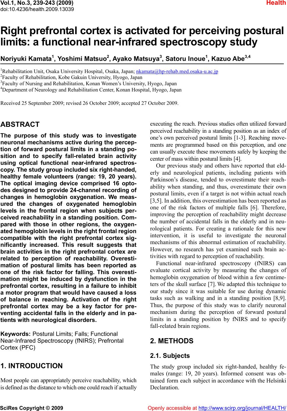 Vol.1, No.3, 239-2 doi:10.4236/health.2009.13039 SciRes Copyright © 2009 Openly accessible at http://www.scirp.org/journal/HEALTH/ 43 (2009) Health Right prefrontal cortex is activated for perceiving postural limits: a functional near-infrared spectroscopy study Noriyuki Kamata1, Yoshimi Matsuo2, Ayako Matsuya3, Satoru Inoue1, Kazuo Abe3,4 1Rehabilitation Unit, Osaka University Hospital, Osaka, Japan; [email protected] 2Faculty of Rehabilitation, Kobe Gakuin University, Hyogo, Japan 3Faculty of Nursing and Rehabilitation, Konan Women’s University, Hyogo, Japan 4Department of Neurology and Rehabilitation Center, Konan Hospital, Hyogo, Japan Received 25 September 2009; revised 26 October 2009; accepted 27 October 2009. ABSTRACT The purpose of this study was to investigate neuronal mechanisms active during the percep- tion of forward postural limits in a standing po- sition and to specify fall-related brain activity using optical functional near-infrared spectros- copy. The study group included six right-handed, healthy female volunteers (range: 19, 20 years). The optical imaging device comprised 16 opto- des designed to provide 24-channel recording of changes in hemoglobin oxygenation. We meas- ured the changes of oxygenated hemoglobin levels in the frontal region when subjects per- ceived reachability in a standing position. Com- pared with those in other regions, the oxygen- ated hemoglobin levels in the right frontal region compatible with the right prefrontal cortex sig- nificantly increased. This result suggests that brain activities in the right prefrontal cortex are related to perception of reachability. Overesti- mation of postural limits has been reported as one of the risk factor for falling. This overesti- mation might be induced by dysfunction in the prefrontal cortex, resulting in a failure to inhibit a motor program that would have caused a loss of balance in reaching. Activation of the right prefrontal cortex may be a key factor for pre- venting accidental falls in the elderly and in pa- tients with neurological disorders. Keywords: Postural Limits; Falls; Functional Near-Infrared Spectroscopy (fNIRS); Prefrontal Cortex (PFC) 1. INTRODUCTION Most people can appropriately perceive reachability, which is defined as the distance to which one could reach if actually executing the reach. Previous studies often utilized forward perceived reachability in a standing position as an index of one’s own perceived postural limits [1-3]. Reaching move- ments are programmed based on this perception, and one can usually execute these movements safely by keeping the center of mass within postural limits [4]. Our previous study and others have reported that eld- erly and neurological patients, including patients with Parkinson’s disease, tended to overestimate their reach- ability when standing, and thus, overestimate their own postural limits, even if a target is not within actual reach [3,5]. In addition, this overestimation has been reported as one of the risk factors of multiple falls [6]. Therefore, improving the perception of reachability might decrease the number of accidental falls in the elderly and in neu- rological patients. For creating a rationale for this new intervention, it is useful to investigate the neuronal mechanisms of this abnormal estimation of reachability. However, no research has yet examined such brain ac- tivities with regard to perception of reachability. Functional near-infrared spectroscopy (fNIRS) can evaluate cortical activity by measuring the changes of hemoglobin oxygenation of blood within a few centime- ters of the skull surface [7]. We adapted this technique to our study since it was suitable for use during dynamic tasks such as walking and in a standing position [8,9]. Thus, the purpose of this study was to clarify neuronal mechanism during the perception of forward postural limits in a standing position by fNIRS and to specify fall-related brain regions. 2. METHODS 2.1. Subjects The study group included six right-handed, healthy fe- males (range: 19, 20 years). Informed consent was ob- tained form each subject in accordance with the Helsinki Declaration. 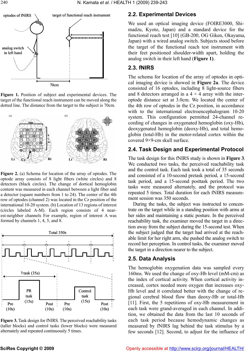 N. Kamata et al. / HEALTH 1 (2009) 239-243 SciRes Copyright © 2009 Openly accessible at http://www.scirp.org/journal/HEALTH/ 240 analog switch in left hand optodes of fNIRStarget of functional reach instrument 70cm analog switch in left hand optodes of fNIRStarget of functional reach instrument 70cm Figure 1. Position of subject and experimental devices. The target of the functional reach instrument can be moved along the dotted line. The distance from the target to the subject is 70cm. 9 9 c c m m 9cm 9cm Rt Lt 24 23 21 22 20 1819 1617 15 14 131112 75 8 64 3 10 9 2 1 Cz RtLt 24 23 21 22 20 1819 1617 15 14 13 1112 75 8 64 3 10 9 2 1 M C H B D G I F L E J A K (b)(a) 9 9 c c m m 9cm 9cm Rt Lt 24 23 21 22 20 1819 1617 15 14 131112 75 8 64 3 10 9 2 1 24 23 21 22 20 1819 1617 15 14 131112 75 8 64 3 10 9 2 1 Cz RtLt 24 23 21 22 20 1819 1617 15 14 13 1112 75 8 64 3 10 9 2 1 24 23 21 22 20 1819 1617 15 14 13 1112 75 8 64 3 10 9 2 1 M C H B D G I F L E J A KM C H B D G I F L E J A K (b)(a) Figure 2. (a) Schema for location of the array of optodes. The optode array consists of 8 light fibers (white circles) and 8 detectors (black circles). The change of dortical hemoglobin content was measured in each channel between a light fiber and a detector (square numbers from 1 to 24). The center of the 4th row of optodes (channel 2) was located in the Cz position of the international 10-20 system. (b) Location of 13 regions of interest (circles labeled A-M). Each region consists of 4 near- est-neighbor channels For example, region of interest A was formed by channels 1, 4, 5, and 8. 1task (35s) Pre (10s) PR task (15s) Post (10s) Pre (10s) Control task (15s) Post (10s) Total 350s 1task (35s) Pre (10s) PR task (15s) Post (10s) Pre (10s) Control task (15s) Post (10s) Total 350s Figure 3. Task design for fNIRS. The perceived reachability tasks (taller blocks) and control tasks (lower blocks) were measured alternately and repeated continuously 5 times. 2.2. Experimental Devices We used an optical imaging device (FOIRE3000, Shi- madzu, Kyoto, Japan) and a standard device for the functional reach test [10] (GB-200, OG Giken, Okayama, Japan) with a wired analog switch. Subjects stood before the target of the functional reach test instrument with their feet positioned shoulder-width apart, holding the analog switch in their left hand (Figure 1). 2.3. fNIRS The schema for location of the array of optodes in opti- cal imaging device is showed in Figure 2a. The device consisted of 16 optodes, including 8 light-source fibers and 8 detectors arranged in a 4 × 4 array with the inter- optode distance set at 3.0cm. We located the center of the 4th row of optodes in the Cz position, in accordance with to the international electroencephalogram 10-20 system. This configuration permitted 24-channel re- cording of changes in oxygenated hemoglobin (oxy-Hb), deoxygenated hemoglobin (deoxy-Hb), and total hemo- globin (total-Hb) in the motor-related cortex within the covered 9×9-cm skull surface. 2.4. Task Design and Experimental Protocol The task design for this fNIRS study is shown in Figure 3. We conducted two tasks, the perceived reachability task and the control task. Each task took a total of 35 seconds and consisted of a 10-second pretask period, a 15-second task period, and a 15-second posttask period. The two tasks were measured alternately, and the protocol was repeated 5 times. Total duration for each fNIRS measure- ment session was 350 seconds. During the tasks, the subject was instructed to concen- trate on the target while in a standing position with arms at her sides and maintaining a static posture. In the perceived reachability task, the examiner moved the target in a direc- tion away from the subject during the 15-second test. When the subject judged that the target had arrived at the reach- able limit for her right arm, she pushed the analog switch to record her perception. In control tasks, the examiner moved the target in a direction nearer to the subject. 2.5. Data Analysis The hemoglobin oxygenation data was sampled every 160ms. We used the change of oxy-Hb level (mM-cm) as the index of cortical activity. When cortical activity in- creased, cortex needed more oxygen that increases oxy- Hb level and it correlated better with the change of re- gional cerebral blood flow than deoxy-Hb or total-Hb [11]. First, the 5 repetitions of oxy-Hb measurement in each task were grand-averaged in each channel. In addi- tion, we obtained the data from the last 10 seconds of each task period because hemodynamic changes as measured by fNIRS lag behind the task stimulus by a few seconds [12]. Second, to adjust for the influence of 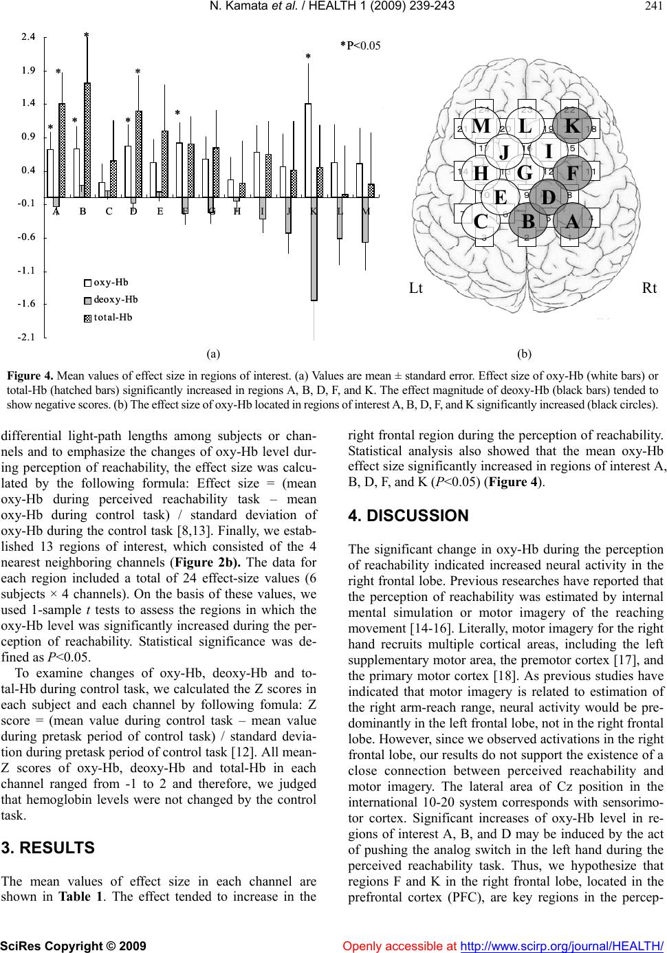 N. Kamata et al. / HEALTH 1 (2009) 239-243 SciRes Copyright © 2009 Openly accessible at http://www.scirp.org/journal/HEALTH/ 241 * 24 23 21 22 20 1819 1617 15 14 13 1112 75 8 64 3 10 9 2 1 M C HG I F L E J K RtLt B D A -2.1 -1.6 -1.1 -0.6 -0.1 0.4 0.9 1.4 1.9 2.4 ABCDEFGHI JKLM oxy-Hb deoxy-Hb total-Hb * * *** *P<0.05 ** * * 24 23 21 22 20 1819 1617 15 14 13 1112 75 8 64 3 10 9 2 1 M C HG I F L E J K RtLt B D A * 24 23 21 22 20 1819 1617 15 14 13 1112 75 8 64 3 10 9 2 1 24 23 21 22 20 1819 1617 15 14 13 1112 75 8 64 3 10 9 2 1 M C HG I F L E J K RtLt B D A -2.1 -1.6 -1.1 -0.6 -0.1 0.4 0.9 1.4 1.9 2.4 ABCDEFGHI JKLM oxy-Hb deoxy-Hb total-Hb * * *** *P<0.05 ** * (a) (b) Figure 4. Mean values of effect size in regions of interest. (a) Values are mean ± standard error. Effect size of oxy-Hb (white bars) or total-Hb (hatched bars) significantly increased in regions A, B, D, F, and K. The effect magnitude of deoxy-Hb (black bars) tended to show negative scores. (b) The effect size of oxy-Hb located in regions of interest A, B, D, F, and K significantly increased (black circles). differential light-path lengths among subjects or chan- nels and to emphasize the changes of oxy-Hb level dur- ing perception of reachability, the effect size was calcu- lated by the following formula: Effect size = (mean oxy-Hb during perceived reachability task – mean oxy-Hb during control task) / standard deviation of oxy-Hb during the control task [8,13]. Finally, we estab- lished 13 regions of interest, which consisted of the 4 nearest neighboring channels (Figure 2b). The data for each region included a total of 24 effect-size values (6 subjects × 4 channels). On the basis of these values, we used 1-sample t tests to assess the regions in which the oxy-Hb level was significantly increased during the per- ception of reachability. Statistical significance was de- fined as P<0.05. To examine changes of oxy-Hb, deoxy-Hb and to- tal-Hb during control task, we calculated the Z scores in each subject and each channel by following fomula: Z score = (mean value during control task – mean value during pretask period of control task) / standard devia- tion during pretask period of control task [12]. All mean- Z scores of oxy-Hb, deoxy-Hb and total-Hb in each channel ranged from -1 to 2 and therefore, we judged that hemoglobin levels were not changed by the control task. 3. RESULTS The mean values of effect size in each channel are shown in Table 1. The effect tended to increase in the right frontal region during the perception of reachability. Statistical analysis also showed that the mean oxy-Hb effect size significantly increased in regions of interest A, B, D, F, and K (P<0.05) (Figure 4). 4. DISCUSSION The significant change in oxy-Hb during the perception of reachability indicated increased neural activity in the right frontal lobe. Previous researches have reported that the perception of reachability was estimated by internal mental simulation or motor imagery of the reaching movement [14-16]. Literally, motor imagery for the right hand recruits multiple cortical areas, including the left supplementary motor area, the premotor cortex [17], and the primary motor cortex [18]. As previous studies have indicated that motor imagery is related to estimation of the right arm-reach range, neural activity would be pre- dominantly in the left frontal lobe, not in the right frontal lobe. However, since we observed activations in the right frontal lobe, our results do not support the existence of a close connection between perceived reachability and motor imagery. The lateral area of Cz position in the international 10-20 system corresponds with sensorimo- tor cortex. Significant increases of oxy-Hb level in re- gions of interest A, B, and D may be induced by the act of pushing the analog switch in the left hand during the perceived reachability task. Thus, we hypothesize that regions F and K in the right frontal lobe, located in the prefrontal cortex (PFC), are key regions in the percep- 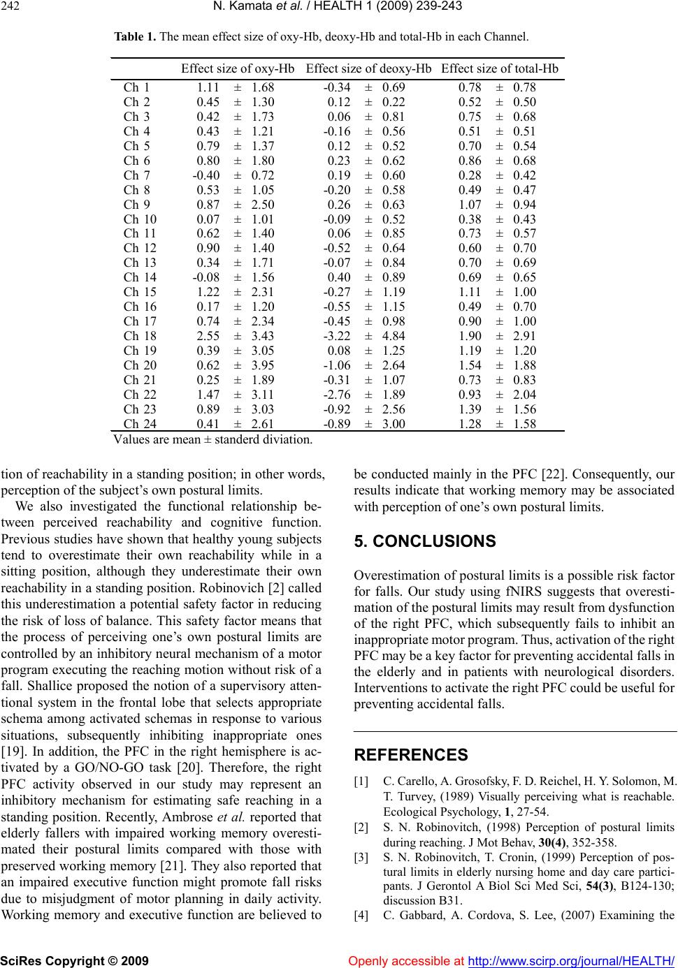 N. Kamata et al. / HEALTH 1 (2009) 239-243 SciRes Copyright © 2009 Openly accessible at http://www.scirp.org/journal/HEALTH/ 242 Table 1. The mean effect size of oxy-Hb, deoxy-Hb and total-Hb in each Channel. Ch11.11± 1.68-0.34± 0.690.78± 0.78 Ch20.45± 1.300.12± 0.220.52± 0.50 Ch30.42± 1.730.06± 0.810.75± 0.68 Ch40.43± 1.21-0.16± 0.560.51± 0.51 Ch50.79± 1.370.12± 0.520.70± 0.54 Ch60.80± 1.800.23± 0.620.86± 0.68 Ch7-0.40± 0.720.19± 0.600.28± 0.42 Ch80.53± 1.05-0.20± 0.580.49± 0.47 Ch90.87± 2.500.26± 0.631.07± 0.94 Ch100.07± 1.01-0.09± 0.520.38± 0.43 Ch110.62± 1.400.06± 0.850.73± 0.57 Ch120.90± 1.40-0.52± 0.640.60± 0.70 Ch130.34± 1.71-0.07± 0.840.70± 0.69 Ch14-0.08± 1.560.40± 0.890.69± 0.65 Ch151.22± 2.31-0.27± 1.191.11± 1.00 Ch160.17± 1.20-0.55± 1.150.49± 0.70 Ch170.74± 2.34-0.45± 0.980.90± 1.00 Ch182.55± 3.43-3.22± 4.841.90± 2.91 Ch190.39± 3.050.08± 1.251.19± 1.20 Ch200.62± 3.95-1.06± 2.641.54± 1.88 Ch210.25± 1.89-0.31± 1.070.73± 0.83 Ch221.47± 3.11-2.76± 1.890.93± 2.04 Ch230.89± 3.03-0.92± 2.561.39± 1.56 Ch240.41± 2.61-0.89± 3.001.28± 1.58 Effect size of oxy-HbEffect size of deoxy-HbEffect size of total-Hb Values are mean ± standerd diviation. tion of reachability in a standing position; in other words, perception of the subject’s own postural limits. We also investigated the functional relationship be- tween perceived reachability and cognitive function. Previous studies have shown that healthy young subjects tend to overestimate their own reachability while in a sitting position, although they underestimate their own reachability in a standing position. Robinovich [2] called this underestimation a potential safety factor in reducing the risk of loss of balance. This safety factor means that the process of perceiving one’s own postural limits are controlled by an inhibitory neural mechanism of a motor program executing the reaching motion without risk of a fall. Shallice proposed the notion of a supervisory atten- tional system in the frontal lobe that selects appropriate schema among activated schemas in response to various situations, subsequently inhibiting inappropriate ones [19]. In addition, the PFC in the right hemisphere is ac- tivated by a GO/NO-GO task [20]. Therefore, the right PFC activity observed in our study may represent an inhibitory mechanism for estimating safe reaching in a standing position. Recently, Ambrose et al. reported that elderly fallers with impaired working memory overesti- mated their postural limits compared with those with preserved working memory [21]. They also reported that an impaired executive function might promote fall risks due to misjudgment of motor planning in daily activity. Working memory and executive function are believed to be conducted mainly in the PFC [22]. Consequently, our results indicate that working memory may be associated with perception of one’s own postural limits. 5. CONCLUSIONS Overestimation of postural limits is a possible risk factor for falls. Our study using fNIRS suggests that overesti- mation of the postural limits may result from dysfunction of the right PFC, which subsequently fails to inhibit an inappropriate motor program. Thus, activation of the right PFC may be a key factor for preventing accidental falls in the elderly and in patients with neurological disorders. Interventions to activate the right PFC could be useful for preventing accidental falls. REFERENCES [1] C. Carello, A. Grosofsky, F. D. Reichel, H. Y. Solomon, M. T. Turvey, (1989) Visually perceiving what is reachable. Ecological Psychology, 1, 27-54. [2] S. N. Robinovitch, (1998) Perception of postural limits during reaching. J Mot Behav, 30(4), 352-358. [3] S. N. Robinovitch, T. Cronin, (1999) Perception of pos- tural limits in elderly nursing home and day care partici- pants. J Gerontol A Biol Sci Med Sci, 54(3), B124-130; discussion B31. [4] C. Gabbard, A. Cordova, S. Lee, (2007) Examining the  N. Kamata et al. / HEALTH 1 (2009) 239-243 SciRes Copyright © 2009 Openly accessible at http://www.scirp.org/journal/HEALTH/ 243 243 effects of postural constraints on estimating reach. J Mot Behav, 39(4), 242-246. [5] N. Kamata, Y. Matsuo, T. Yoneda, H. Shinohara, S. Inoue, K. Abe, (2007) Overestimation of stability limits leads to a high frequency of falls in patients with Parkinson’s disease. Clin Rehabil, 21(4), 357-361. [6] Y. Okada, K. Takatori, K. Ikuno, K. Tsuruta, K. Tokuhisa, K. Nagino, et al., (2008) Perception of postural limits and falls in community-dwelling elderly people. International Journal on Disability and Human Development, 7(1), 73-80. [7] I. Miyai, H. C. Tanabe, I. Sase, H. Eda, I. Oda, I. Konishi, et al., (2001) Cortical mapping of gait in humans: A near-in- frared spectroscopic topography study. Neuroi- mage, 14(5), 1186-1192. [8] M. Suzuki, I. Miyai, T. Ono, K. Kubota, (2008) Activities in the frontal cortex and gait performance are modulated by preparation: An fNIRS study. Neuroimage, 39(2), 600-607. [9] M. Mihara, I. Miyai, M. Hatakenaka, K. Kubota, S. Sa- koda, (2008) Role of the prefrontal cortex in human bal- ance control. Neuroimage, 43(2), 329-336. [10] P. W. Duncan, S. Studenski, J. Chandler, B. Prescott, (1992) Functional reach: Predictive validity in a sample of elderly male veterans. J Gerontol, 47(3), M93-98. [11] Y. Hoshi, N. Kobayashi, M. Tamura, (2001) Interpretation of near-infrared spectroscopy signals: A study with a newly developed perfused rat brain model. J Appl Physiol 90(5), 1657-1662. [12] Y. Otsuka, E. Nakato, S. Kanazawa, M. K. Yamaguchi, S. Watanabe, R. Kakigi, (2007) Neural activation to upright and inverted faces in infants measured by near-infrared spectroscopy. Neuroimage, 34(1), 399-406. [13] M. L. Schroeter, S. Zysset, F. Kruggel, D. Y. von Cramon, (2003) Age dependency of the hemodynamic response as measured by functional near-infrared spectroscopy. Neuroimage, 19(3), 555-564. [14] M. H. Fischer, (2003) Can we correctly perceive the reaching range of others? Br J Psychol, 94(Pt 4), 487-500. [15] M. H. Fischer, (2005) Perceived reachability: The roles of handedness and hemifield. Exp Brain Res, 160(3), 283- 289. [16] C. Lamm, M. H. Fischer, J. Decety, C. Lamm, (2007) Predicting the actions of others taps into one’s own somatosensory representations—a functional MRI study. Neuropsychologia, 45(11), 2480-2491. [17] P. E. Roland, B. Larsen, N. A. Lassen, E. Skinhoj, (1980) Supplementary motor area and other cortical areas in or- ganization of voluntary movements in man. J Neuro- physiol, 43(1), 118-136. [18] C. A. Porro, M. P. Francescato, V. Cettolo, M. E. Diamond, P. Baraldi, C. Zuiani, et al., (1996) Primary motor and sensory cortex activation during motor performance and motor imagery: A functional magnetic resonance imaging study. J Neurosci, 16(23), 7688-7698. [19] T. Shallice, (1982) Specific impairments of planning. Philos Trans R Soc Lond B Biol Sci, 298(1089), 199-209. [20] R. Kawashima, K. Satoh, H. Itoh, S. Ono, S. Furumoto, R. Gotoh, et al., (1996) Functional anatomy of GO/NO-GO discrimination and response selection—a PET study in man. Brain Res, 728(1), 79-89. [21] T. Liu-Ambrose, Y. Ahamed, P. Graf , F. Feldman, S. N. Robinovitch, (2008) Older fallers with poor working memory overestimate their postural limits. Arch Phys Med Rehabil, 89(7), 1335-1340. [22] M. D’Esposito, J. A. Detre, D. C. Alsop, R. K. Shin, S. Atlas, M. Grossman, (1995) The neural basis of the central executive system of working memory. Nature, 378(6554), 279-81. |