

Paper Menu >>
Journal Menu >>
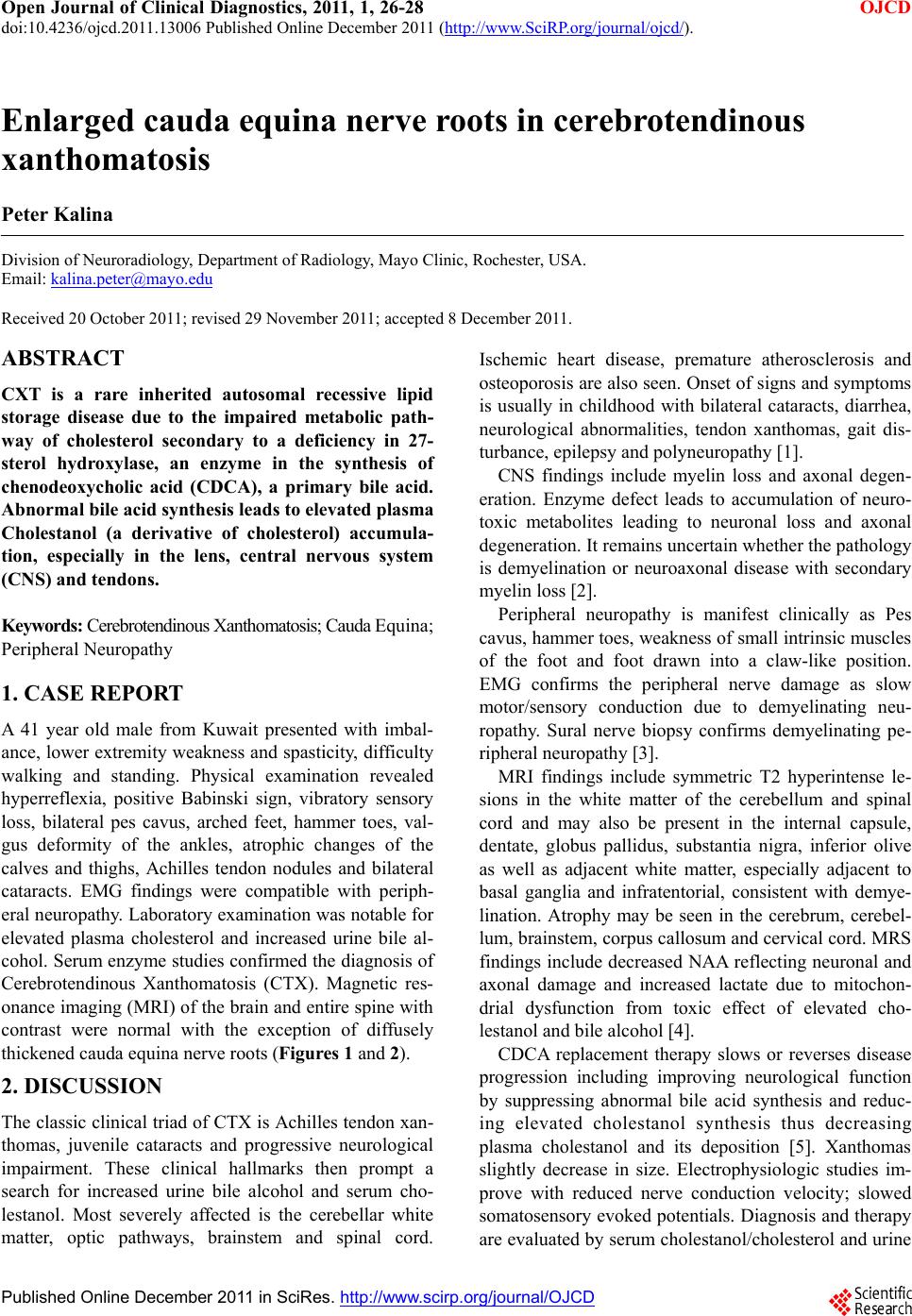 Open Journal of Clinical Diagnostics, 2011, 1, 26-28 OJCD doi:10.4236/ojcd.2011.13006 Published Online December 2011 (http://www.SciRP.org/journal/ojcd/). Published Online December 2011 in SciRes. http://www.scirp.org/journal/OJCD Enlarged cauda equina nerve roots in cerebrotendinous xanthomatosis Peter Kalina Division of Neuroradiology, Department of Radiology, Mayo Clinic, Rochester, USA. Email: [email protected] Received 20 October 2011; revised 29 November 2011; accepted 8 December 2011. ABSTRACT CXT is a rare inherited autosomal recessive lipid storage disease due to the impaired metabolic path- way of cholesterol secondary to a deficiency in 27- sterol hydroxylase, an enzyme in the synthesis of chenodeoxycholic acid (CDCA), a primary bile acid. Abnormal bile acid synthesis leads to elevated plasma Cholestanol (a derivative of cholesterol) accumula- tion, especially in the lens, central nervous system (CNS) and tend ons. Keywords: Cerebrotendinous X anthomat osis; Cauda Equina; Peripheral Neuropathy 1. CASE REPORT A 41 year old male from Kuwait presented with imbal- ance, lower extremity weakness and spasticity, difficulty walking and standing. Physical examination revealed hyperreflexia, positive Babinski sign, vibratory sensory loss, bilateral pes cavus, arched feet, hammer toes, val- gus deformity of the ankles, atrophic changes of the calves and thighs, Achilles tendon nodules and bilateral cataracts. EMG findings were compatible with periph- eral neuropathy. Laboratory examination was notable for elevated plasma cholesterol and increased urine bile al- cohol. Serum enzyme studies confirmed the diagnosis of Cerebrotendinous Xanthomatosis (CTX). Magnetic res- onance imaging (MRI) of the brain and entire spin e with contrast were normal with the exception of diffusely thickened caud a equi na ne rv e roots (Figures 1 and 2). 2. DISCUSSION The classic clinical triad of CTX is Achilles tendon xan- thomas, juvenile cataracts and progressive neurological impairment. These clinical hallmarks then prompt a search for increased urine bile alcohol and serum cho- lestanol. Most severely affected is the cerebellar white matter, optic pathways, brainstem and spinal cord. Ischemic heart disease, premature atherosclerosis and osteoporosis are also seen. Onset of signs and symptoms is usually in childhood with bilateral cataracts, diarrhea, neurological abnormalities, tendon xanthomas, gait dis- turbance, epilepsy and polyneuropathy [1]. CNS findings include myelin loss and axonal degen- eration. Enzyme defect leads to accumulation of neuro- toxic metabolites leading to neuronal loss and axonal degeneration. It remains uncertain whether the pathology is demyelination or neuroaxonal disease with secondary myelin loss [2]. Peripheral neuropathy is manifest clinically as Pes cavus, hammer toes, weakness of small intrinsic muscles of the foot and foot drawn into a claw-like position. EMG confirms the peripheral nerve damage as slow motor/sensory conduction due to demyelinating neu- ropathy. Sural nerve biopsy confirms demyelinating pe- ripheral neuropathy [3]. MRI findings include symmetric T2 hyperintense le- sions in the white matter of the cerebellum and spinal cord and may also be present in the internal capsule, dentate, globus pallidus, substantia nigra, inferior olive as well as adjacent white matter, especially adjacent to basal ganglia and infratentorial, consistent with demye- lination. Atrophy may be seen in the cerebrum, cerebel- lum, brainstem, corpus callosum and cervical cord. MRS findings include decreased NAA reflecting neuronal and axonal damage and increased lactate due to mitochon- drial dysfunction from toxic effect of elevated cho- lestanol and bile alcohol [4]. CDCA replacement therapy slows or reverses disease progression including improving neurological function by suppressing abnormal bile acid synthesis and reduc- ing elevated cholestanol synthesis thus decreasing plasma cholestanol and its deposition [5]. Xanthomas slightly decrease in size. Electrophysiologic studies im- prove with reduced nerve conduction velocity; slowed somatosensory evoked potentials. Diag nosis and therapy are evaluated by serum cholestanol/cholesterol and ur ine 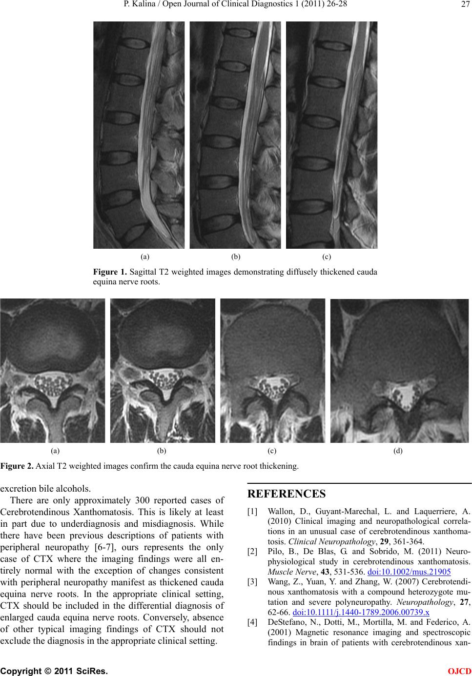 P. Kalina / Open Journal of Clinical Diagnostics 1 (2011) 26-28 27 (a) (b) (c) Figure 1. Sagittal T2 weighted images demonstrating diffusely thickened cauda equina nerve roots. (a) (b) (c) (d) Figure 2. Axial T2 weighted images confirm the cauda equina nerve root thickening. excretion bile alcohols. There are only approximately 300 reported cases of Cerebrotendinous Xanthomatosis. This is likely at least in part due to underdiagnosis and misdiagnosis. While there have been previous descriptions of patients with peripheral neuropathy [6-7], ours represents the only case of CTX where the imaging findings were all en- tirely normal with the exception of changes consistent with peripheral neuropathy manifest as thickened cauda equina nerve roots. In the appropriate clinical setting, CTX should be included in the differential diagnosis of enlarged cauda equina nerve roots. Conversely, absence of other typical imaging findings of CTX should not exclude the diagnosis in the appropriate clinical setting. REFERENCES [1] Wallon, D., Guyant-Marechal, L. and Laquerriere, A. (2010) Clinical imaging and neuropathological correla- tions in an unusual case of cerebrotendinous xanthoma- tosis. Clinical Neuropathology, 29, 361-364. [2] Pilo, B., De Blas, G. and Sobrido, M. (2011) Neuro- physiological study in cerebrotendinous xanthomatosis. Muscle Nerve, 43, 531-536. doi:10.1002/mus.21905 [3] Wang, Z., Yuan, Y. and Zhang, W. (2007) Cerebrotendi- nous xanthomatosis with a compound heterozygote mu- tation and severe polyneuropathy. Neuropathology, 27, 62-66. doi:10.1111/j.1440-1789.2006.00739.x [4] DeStefano, N., Dotti, M., Mortilla, M. and Federico, A. (2001) Magnetic resonance imaging and spectroscopic findings in brain of patients with cerebrotendinous xan- C opyright © 2011 SciRes. OJCD  P. Kalina / Open Journal of Clinical Diagnostics 1 (2011) 26-28 28 thomatosis. Brain, 124, 121-131. doi:10.1093/brain/124.1.121 [5] Federico, A. and Dotti, M. (2003) Cerebrotendinous xan- thomatosis: Clinical manifestations, diagnostic criteria, pathogenesis and therapy. Journal of Child Neurology, 18, 633-638. doi:10.1177/08830738030180091001 [6] Rafiq, M., Sharrack, N., Shaw, P. and Hadjivassiliou, M. (2011) A neurological rarity not to be missed: Cerebro- tendinous xanthomatosis. Practice Neurology, 11 , 296-300. doi:10.1136/practneurol-2011-000003 [7] Chen, S., Tsai, N., Chang, C., et al. (2011) Neuromuscu- lar abnormality and autonomic dysfunction in patients with cerebrotendinous xanthomatosis. Bio Medical Cen- tral Neurology, 11 , 63-72. C opyright © 2011 SciRes. OJCD |