

Paper Menu >>
Journal Menu >>
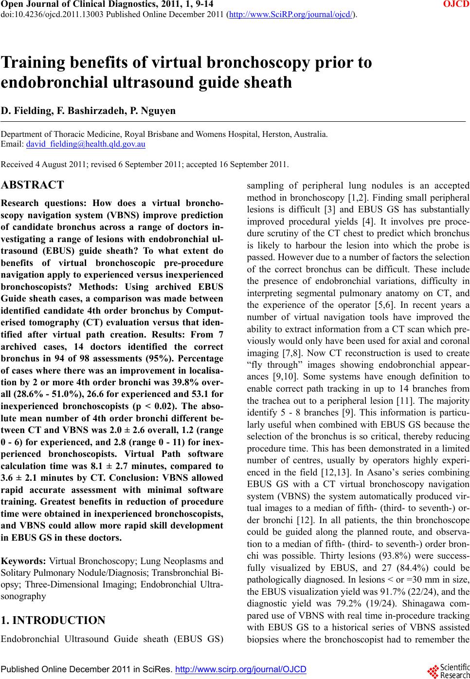 Open Journal of Clinical Diagnostics, 2011, 1, 9-14 doi:10.4236/ojcd.2011.13003 Published Online December 2011 (http://www.SciRP.org/journal/ojcd/ OJCD ). Published Online December 2011 in SciRes. http://www.scirp.org/journal/OJCD Training benefits of virtual bronchoscopy prior to endobronchial ultrasound guide sheath D. Fielding, F. Bashirzadeh, P. Nguyen Depart ment of Thoracic Medicine , Royal Brisbane an d Womens Hospital, Herst o n, Australia. Email: [email protected] Received 4 August 2011; revised 6 September 2011; accepted 16 September 2011. ABSTRACT Research questions: How does a virtual broncho- scopy navigation system (VBNS) improve prediction of candidate bronchus across a range of doctors in- vestigating a range of lesions with endobronchial ul- trasound (EBUS) guide sheath? To what extent do benefits of virtual bronchoscopic pre-procedure navigation apply to experienced versus inexperienced bronchoscopists? Methods: Using archived EBUS Guide sheath cases, a comparison was made between identified candidate 4th order bronchus by Comput- erised tomography (CT) evaluation versus that iden- tified after virtual path creation. Results: From 7 archived cases, 14 doctors identified the correct bronchus in 94 of 98 assessments (95%). Percentage of cases where there was an improvement in localisa- tion by 2 or more 4th order bronchi was 39.8% over- all (28.6% - 51.0%), 26.6 for experienced and 53.1 for inexperienced bronchoscopists (p < 0.02). The abso- lute mean number of 4th order bronchi different be- tween CT and VBNS was 2.0 ± 2.6 overall, 1.2 (range 0 - 6) for experienced, and 2.8 (range 0 - 11) for inex- perienced bronchoscopists. Virtual Path software calculation time was 8.1 ± 2.7 minutes, compared to 3.6 ± 2.1 minutes by CT. Conclusion: VBNS allowed rapid accurate assessment with minimal software training. Greatest benefits in reduction of procedure time were obtained in inexperienced bronchoscopists, and VBNS could allow more rapid skill development in EBUS GS in these doctors. Keywords: Virtual Bronchoscopy; Lung Neoplasms and Solitary Pulmonary Nodule/Diagnosis; Transbronchial Bi- opsy; Three-Dimensional Imaging; Endobronchial Ultra- sonography 1. INTRODUCTION Endobronchial Ultrasound Guide sheath (EBUS GS) sampling of peripheral lung nodules is an accepted method in bronchoscopy [1,2]. Finding small peripheral lesions is difficult [3] and EBUS GS has substantially improved procedural yields [4]. It involves pre proce- dure scrutiny of the CT chest to predict which bronchus is likely to harbour the lesion into which the probe is passed. However due to a number of factors the selection of the correct bronchus can be difficult. These include the presence of endobronchial variations, difficulty in interpreting segmental pulmonary anatomy on CT, and the experience of the operator [5,6]. In recent years a number of virtual navigation tools have improved the ability to extract info rmation from a CT scan which pre- viously would only have been used for axial and coronal imaging [7,8]. Now CT reconstruction is used to create “fly through” images showing endobronchial appear- ances [9,10]. Some systems have enough definition to enable correct path tracking in up to 14 branches from the trachea out to a peripheral lesion [11]. The majority identify 5 - 8 branches [9]. This information is particu- larly useful when combined with EBUS GS because the selection of the bronchus is so critical, thereby reducing procedure time. This has been demonstrated in a limited number of centres, usually by operators highly experi- enced in the field [12,13]. In Asano’s series combining EBUS GS with a CT virtual bronchoscopy navigation system (VBNS) the system automatically produced vir- tual images to a median of fifth- (third- to seventh-) or- der bronchi [12]. In all patients, the thin bronchoscope could be guided along the planned route, and observa- tion to a median of fifth- (third- to seventh-) order bron- chi was possible. Thirty lesions (93.8%) were success- fully visualized by EBUS, and 27 (84.4%) could be pathologically diagnosed. In lesions < or =30 mm in size, the EBUS visualization yield was 91.7% (22/24), and the diagnostic yield was 79.2% (19/24). Shinagawa com- pared use of VBNS with real time in-procedure tracking with EBUS GS to a historical series of VBNS assisted biopsies where the bronchoscopist had to remember the 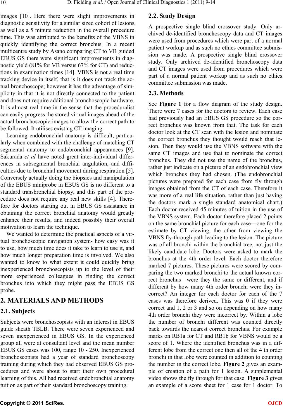 D. Fielding et al. / Open Journal of Clinical Diagnostics 1 (2011) 9-14 10 images [10]. Here there were slight improvements in diagnostic sensitivity for a similar sized cohort of lesions, as well as a 5 minute reduction in the overall procedure time. This was attributed to the benefits of the VBNS in quickly identifying the correct bronchus. In a recent multicentre study by Asan o comparing CT to VB guided EBUS GS there were significant improvements in diag- nostic yield (81% for VB versus 67% for CT) and reduc- tions in examination times [14]. VBNS is not a real time tracking device in itself, that is it does not track the ac- tual bronchoscope; however it has the advantage of sim- plicity in that it is not directly connected to the patient and does not require additional bronch oscopic hardware. It is almost real time in the sense that the proceduralist can easily progress the stored virtual images ahead of the actual bronchoscopic images to allow the correct path to be followed. It utilises existing CT imaging. Learning endobronchial anatomy is difficult, particu- larly when combined with the challeng e of matching CT segmental anatomy to endobronchial appearances [9]. Sakurada et al have noted great inter-individual differ- ences in subsegmental bronchial angulation, and diffi- culties due to bronchial movement during respiration [5]. Conversely actually doing the biopsies and manipulation of the EBUS miniprobe in EBUS GS is no different to a standard transbronchial biopsy, and this part of the pro- cedure does not require any real new skills [4]. There- fore for doctors starting out in EBUS GS assistance in obtaining the correct bronchial anatomy would greatly enhance their results, and indeed possibly their overall motivation to learn the technique. We wanted to determine the practical aspects of a vir- tual bronchoscopic navigation system- how easy was it to use, how much time does it take to learn to use it, and how much longer preparation time is involved. We also wanted to know to what extent it could quickly bring inexperienced bronchoscopists up to the level of their more experienced colleagues in finding the correct bronchus into which they might pass the EBUS GS probe. 2. MATERIALS AND METHODS 2.1. Subjects Subjects were bronchoscopists with an interest in EBUS guide sheath TBLB. There were seven experienced and seven inexperienced in EBUS GS. In the experienced group all were at consultant level and the mean number EBUS GS cases was 100, range 10 - 250. Inexperienced bronchoscopists had a year of standard bronchoscopy training during which they had observed EBUS GS pro- cedures and were about to start their own procedural learning of this. All had received endobronchial anatomy tuition as part of their standard bronchoscopy training. 2.2. Study Design A prospective single blind crossover study. Only ar- chived de-identified bronchoscopy data and CT images were used from procedures which were part of a normal patient workup and as such no ethics committee submis- sion was made. A prospective single blind crossover study. Only archived de-identified bronchoscopy data and CT images were used from procedures which were part of a normal patient workup and as such no ethics committee submission was made. 2.3. Methods See Figure 1 for a flow diagram of the study design. There were 7 cases for the doctors to review. Each case had previously had an EBUS GS procedure so the cor- rect bronchus was known from that. The task for each doctor look at the CT scan with the lesion and nominate the correct bronchus they thought would reach that le- sion. Then they would use the VBNS software with the same CT images and use that to nominate the correct bronchus. They did not use the name of the bronchus, rather just indicate on a picture of an endobronchial view which bronchus they had chosen. (The endobronchial pictures were prepared for each case from fly through images obtained from the CT of each case. Therefore it was more of a real life situation, rather than just having the doctors mark a single standard anatomical chart.) Each doctor received 45 minutes of tuition in the use of the VBNS system. Each doctor therefore placed 2 points on the same bronchial picture for each case—one for the estimate by CT viewing, the other from viewing the VBNS fly-through path leading to th e lesion. Th e pictur e was of all bronchi within the bronchial tree, not just the likely candidate lobe. Doctors were asked to mark the bronchus at the 4th order level. Each doctor therefore marked 7 pictures. These pictures were scored by com- paring the two marked bronchi to the actual known cor- rect bronchus—were they the same or different, and if different by how many 4th order bronchi were they in- correct? An integer for each doctor for each of the 7 cases was therefore derived. This was 0 if they were correct and 1, 2 or 3 and so on depending on how many 4th order bronchi they were incorrect by. Within a lobe the number of bronchi different was counted directly back towards the nearest correct bronchus. For example marks on RB1a for CT and RB1b for VBNS would be a score of 1. Where the identified bronchus was in a dif- ferent lobe from the correct one then all of the 4 th order bronchi in that lobe were counted in addition to co unting the number in the correct lobe. Figure 2 gives an exam- ple of creation of a path for 1 lesion. A supplemental video shows the fly through for that case. Figure 3 gives an example of a score sheet for 1 case for 1 doctor. To C opyright © 2011 SciRes. OJCD 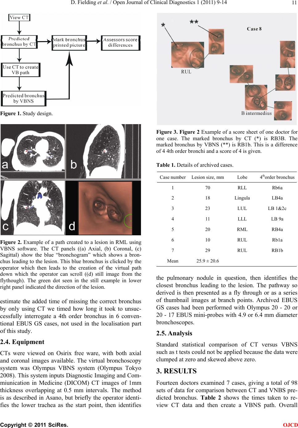 D. Fielding et al. / Open Journal of Clinical Diagnostics 1 (2011) 9-14 11 Figure 1. Study design. Figure 2. Example of a path created to a lesion in RML using VBNS software. The CT panels ((a) Axial, (b) Coronal, (c) Sagittal) show the blue “bronchogram” which shows a bron- chus leading to the lesion. This blue bronchus is clicked by the operator which then leads to the creation of the virtual path down which the operator can scroll ((d) still image from the flythough). The green dot seen in the still example in lower right panel indicated the direction of the lesion. estimate the added time of missing the correct bronchus by only using CT we timed how long it took to unsuc- cessfully interrogate a 4th order bronchus in 6 conven- tional EBUS GS cases, not used in the localisation part of this study. 2.4. Equipment CTs were viewed on Osirix free ware, with both axial and coronal images available. The virtual bronchoscopy system was Olympus VBNS system (Olympus Tokyo 2008). This system inputs Diagnostic Imaging and Com- miunication in Medicine (DICOM) CT images of 1mm thickness overlapping at 0.5 mm intervals. The method is as described in Asano, but briefly the operator identi- fies the lower trachea as the start point, then identifies Figure 3. Figure 2 Example of a score sheet of one doctor for one case. The marked bronchus by CT (*) is RB3B. The marked bronchus by VBNS (**) is RB1b. This is a difference of 4 4th order bronchi and a score of 4 is given. Table 1. Details of archived cases. Case numberLesion size, mmLobe 4thorder bronchus 1 70 RLL Rb6a 2 18 Lingula LB4a 3 23 LUL LB 1&2c 4 11 LLL LB 9a 5 20 RML RB4a 6 10 RUL Rb1a 7 29 RUL RB1b Mean 25.9 ± 20.6 the pulmonary nodule in question, then identifies the closest bronchus leading to the lesion. The pathway so derived is then presented as a fly through or as a series of thumbnail images at branch points. Archived EBUS GS cases had been performed with Olympus 20 - 20 or 20 - 17 EBUS mini-probes with 4.9 or 6.4 mm diameter bronchoscopes. 2.5. Analysis Standard statistical comparison of CT versus VBNS such as t tests could not be applied because the data were clumped at zero and skewed above zero. 3. RESULTS Fourteen doctors examined 7 cases, giving a total of 98 sets of data for comparison between CT and VNBS pre- dicted bronchus. Table 2 shows the times taken to re- view CT data and then create a VBNS path. Overall C opyright © 2011 SciRes. OJCD 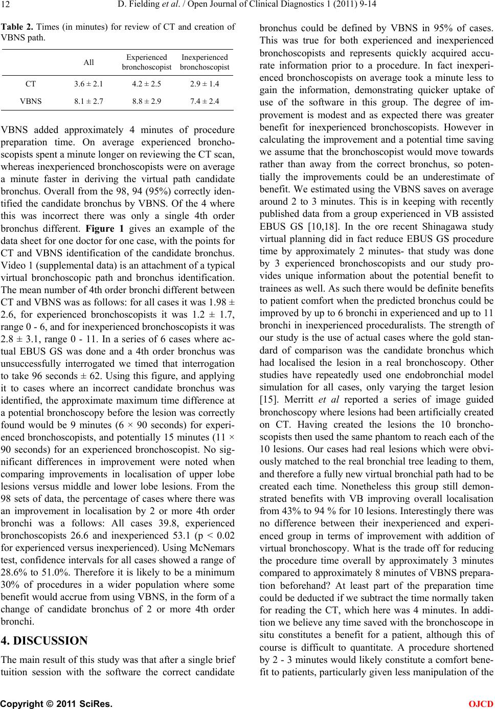 D. Fielding et al. / Open Journal of Clinical Diagnostics 1 (2011) 9-14 12 Table 2. Times (in minutes) for review of CT and creation of VBNS path. All Experienced bronchoscopist Inexperienced bronchoscopist CT 3.6 ± 2.1 4.2 ± 2.5 2.9 ± 1.4 VBNS 8.1 ± 2.7 8.8 ± 2.9 7.4 ± 2.4 VBNS added approximately 4 minutes of procedure preparation time. On average experienced broncho- scopists spent a minute long er on reviewing the CT scan , whereas inexperienced bronchoscopists were on average a minute faster in deriving the virtual path candidate bronchus. Overall from the 98, 94 (95%) correctly iden- tified the candidate bronchus by VBNS. Of the 4 where this was incorrect there was only a single 4th order bronchus different. Figure 1 gives an example of the data sheet for one doctor for one case, with the points for CT and VBNS identification of the candidate bronchus. Video 1 (supplemental data) is an attachment of a typical virtual bronchoscopic path and bronchus identification. The mean number of 4th order bronchi different between CT and VBNS was as follows: for all cases it was 1.98 ± 2.6, for experienced bronchoscopists it was 1.2 ± 1.7, range 0 - 6, and fo r inex p erien ced br onch osco p ists it was 2.8 ± 3.1, range 0 - 11. In a series of 6 cases where ac- tual EBUS GS was done and a 4th order bronchus was unsuccessfully interrogated we timed that interrogation to take 96 seconds ± 62. Using this figure, and applying it to cases where an incorrect candidate bronchus was identified, the approximate maximum time difference at a potential bronchoscopy before the lesion was correctly found would be 9 minutes (6 × 90 seconds) for experi- enced bronchoscopists, and potentially 15 minutes (11 × 90 seconds) for an experienced bronchoscopist. No sig- nificant differences in improvement were noted when comparing improvements in localisation of upper lobe lesions versus middle and lower lobe lesions. From the 98 sets of data, the percentage of cases where there was an improvement in localisation by 2 or more 4th order bronchi was a follows: All cases 39.8, experienced bronchoscopists 26.6 and inexperienced 53.1 (p < 0.02 for experienced versus inexperienced). Using McNemars test, confidence intervals for all cases showed a range of 28.6% to 51.0%. Therefore it is likely to be a minimum 30% of procedures in a wider population where some benefit would accrue from using VBNS, in the form of a change of candidate bronchus of 2 or more 4th order bronchi. 4. DISCUSSION The main result of this study was that after a single brief tuition session with the software the correct candidate bronchus could be defined by VBNS in 95% of cases. This was true for both experienced and inexperienced bronchoscopists and represents quickly acquired accu- rate information prior to a procedure. In fact inexperi- enced bronchoscopists on average took a minute less to gain the information, demonstrating quicker uptake of use of the software in this group. The degree of im- provement is modest and as expected there was greater benefit for inexperienced bronchoscopists. However in calculating the improvement and a potential time saving we assume that the bronchoscopist would move towards rather than away from the correct bronchus, so poten- tially the improvements could be an underestimate of benefit. We estimated using the VBNS saves on average around 2 to 3 minutes. This is in keeping with recently published data from a group experienced in VB assisted EBUS GS [10,18]. In the ore recent Shinagawa study virtual planning did in fact reduce EBUS GS procedure time by approximately 2 minutes- that study was done by 3 experienced bronchoscopists and our study pro- vides unique information about the potential benefit to trainees as well. As such there would be definite benefits to patient comfort when the predicted bronchus could be improved by up to 6 bronchi in experienced and up to 11 bronchi in inexperienced proceduralists. The strength of our study is the use of actual cases where the gold stan- dard of comparison was the candidate bronchus which had localised the lesion in a real bronchoscopy. Other studies have repeatedly used one endobronchial model simulation for all cases, only varying the target lesion [15]. Merritt et al reported a series of image guided bronchoscopy where lesions had been artificially created on CT. Having created the lesions the 10 broncho- scopists then used the same phantom to reach each of the 10 lesions. Our cases had real lesions which were obvi- ously matched to the real bron chial tree leading to them, and therefore a fully new virtual bronchial path had to be created each time. Nonetheless this group still demon- strated benefits with VB improving overall localisation from 43% to 94 % for 10 lesions. Interestingly there was no difference between their inexperienced and experi- enced group in terms of improvement with addition of virtual bronchoscopy. What is the trade off for reducing the procedure time overall by approximately 3 minutes compared to approximately 8 minutes of VBNS prepara- tion beforehand? At least part of the preparation time could be deducted if we subtract the time normally taken for reading the CT, which here was 4 minutes. In addi- tion we believe any time saved with the bronchoscop e in situ constitutes a benefit for a patient, although this of course is difficult to quantitate. A procedure shortened by 2 - 3 minutes would likely constitute a comfort bene- fit to patients, particularly given less manipulation of the C opyright © 2011 SciRes. OJCD  D. Fielding et al. / Open Journal of Clinical Diagnostics 1 (2011) 9-14 13 EBUS GS probe in the bronchus searching for the cor- rect bronchus. Overall our inexperienced candidates scored very well on their knowledge of CT and endobronchial anatomy before applying VBNS. This may in part reflect their exposure to others performing the procedure and better learning tools now available for training bronchoscopists [16]. In one other series of both experienced and inexpe- rienced bronchoscopists Dolina reported a simulation study where 10 lesions were artificially created at endo- bronchial sites between 3rd and 5th order bronchi [17]. These lesions were therefore considerably easier to iden- tify and locate than the lesions in this study which were placed at non bronchoscopically visible locations typi- cally at 8th order bronchus level. Similar to our study bronchoscopists recorded their results on a paper deci- sion sheet in this case it was the bronchoscope path to the lesion. Results were that without VB assistance the correct path was only found in 14%, which improved to 49% with the addition of VB assistance. With a com- puter tracking tool, perhaps more accurate in terms of the subjects’ decisions, there was an improvement from 40% to 96% in reaching the target. With either method the basic results for their subjects withou t the aid of vir- tual bronchoscopy were therefore lower than our group. A limitations of our study was that only two of the cases had endobronchial variations, and cases where more variations had been present may well have shown a bigger improvement with VBNS. Also we were not test- ing manipulation of a bronchoscope to arrive at a certain bronchus as has been done in other mannequin studies. Sakurada et al. [5] noted that some bronchi are more difficult to gain access to such as RB3a and LB 1 & 2c, however we did not include manual dexterity for access in this study. In terms of the input of CT data into the VBNS system a significant number of cases could not be performed because the CT had not been acquired with thin enough slices, thereby severely reducing definition of virtual images. In summary we have showed virtual bronchoscopy is an easily aquired skill which improves bronchial local- isation, even with a high baseline knowledge of anatomy. Inexperienced bronchoscopists quickly acquire the skill and this affords them greater potential in-procedure benefit. This is in line with the use of virtual tools for a range of bronchoscopic skills [18]. We believe this sim- ple method should be available to all proceduralists do- ing EBUS GS, but particularly inexperienced broncho- scopists as a compliment to their learning of the EBUS GS method. 5. ACKNOWLEDGEMENTS Supply of VNS software prototype by Olympus Medical Systems Tokyo. REFERENCES [1] Herth, F.J., Ernst, A. and Becker, H.D. (2002) Endo- bronchial ultrasound-guided transbronchial lung biopsy in solitary pulmonary nodules and peripheral lesions. European Respiratory Journal, 20, 972-974. doi:10.1183/09031936.02.00032001 [2] Shirakawa, T., Imamura, F., Hamamoto, J., Honda, I., Fukushima, K., Sugimoto, M., et al. (2004) Usefulness of endobronchial ultrasonography for transbronchial lung biopsies of peripheral lung lesions. Respiration, 71, 260-268. doi:10.1159/000077424 [3] Rivera, M.P. and Mehta, A.C. (2007) Initial diagnosis of lung cancer: ACCP evidence-based clinical practice gui- delines, 2nd Edition, Chest, 132, S131-148. doi:10.1378/chest.07-1357 [4] Kurimoto, N., Miyazawa, T., Okimasa, S., Maeda, A., Olwa, H., Miyazy, Y., et al. (2004) Endobronchial ultra- sonography using a guide sheath increases the ability to diagnose peripheral pulmonary lesions endoscopically. Chest, 126, 959-965. doi:10.1378/chest.126.3.959 [5] Sakurada, A., Takahashi, N., Sato, M., Miyagawa, Y., Matsumura, H. and Murakami, G. (2005) Are difficulties during transbronchial lung biopsy/brush through a fiber- optic bronchoscope based on the bronchial anatomy? Surgical and Radiologic Anatomy, 27, 94-99. doi:10.1007/s00276-004-0297-0 [6] Minami, H., Ando, Y., Nomura, F., Sakai, S. and Shimo- kata, K. (1994) Interbronchoscopist variability in the di- agnosis of lung cancer by flexible bronchoscopy. Chest, 105, 1658-1662. doi:10.1378/chest.105.6.1658 [7] Vining, D.J., Liu, K., Choplin, R.H. and Haponik, E.F. (1996) Virtual bronchoscopy, relationships of virtual re- ality endobronchial simulations to actual bronchoscopic findings. Chest, 109, 549-553. doi:10.1378/chest.109.2.549 [8] Brillet, P.Y., Fetita, C.I., Biegelman-Aubry, C., Prêteux, F. and Grenier, P.A. (2007) Quantification of bronchial di- mensions at MDCT using dedicated software. European Radiology, 17, 1483-1489. doi:10.1007/s00330-006-0496-7 [9] Asano, F. (2010) Virtual bronchoscopic navigation. Clin- ics in Chest Medicine, 31, 75-85. doi:10.1016/j.ccm.2009.08.007 [10] Shinagawa, N., Yamazaki, K., Onodera, Y., Asahina, H., Kikuchi, E., Asano, F., et al. (2007) Virtual broncho- scopic navigation system shortens the examination ti- me—feasibility study of virtual bronchoscopic naviga- tio n system. Lung Cancer, 56, 201-206. doi:10.1016/j.lungcan.2006.12.005 [11] Yu, K.C., Gibbs, J.D., Graham, M.W. and Higgins, W.E. (2010) Image based reporting for bronchoscopy. Journal of Digital Imaging, 23, 39-50. doi:10.1007/s10278-008-9170-8 [12] Asano, F., Matsuno, Y., Tsuzuku, A., Anzai, M., Shina- gawa, N., et al. (2008) Diagnosis of peripheral pulmo- nary lesions using a bronchoscope insertion guidance system combined with endobronchial ultrasonography with a guide sheath. Lung Cancer, 60, 366-373. doi:10.1016/j.lungcan.2007.10.022 C opyright © 2011 SciRes. OJCD  D. Fielding et al. / Open Journal of Clinical Diagnostics 1 (2011) 9-14 Copyright © 2011 SciRes. 14 OJCD [13] Okisama, S. (2007) Endobronchial ultrasonography with a guide sheath and virtual bronchoscopy navigation aids management of peripheral pulmonary nodules. Hiros- hima journal of medical sciences, 56, 19-22. [14] Asano, F. , Yamazaki, K. and Ishida, T. (2008) Usefulness of virtual bronchoscopic navigation in transbronchial bi- opsy for small pulmonary peripheral lesions: A multi- center, randomized trial. Programs and abstracts of the 15th World Congress for Bronchology, Tokyo, 32. [15] Merritt, S.A., Gibbs, J.D., Yu, K.C., Patel, V., Rai, L., et al. (2008) Image-guided bronchoscopy for peripheral lung lesions: A phantom study. Chest, 134, 1017-1026. doi:10.1378/chest.08-0603 [16] Colt, H.G., Davoudi, M. and Quadrelli, S. (2007) Pilot study of web-based bronchoscopy education using the essential bronchoscopist in developing countries. Respi- ration, 74, 358-359. [17] Dolina, M.Y., Cornish, D.C., Merri tt, S.A., Rai, L., Mah- raj, R., et al. (2008) Interbronchoscopist variability in endobronchial path selection: A simulation study. Chest, 133, 897-905. doi:10.1378/chest.07-2540 [18] Ishida, T., Asano, F., Yamazaki, K., et al. (2011) Virtual bronchoscopic navigation combined with endobronchial ultrasound to diagnose small peripheral pulmonary le- sions: A randomised trial. Thorax. |