

Paper Menu >>
Journal Menu >>
 Open Journal of Me di cal Imaging, 2011, 1, 21-25 doi:10.4236/ojmi.2011.12004 Published Online December 2011 (http://www.SciRP.org/journal/ojmi) Copyright © 2011 SciRes. OJMI Adenosine Stress Perfusion Cardiac MRI: Improving Image Quality Using a 32-Channel Surface Coil Thomas R. Burchell, Redha Boubertakh, Saidi Mohiddin, Marc E. Miquel, Mark A. Westwood, Anthony Mathur, L. Ceri Davies The London Chest Hospital, London, UK E-mail: [email protected] Received September 14, 2011; revised November 8, 2011; accepted N ovember 18, 2011 Abstract Purpose: Adenosine stress CMR is commonly used to assess myocardial ischaemia. Obtaining high quality images requires maximising signal to noise ratio (SNR) over a large double-oblique field of view (FOV) whilst minimising artefacts. A 32-channel surface coil may provide a higher SNR over a larger FOV com- pared to standard coils, possibly leading to improved image quality. Materials and Methods: 50 adenosine perfusion CMR scans were performed on a Philips Achieva CV 1.5T, with either a 5 or 32-channel coil (25 patients each) using standardised acquisition protocols. 3 short axis slices were acquired per cardiac cycle and the resulting cine images were scored by two blinded CMR specialists on a quality scale of 1 to 5. Phan- tom studies were performed using similar acquisition parameters and the SNR was calculated and compared across a range of acceleration factors. Results: The mean patient age was 62 ± 11 years and 50% of patients were male. The image quality scores were higher using the 32-channel coil (mean 3.8 ± 0.7 vs 3.2 ± 0.9 p = 0.002). The average phantom SNR was greater for the 32-element coil across the range of acceleration fac- tors measured (103 vs 86 p = <0.001). Conclusions: The 32-channel coil produces significantly higher qual- ity images and a higher SNR than the 5-channel coil in routine perfusion CMR. Keywords: Cardiovascular MRI, Myocardial Perfusion, 32-Channel Coil, Adenosine 1. Introduction Perfusion Imaging First pass stress perfusion cardiac magnetic resonance imaging (Stress Perfusion CMR) has been comprehen- sively shown to be a safe, reliable and reproducible method of identifying areas of inducible myocardial is- chaemia without the use of ionising radiation [1-5]. It has also been shown to predict cardiovascular morbidity and mortality in patients with known coronary disease [6]. Acquiring high quality diagnostic images in a routine clinical setting can be technically challenging due to the limitations of the current hardware and acquisition pro- tocols. These limitations can lead to a low in-plan e reso- lution, resulting in dark rim Gibbs artefact [7], non-uni- form sensitivity over the FOV, poor SNR and long ac- quisition times. Various techniques have been used in an attempt to overcome these limitations: A combination of highly accelerated parallel imaging and temporal encoding (e.g. k-t SENSE) can be used to increase spatial resolution [8,9] however, using temporal information requires post processing and is prone to movement artefacts, which cannot be identified until after the perfusion scan has been completed. Higher field strengths to increase contrast to noise ratio (CNR) have shown promise using 3T but CNR inhomogeneities occur across the left ventricle, particularly in the inferior wall [10]. Also 3T magnets are not yet widely available in the clinical setting. Increasing the number of surface coils has the poten- tial advantages of increasing the SNR [11-13] and pro- vides a better field of view coverage, allowing higher acceleration factors and hence higher spatial resolution or faster acquisition times [14]. We assessed the difference in visual quality and SNR between dedicated cardiac 32-element and standard 5- element surface coils in routine clinical myocardial per- fusion imaging using an identical acquisition proto col for both coils. 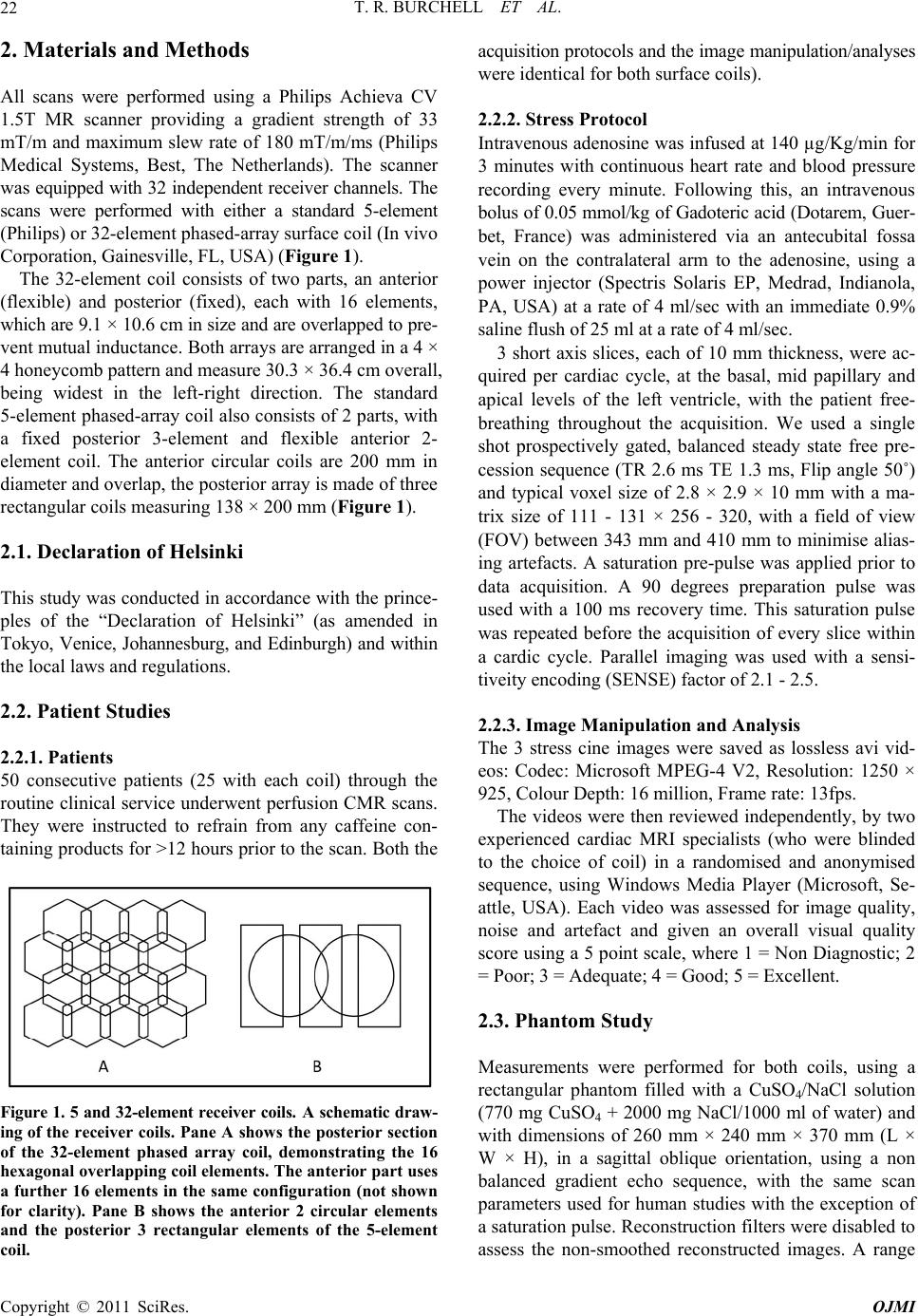 T. R. BURCHELL ET AL. 22 2. Materials and Methods All scans were performed using a Philips Achieva CV 1.5T MR scanner providing a gradient strength of 33 mT/m and maximum slew rate of 180 mT/m/ms (Philips Medical Systems, Best, The Netherlands). The scanner was equipped with 32 independent receiver channels. The scans were performed with either a standard 5-element (Philips) or 32-element phased-array surface coil (In vivo Corporation, Gainesville, FL, USA) (Figure 1). The 32-element coil consists of two parts, an anterior (flexible) and posterior (fixed), each with 16 elements, which are 9.1 × 10.6 cm in size and are overlapped to pre- vent mutual inductance. Both arrays are arranged in a 4 × 4 honeycomb pattern and measure 30.3 × 36.4 cm overall, being widest in the left-right direction. The standard 5-element phased-array co il also consists of 2 parts, with a fixed posterior 3-element and flexible anterior 2- element coil. The anterior circular coils are 200 mm in diameter and overlap, the posterior array is made of three rectangular coils measuring 138 × 200 mm (Figure 1). 2.1. Declaration of Helsinki This study was conducted in accordance with the prince- ples of the “Declaration of Helsinki” (as amended in Tokyo, Venice, Johannesburg, and Edinburgh) and within the local laws and regulations. 2.2. Patient Studies 2.2.1. Patient s 50 consecutive patients (25 with each coil) through the routine clinical service underwent perfusion CMR scans. They were instructed to refrain from any caffeine con- taining products for >1 2 hours prior to the scan. Both the Figure 1. 5 and 32-element receiver coils. A schematic draw- ing of the receiver coils. Pane A shows the posterior section of the 32-element phased array coil, demonstrating the 16 hexagonal overlapping coil elements. The anterior part uses a further 16 elements in the same configuration (not shown for clarity). Pane B shows the anterior 2 circular elements and the posterior 3 rectangular elements of the 5-element coil. acquisition protocols and the image manipulation/analyses were identical for both surface coils). 2.2.2. Stress P rotocol Intravenous aden osine was infused at 140 µg/Kg/min for 3 minutes with continuous heart rate and blood pressure recording every minute. Following this, an intravenous bolus of 0.05 mmol/kg of Gadoteric acid (Dotarem, Guer- bet, France) was administered via an antecubital fossa vein on the contralateral arm to the adenosine, using a power injector (Spectris Solaris EP, Medrad, Indianola, PA, USA) at a rate of 4 ml/sec with an immediate 0.9% saline flush of 25 ml at a rate of 4 ml/sec. 3 short axis slices, each of 10 mm thickness, were ac- quired per cardiac cycle, at the basal, mid papillary and apical levels of the left ventricle, with the patient free- breathing throughout the acquisition. We used a single shot prospectively gated, balanced steady state free pre- cession sequence (TR 2.6 ms TE 1.3 ms, Flip angle 50˚) and typical voxel size of 2.8 × 2.9 × 10 mm with a ma- trix size of 111 - 131 × 256 - 320, with a field of view (FOV) between 343 mm and 410 mm to minimise alias- ing artefacts. A saturation pre-pulse was applied prior to data acquisition. A 90 degrees preparation pulse was used with a 100 ms recovery time. This saturation pulse was repeated before the acquisition of every slice within a cardic cycle. Parallel imaging was used with a sensi- tiveity encoding (SENSE) factor of 2.1 - 2.5. 2.2.3. Image Ma nipul a ti o n and Analysis The 3 stress cine images were saved as lossless avi vid- eos: Codec: Microsoft MPEG-4 V2, Resolution: 1250 × 925, Colour Depth: 16 million, Frame rate: 13fps. The videos were then reviewed independently, by two experienced cardiac MRI specialists (who were blinded to the choice of coil) in a randomised and anonymised sequence, using Windows Media Player (Microsoft, Se- attle, USA). Each video was assessed for image quality, noise and artefact and given an overall visual quality score using a 5 point scale, where 1 = Non Diagnostic; 2 = Poor; 3 = Adequate; 4 = Good; 5 = Excellent. 2.3. Phantom Study Measurements were performed for both coils, using a rectangular phantom filled with a CuSO4/NaCl solution (770 mg CuSO4 + 2000 mg NaCl/1000 ml of water) and with dimensions of 260 mm × 240 mm × 370 mm (L × W × H), in a sagittal oblique orientation, using a non balanced gradient echo sequence, with the same scan parameters used for human studies with the exception of a saturation pulse. Reconstruction filters were disabled to assess the non-smoothed reconstructed images. A range Copyright © 2011 SciRes. OJMI 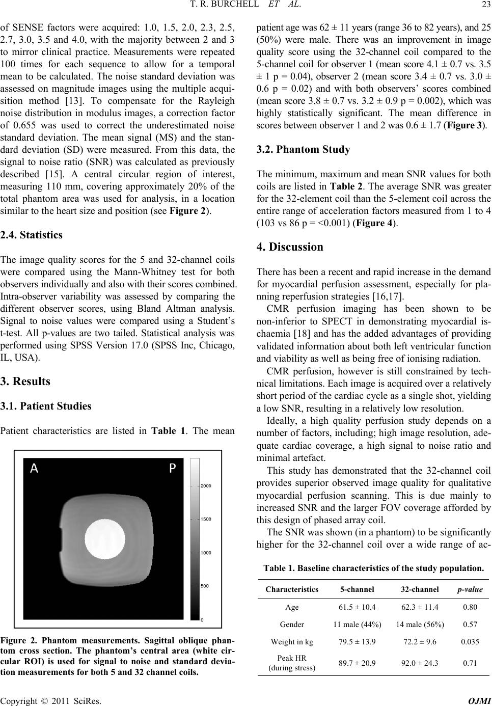 T. R. BURCHELL ET AL.23 of SENSE factors were acquired: 1.0, 1.5, 2.0, 2.3, 2.5, 2.7, 3.0, 3.5 and 4.0, with the majority between 2 and 3 to mirror clinical practice. Measurements were repeated 100 times for each sequence to allow for a temporal mean to be calculated. The noise standard deviation was assessed on magnitude images using the multiple acqui- sition method [13]. To compensate for the Rayleigh noise distribution in modulus images, a correction factor of 0.655 was used to correct the underestimated noise standard deviation. The mean signal (MS) and the stan- dard deviation (SD) were measured. From this data, the signal to noise ratio (SNR) was calculated as previously described [15]. A central circular region of interest, measuring 110 mm, covering approximately 20% of the total phantom area was used for analysis, in a location similar to the heart size and position (see Figure 2). 2.4. Statistics The image quality scores for the 5 and 32-channel coils were compared using the Mann-Whitney test for both observers individually and also with their scores combined. Intra-observer variability was assessed by comparing the different observer scores, using Bland Altman analysis. Signal to noise values were compared using a Student’s t-test. All p-values are two tailed. Statistical analysis was performed using SPSS Version 17.0 (SPSS Inc, Chicago, IL, USA). 3. Results 3.1. Patient Studies Patient characteristics are listed in Table 1. The mean Figure 2. Phantom measurements. Sagittal oblique phan- tom cross section. The phantom’s central area (white cir- cular ROI) is used for signal to noise and standard devia- tion measurements for both 5 and 32 channel coils. patient age was 62 ± 11 years (range 36 to 82 years), and 25 (50%) were male. There was an improvement in image quality score using the 32-channel coil compared to the 5-channel coil for observer 1 (mean score 4.1 ± 0.7 vs. 3.5 ± 1 p = 0.04), observer 2 (mean score 3.4 ± 0.7 vs. 3.0 ± 0.6 p = 0.02) and with both observers’ scores combined (mean score 3.8 ± 0.7 vs. 3.2 ± 0.9 p = 0.002), which was highly statistically significant. The mean difference in scores between observer 1 and 2 was 0.6 ± 1.7 (Fig ure 3). 3.2. Phantom Study Th e min imu m, max imu m an d mea n S NR v alue s fo r bo th coils are listed in Table 2. The average SNR was greater for the 32-element coil than the 5-element coil across the entire range of acceleration factors measured from 1 to 4 (103 vs 86 p = <0.001) (Figure 4). 4. Discussion There has been a recent and rapid increase in the demand for myocardial perfusion assessment, especially for pla- nning reperfusion strategies [16,17]. CMR perfusion imaging has been shown to be non-inferior to SPECT in demonstrating myocardial is- chaemia [18] and has the added advantages of providing validated information about both left ventricular function and viability as well as being free of ionising radiation. CMR perfusion, however is still constrained by tech- nical limitations. Each image is acquired over a relatively short period of the cardiac cycle as a single shot, yielding a low SNR, resulting in a relatively low resolutio n. Ideally, a high quality perfusion study depends on a number of factors, including; high image resolution, ade- quate cardiac coverage, a high signal to noise ratio and minimal artefact. This study has demonstrated that the 32-channel coil provides superior observed image quality for qualitative myocardial perfusion scanning. This is due mainly to increased SNR and the larger FOV coverage afforded by this design of phased array coil. The SNR was shown (in a phantom) to be significantly higher for the 32-channel coil over a wide range of ac- Table 1. Baseline characteristics of the study population. Characteristics5-channel 32-channel p-value Age 61.5 ± 10.4 62.3 ± 11.4 0.80 Gender 11 male (44%) 14 male (56%)0.57 Weight in kg 79.5 ± 13.9 72.2 ± 9.6 0.035 Peak HR (during stress) 89.7 ± 20.9 92.0 ± 24.3 0.71 Copyright © 2011 SciRes. OJMI 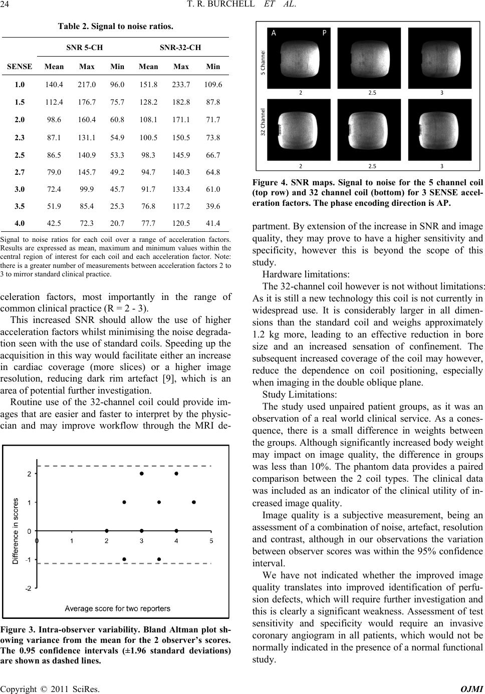 T. R. BURCHELL ET AL. 24 Table 2. Signal to noise ratios. SNR 5-CH SNR-32-CH SENSE Mean Max MinMean Max Min 1.0 140.4 217.0 96.0151.8 233.7109.6 1.5 112.4 176.7 75.7128.2 182.887.8 2.0 98.6 160.4 60.8108.1 171.171.7 2.3 87.1 131.1 54.9100.5 150.573.8 2.5 86.5 140.9 53.398.3 145.966.7 2.7 79.0 145.7 49.294.7 140.364.8 3.0 72.4 99.9 45.7 91.7 133.461.0 3.5 51.9 85.4 25.3 76.8 117.239.6 4.0 42.5 72.3 20.7 77.7 120.541.4 Signal to noise ratios for each coil over a range of acceleration factors. Results are expressed as mean, maximum and minimum values within the central region of interest for each coil and each acceleration factor. Note: there is a gr eater number of measu remen ts between acc elerat ion facto rs 2 to 3 to mirror standard clinical practice. celeration factors, most importantly in the range of common clinical practice (R = 2 - 3). This increased SNR should allow the use of higher acceleration factors whilst minimising the noise degrada- tion seen with the use of standard coils. Speeding up the acquisition in this way would facilitate either an increase in cardiac coverage (more slices) or a higher image resolution, reducing dark rim artefact [9], which is an area of potential further investigation. Routine use of the 32-channel coil could provide im- ages that are easier and faster to interpret by the physic- cian and may improve workflow through the MRI de- Figure 3. Intra-observer variability. Bland Altman plot sh- owing variance from the mean for the 2 observer’s scores. The 0.95 confidence intervals (±1.96 standard deviations) are shown as dashed lines. Figure 4. SNR maps. Signal to noise for the 5 channel coil (top row) and 32 channel coil (bottom) for 3 SENSE accel- eration factors. The phase encoding direction is AP. partment. By extension of the increase in SNR and image quality, they may prove to have a higher sensitivity and specificity, however this is beyond the scope of this study. Hardware limitations: The 32-channel coil howev er is not without limitations: As it is still a new technology this coil is no t currently in widespread use. It is considerably larger in all dimen- sions than the standard coil and weighs approximately 1.2 kg more, leading to an effective reduction in bore size and an increased sensation of confinement. The subsequent increased coverage of the coil may however, reduce the dependence on coil positioning, especially when imaging in the double oblique plane. Study Limi ta tions: The study used unpaired patient groups, as it was an observation of a real world clinical service. As a cones- quence, there is a small difference in weights between the groups. Although significantly in creased body weight may impact on image quality, the difference in groups was less than 10%. The phantom data provides a paired comparison between the 2 coil types. The clinical data was included as an indicator of the clinical utility of in- creased image quality. Image quality is a subjective measurement, being an assessment of a combination of noise, artefact, resolution and contrast, although in our observations the variation between observer scores was within the 95% confidence interval. We have not indicated whether the improved image quality translates into improved identification of perfu- sion defects, which will require further investigation and this is clearly a significant weakness. Assessment of test sensitivity and specificity would require an invasive coronary angiogram in all patients, which would not be normally indicated in the presence of a normal functional study. Copyright © 2011 SciRes. OJMI  T. R. BURCHELL ET AL. Copyright © 2011 SciRes. OJMI 25 In conclusion, the 32-channel phased array coil pro- duces both a high er SNR and significantly higher quality images than the standard 5-channel coil in routine stress CMR despite using near identical acquisition protocols for both coi l s. 5. References [1] N. Al-Saadi, E. Nagel, M. Gross, et al., “Noninvasive Detection of Myocardial Ischemia from Perfusion Re- serve Based on Cardiovascular Magnetic Resonance,” Circulation, Vol. 101, No. 12, 2000, pp. 1379-1383. [2] T. Giang, D. Nanz, R. Coulden, et al., “Detection of Coronary Artery Disease by Magnetic Resonance Myo- cardial Perfusion Imaging with Various Contrast Medium Doses: First European Multi-Centre Experience,” Euro- pean Heart Journal, Vol. 25, No. 18, 2004, pp. 1657- 1665. doi:10.1016/j.ehj.2004.06.037 [3] E. Nagel, C. Klein, I. Paetsch, et al., “Magnetic Reso- nance Perfusion Measurements for the Noninvasive De- tection of Coronary Artery Disease,” Circulation, Vol. 108, No. 4, 2003, pp. 432-437. doi:10.1161/01.CIR.0000080915.35024.A9 [4] J. Schwitter, D. Nanz, S. Kneifel, et al., “Assessment of Myocardial Perfusion in Coronary Artery Disease by Magnetic Resonance a Comparison with Positron Emis- sion Tomography and Coronary Angiography,” Circula- tion, Vol. 103, No. 18, 2001, pp. 2230-2235. [5] N. Wilke, M. Jerosch-Herold, A. Zenovich and A. Still- man, “Magnetic Resonance First-Pass Myocardial Perfu- sion Imaging: Clinical Validation and Future Applica- tions,” Journal of Magnetic Resonance Imaging, Vol. 10, No. 5, 1999, pp. 676-685. doi:10.1002/(SICI)1522-2586(199911)10:5<676::AID-J MRI10>3.0.CO;2-L [6] C. Jahnke, E. Nagel, R. Gebker, et al., “Prognostic Value of Cardiac Magnetic Resonance Stress Tests: Adenosine Stress Perfusion and Dobutamine Stress Wall Motion Imaging,” Circulation, Vol. 115, No. 13, 2007, pp. 1769- 1776. doi:10.1161/CIRCULATIONAHA.106.652016 [7] E. Di Bella, D. Parker and A. Sinusas, “On the Dark Rim Artifact in Dynamic Contrast-Enhanced MRI Myocardial Perfusion Studies,” Magnetic Resonance in Medicine, Vol. 54, No. 5, 2005, pp. 1295-1299. [8] T. Niendorf and D. Sodickson, “Highly Accelerated Car- diovascular MR Imaging Using Many Channel Technol- ogy: Concepts and Clinical Applications,” European Ra- diology, Vol. 18, No. 1, 2008, pp. 87-102. doi:10.1007/s00330-007-0692-0 [9] S. Plein, S. Kozerke, D. Suerder, et al., “High Spatial Resolution Myocardial Perfusion Cardiac Magnetic Resonance for the Detection of Coronary Artery Dis- ease,” European Heart Journal, Vol. 29, No. 17, 2008, pp. 2148- 2155. [10] P. Araoz, J. Glockner, K. McGee, et al., “3 Tesla MR Imaging Provides Improved Contrast in First-Pass Myo- cardial Perfusion Imaging over a Range of Gadolinium Doses,” Journal of Cardiovascular Magnetic Resonance, Vol. 7, No. 3, 2005, pp. 559-564. doi:10.1081/JCMR-200060622 [11] C. J. Hardy, H. E. Cline, R. O. Giaquinto, et al., “32- Element Receiver-Coil Array for Cardiac Imaging,” Mag- netic Resonance in Medicine, Vol. 55, No. 5, 2006, pp. 1142-1149. doi:10.1002/mrm.20870 [12] Y. Zhu, C. J. Hardy, et al., “Highly Parallel Volumetric Imaging with a 32-Element RF Coil Array,” Magnetic Resonance in Medicine, Vol. 52, No. 4, 2004, pp. 869- 877. doi:10.1002/mrm.20209 [13] S. Reeder, B. Wintersperger, O. Dietrich, et al., “Practical Approaches to the Evaluation of Signal-to-Noise Ratio Performance with Parallel Imaging: Application with Cardiac Imaging and a 32-Channel Cardiac Coil,” Mag- netic Resonance in Medicine, Vol. 54, No. 3, 2005, pp. 748-754. doi:10.1002/mrm.20636 [14] M. Ohliger and D. Sodickson, “An Introduction to Coil Array Design for Parallel MRI,” NMR in Biomedicine, Vol. 19, No. 3, 2006, pp. 300-315. doi:10.1002/nbm.1046 [15] M. Fenchel, V. Deshpande, K. Nael, et al., “Cardiac Cine Imaging at 3 Tesla: Initial Experience With a 32-Element Body-Array Coil,” Investigative Radiology, Vol. 41, No. 8, 2006, pp. 601-608. doi:10.1097/01.rli.0000223896.70095.49 [16] L. Shaw, D. Berman, D. Maron, et al., “Optimal Medical Therapy with or without Percutaneous Coronary Inter- vention to Reduce Ischemic Burden: Results from the Clinical Outcomes Utilizing Revascularization and Ag- gressive Drug Evaluation (COURAGE) Trial Nuclear substudy,” Circulation, Vol. 117, No. 10, 2008, pp. 1283- 1291. doi:10.1161/CIRCULATIONAHA.107.743963 [17] P. Tonino, B. De Bruyne, N. Pijls, et al., “Fractional Flow Reserve versus Angiography for Guiding Percuta- neous Coronary Intervention,” The New England Journal of Medicine, Vol. 360, No. 3, 2009, pp. 213-224. doi:10.1056/NEJMoa0807611 [18] J Schwitter, C. Wacker, A. van Rossum, et al., “MR- IMPACT: Comparison of Perfusion-Cardiac Magnetic Resonance with Single-Photon Emission Computed To- mography for the Detection of Coronary Artery Disease in a Multicentre, Multivendor, Randomized Trial,” Euro- pean Heart Journal, Vol. 29, No. 4, 2008, pp. 480-489. doi:10.1093/eurheartj/ehm617 |