

Paper Menu >>
Journal Menu >>
 Journal of Biomaterials and Nanobiotechnology, 2011, 2, 477-484 doi:10.4236/jbnb.2011.225058 Published Online December 2011 (http://www.scirp.org/journal/jbnb) Copyright © 2011 SciRes. JBNB 477 Controlling Drug Release from Titania Nanotube Arrays Using Polymer Nanocarriers and Biopolymer Coating Moom Sinn Aw, Karan Gulati, Dusan Losic* Ian Wark Research Institute, The University of South Australia, Adelaide, Australia. E-mail: *[email protected] Received September 9th, 2011; revised October 14th, 2011; accepted November 10th, 2011. ABSTRACT Titania nanotube arrays (TNT) prepared by self-ordering electrochemical anodization have attracted considerable at- tention for the development of new devices for local drug delivery applications. Two approaches to extend drug re- lease of water insoluble drugs by integrating TNTs with polymeric micelles and biopolymer coatings are presented in this work. The proposed strategies emphasized on remarkable properties of these materials and their unique combina- tion to design local drug delivery system with advanced performance. The first concept integrates TNTs with drug loaded polymeric micelles (Pluronic F127) as drug nanocarrier, until the second concept includes polymer coating of drug loaded TNT with biodegradable polymer (chitosan). The water insoluble, anti-inflammatory drug, indomethacin was used as a model drug. Both approaches showed a significant improvement of the drug release cha racteristics, with reduced burst release (from 77% to 39%) and extended overall release from 9 days to more than 28 days. These results suggest the capability o f TNT based systems to be applied for local drug delivery d eliver over an extended period with predictable kinetics that is particularly important for bone implant therapies. Keywords: Drug Delivery, Titania Nanotubes, Polymeric Micelles, Pluronic F127, Chitosan, Indomethacin 1. Introduction To overcome limitations of the systemic therapy and conventional drug delivery systems such as limited drug solubility which leads to poor biodistribution, poor targeting and efficacy, uncontrolled pharmacokinetics, and serious side effects in non-target tissues, local drug delivery using implantable systems has become accept- able as an attractive solution to address these challenges [1,2]. The majority of drugs like antibiotics, anti-cancer and anti-inflammatory drugs are insoluble in water or be- come unstable during their transport to the targeted sites, hence they require special delivery systems [3,4]. Porous materials have been demonstrated as excellent candidates for the design of therapeutic implants, not only because porous structures support tissue integration, but also be- cause pores act as remarkable reservoirs for slow drug elution over extended time periods [5]. Advances in nanoscience and nanotechnology have led to the devel- opment of several new nanoporous materials for local drug delivery administrations. Among them, vertically aligned titania nanotube (TNT) arrays on Ti surfaces prepared by self-ordering electrochemical anodization has been recognized as one of the most promising solu- tions for the development of advanced implantable and local drug delivery systems [6,7]. TNTs have excellent biocompatibility, thermal and chemical stability, controllable dimensions, tunable sur- face chemistry and high surface-to-volume ratio due to their long and well aligned hollow nanotube structures. These advantages render favourable for bone cell growth, cell differentiation, and apatite-forming abilities, which make TNTs ideal platforms for bone drug delivery ap- plications [8-10]. The application of TNT films for im- plantable medical devices such as orthopaedic implants, vascular stents, immunoisolation capsules, dental im- plants, for the prevention of bacterial adhesion, and growth support for bone and stem cells has been recently reported [11-15]. For drug delivery applications of TNTs, it is important to achieve controlled and sustained release pattern with uniform drug elution over time. Controlling the pore diameters and lengths of TNTs by anodization process is used as a simple method to control drug re- 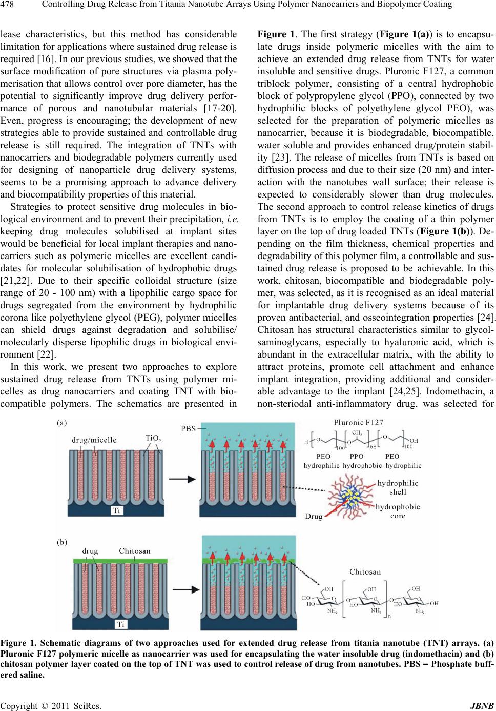 Controlling Drug Release from Titania Nanotube Arrays Using Polymer Nanocarriers and Biopolymer Coating 478 lease characteristics, but this method has considerable limitation for applications where sustained drug release is required [16]. In our previous studies, we showed that the surface modification of pore structures via plasma poly- merisation that allows control over pore diameter, has the potential to significantly improve drug delivery perfor- mance of porous and nanotubular materials [17-20]. Even, progress is encouraging; the development of new strategies able to provide sustained and controllable drug release is still required. The integration of TNTs with nanocarriers and biodegradable polymers currently used for designing of nanoparticle drug delivery systems, seems to be a promising approach to advance delivery and biocompatibility properties of this material. Strategies to protect sensitive drug molecules in bio- logical environment and to prevent their precipitation, i.e. keeping drug molecules solubilised at implant sites would be beneficial for local implant therapies and nano- carriers such as polymeric micelles are excellent candi- dates for molecular solubilisation of hydrophobic drugs [21,22]. Due to their specific colloidal structure (size range of 20 - 100 nm) with a lipophilic cargo space for drugs segregated from the environment by hydrophilic corona like polyethylene glycol (PEG), polymer micelles can shield drugs against degradation and solubilise/ molecularly disperse lipophilic drugs in biological envi- ronment [22]. In this work, we present two approaches to explore sustained drug release from TNTs using polymer mi- celles as drug nanocarriers and coating TNT with bio- compatible polymers. The schematics are presented in Figure 1. The first strategy (Figure 1(a)) is to encapsu- late drugs inside polymeric micelles with the aim to achieve an extended drug release from TNTs for water insoluble and sensitive drugs. Pluronic F127, a common triblock polymer, consisting of a central hydrophobic block of polypropylene glycol (PPO), connected by two hydrophilic blocks of polyethylene glycol PEO), was selected for the preparation of polymeric micelles as nanocarrier, because it is biodegradable, biocompatible, water soluble and provides enhanced drug/protein stabil- ity [23]. The release of micelles from TNTs is based on diffusion process and due to their size (20 nm) and inter- action with the nanotubes wall surface; their release is expected to considerably slower than drug molecules. The second approach to control release kinetics of drugs from TNTs is to employ the coating of a thin polymer layer on the top of drug loaded TNTs (Figure 1(b) ). De- pending on the film thickness, chemical properties and degradability of this polymer film, a controllable and sus- tained drug release is proposed to be achievable. In this work, chitosan, biocompatible and biodegradable poly- mer, was selected, as it is recognised as an ideal material for implantable drug delivery systems because of its proven antibacterial, and osseointegration properties [24]. Chitosan has structural characteristics similar to glycol- saminoglycans, especially to hyaluronic acid, which is abundant in the extracellular matrix, with the ability to attract proteins, promote cell attachment and enhance implant integration, providing additional and consider- able advantage to the implant [24,25]. Indomethacin, a non-steriodal anti-inflammatory drug, was selected for Figure 1. Schematic diagrams of two approaches used for extended drug release from titania nanotube (TNT) arrays. (a) Pluronic F127 polymeric micelle as nanocarrier was used for encapsulating the water insoluble drug (indomethacin) and (b) chitosan polymer layer coated on the top of TNT was used to co ntrol release of drug from nanotubes. PBS = Phosphate bu ff- red saline. e Copyright © 2011 SciRes. JBNB  Controlling Drug Release from Titania Nanotube Arrays Using Polymer Nanocarriers and Biopolymer Coating479 this study as the model of water insoluble drugs [26]. The drug release characteristics of these two systems are ex- plored to demonstrate their potential for the development of TNTs as drug eluting implants with extended drug release characteristics. 2. Materials and Methods 2.1. Materials Titanium foil, Ti (99.9% purity), with a thickness of 0.25 mm, was supplied by Alfa Aesar (USA). Ethylene glycol, ammonium fluoride, chitosan (low molecular weight, 75% - 85% deacetylation), and indomethacin (>99% TLC), Pluronic® F127 (Bioreagent, average Mw~12,600) were obtained from Sigma Aldrich Co. (Australia). Dialy- sis sacks (avg. flat width of 35 mm) were ordered form the same company. High purity water, ultra-pure Milli-Q grade (18.2 M·Ω·cm) with a final filtering step through a 0.22 m filter was used. All chemicals were of analytic reagent grade and used without further purification. 2.2. The Fabrication of Titania Nanotube (TNT) Arrays on Ti To prepare a TNT layer on Ti foil (TNT-Ti), two anodi- zation steps were performed using a specially designed electrochemical cell and computer controlled power sup- ply (Agilent), using previously described procedures [19, 27,28]. A specially designed electrode holder permitted only a circular area of diameter 1 cm of Ti metal available for anodization. In the first anodization step, a constant voltage of 100 V was applied for 2 hours in NH4F/ethy- leneglycol electrolyte (3% water and 0.3% NH4F) at a room temperature of 20˚C. The resultant TNT layer was removed (mechanically followed by sonication), leaving the nanotextured titanium surface for the second anodi- zation. The second anodization step was performed using the same conditions (100 V) as the first anodization, with an anodization time of 1 hour to make about 50 µm thick TNTs layer on Ti with perfectly ordered nanotube struc- tures. The voltage/current, voltage-time, and current-time signals were adjusted and continuously recorded (Lab- view, National Instruments) during the anodization proc- ess to assure the reproducibility of the fabrication process. 2.3. Synthesis of Pluronic Micelles and Drug Encapsulation Pluronic F127 micelles was synthesised using the direct dissolution technique as described previously [29]. 15 mg of micelles were dissolved in 5 ml chloroform and upon solvent evaporation in a rotary vacuum evaporator; micelles were dispersed in 20 ml of Milli-Q water. Samples were then dialysed against Milli-Q water for 2 days to produce the micelle suspensions. The drug (indo- methacin) was dissolved in chloroform prior to drying and then added to the micelle solution (10 ml of Milli-Q water containing 50 mg of micelles) under moderate magnetic stirring. The remaining chlroform is removed under reduced pressure via osmosis effect using regene- rated cellulose membrane (Spectrum Labs, Inc.) of 15 mm flat width with 20 cm long tubing. The size of the micelles (Pluronic and Pluronic-Ind) before and after loading were determined by laser light scattering using Zetasizer Nano (Malvern Instruments) from 5 repeated measurements. 2.4. Loading of Drug and Drug-Loaded Micelles into Titania Nanotubes The prepared TNT-Ti substrates (12 mm ×12 mm) were loaded with drug and drug-loaded micelles via a simp- lified lyophilisation method. Micelle dispersion (10 μl) was pipetted onto the substrate surface and gently spread to ensure an even coverage. The surfaces were then allowed to dry under vacuum at room temperature for 2 h. After drying, the loading step was repeated 20 times until the appropriate amount of micelles was present in the nanopore array. The top of the substrates on the end was carefully cleaned to remove adsorbed micelles from the surface. A solution of indomethacin (1% w/v) was pre- pared in ethanol and 10 µl of the drug solution was loaded into the nanotube surface and allowed to dry in air for 15 - 20 min. After drying, the TNT surface was wiped with a soft tissue to remove excess drug from the TNT surface. The sample was placed in a vacuum dessicator under vacuum for 1 h to remove the solvent and water. The cycles of loading, drying and wiping were repeated 25 times to load a sufficient amount of indomethacin drug into the nanotubes. 2.5. Chitosan Coating of Drug Loaded Titania Nanotubes Polymer solutions of chitosan (1% (w/v), chitosan + 0.8% (v/v) acetic acid in deionised water) was prepared. The dip-coating process was performed by dipping the TNT-Ti implant into the polymer solution, followed by drying in an oven at 70˚C for 10 min. The number of dipping can control the thickness of the deposited polymer and for this study; we did 5 times of dipping to make a thicker polymer film. The film thickness of the chitosan coat of prepared samples (Chit-Ind-TNT-Ti) was measured using an ellipsometer (Si wafer as control sample) and cross-sectional SEM imaging. 2.6. Drug and Drug-Micelles Loading and Release Characterization To determine the amount of loaded and released drug and drug-micelles in TNT samples, thermogravimetric ana- Copyright © 2011 SciRes. JBNB  Controlling Drug Release from Titania Nanotube Arrays Using Polymer Nanocarriers and Biopolymer Coating 480 lysis (TGA) was performed (Hi-Res Modulated TGA 2950) after drug and drug-micelles loading, after poly- mer coating and at the end of the release experiment. In order to find the correct range of the drug decomposition, 20 - 25 mg drug (indomethacin) was mounted onto the platinum pan, then heated up to 800˚C at a scanning rate of 10˚C/min under a nitrogen gas flow of 50 ml/min. This was performed to obtain its weight loss peak. It was followed by drug-, (Ind-TNT-Ti) drug-loaded micelle (Pluronic-Ind-TNT-Ti) and chitosancoated TNTs. The thermograms were analyzed using TGA data analyzing software (Universal Analysis, 2000), to calculate the loading capacity. In vitro drug release from all prepared TNT samples (drug and drug-loaded micelles with and without chi- tosan coating) was investigated by immersing the sam- ples in 5 ml phosphate buffer (PBS) at pH = 7.4, where the amount of released drug was measured via UV-Vis spectroscopy using Varian UV-Vis spectrophotometer. Measurements were taken at short intervals (every 5 - 15 min) during the first 6 hours to monitor the initial burst release, followed by testing every 24 h to observe the delayed release until the total amount of drug was released into the surrounding PBS. Absorbance was mea- sured at 320 nm for drug and at 760 nm for drug-loaded micelles. The corresponding drug concentration was calculated based on the calibration curve obtained. The final release profile of each experimental set was ex- pressed for burst (first 6 hours) and delayed release (7 - 28 days) in a graph with cumulative release (percentage) vs. time. The release of drug (indomethacin) from TNT without micelles and chitosan coating is used as the control for comparing how release can be extended by these methods. 2.7. Structural Characterization Structural characterization of the prepared TNT-Ti sub- strates before and after drug loading and after polymer coating and drug release were performed using a field emission scanning electron microscope (SEM) (Philips XL 30). The samples were cut into small (approximately 10 × 10 mm) pieces, mounted on a holder with double- sided conductive tape and coated with a layer of platinum 3-5 nm thick. Images with a range of scan sizes at normal incidence and at a 30 degree angle were acquired from the top, the bottom surface and cross-sections. 3. Results and Discussion 3.1. Structure and Morphology of Prepared Titania Nanotube (TNT) Arrays The structure and morphology of the prepared TNT-Ti substrates was characterised by SEM and is summarised in Figure 2. A typical cross-sectional image of free- standing TNT structures, after removing them from the Ti substrate (for imaging purposes) is presented in Fig- ure 2(a). The thickness of the TNT layer was about 50 µm, which was controlled by selecting the appropriate voltage (100 V) and anodization time (1 hour). A whole TNT layer with a diameter of 1 cm, formed on a Ti foil substrate, is shown in Figure 2(a) (inset). SEM images of the top nanotube surface (Figure 2(b)) show pores with diameters of 120 ± 20 nm. A high-resolution image of the cross-sectional SEM image of the TNT layer shows a vertically aligned and densely packed array of nanotubes across the entire structure (Figure 2(c)). The bottom surface of the TNT nanotube layer, after the de- tachment from the Ti substrate (Figure 2(d)) shows that the nanotubes were closed with spherical oxide barrier layer. For this study, titania nanotubes with pore diameters ~120 nm and a length of 50 µm were prepared, in order to maximise their drug loading capacity 3.2. Characterisation of Polymeric Micelles (Pluronic F127) before and after Drug Encapsulation The average size of prepared Pluronic F127 micelles was determined to be 20.0 ± 0.7 nm using dynamic light scattering measurements (Figure 3). When drug indome- thacin was encapsulated inside the micelle (hydrophobic core), the diameter was only slightly increased to 23.0 ± 1.4 nm (Figure 3), showing a minor difference in size between the drug-loaded and drug-free micelle (~3 - 5 nm). The micelle size was found to be consistent over a 6-month monitoring period, indicating long term stability Figure 2. SEM image of titania nanotube arrays prepared on Ti foil by electrochemical anodization showing, (a) side view (whole structure is shown in inset); (b) the top surface with open pores; (c) cross-sectional image showing hollow nanotube structures, and (d) the bottom surface with closed pores. Copyright © 2011 SciRes. JBNB 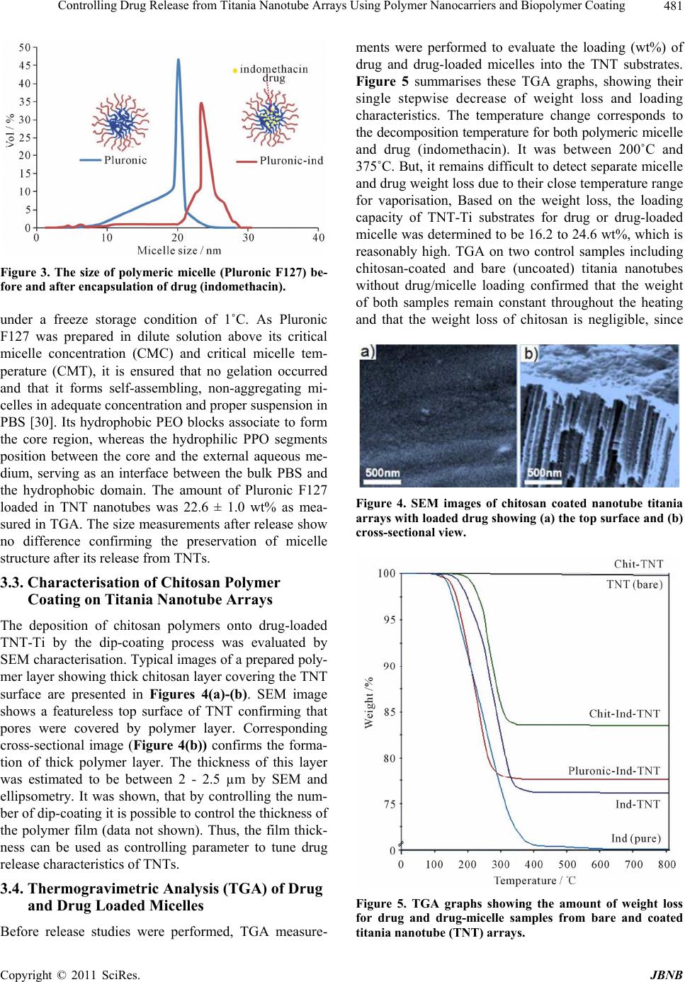 Controlling Drug Release from Titania Nanotube Arrays Using Polymer Nanocarriers and Biopolymer Coating481 Figure 3. The size of polymeric micelle (Pluronic F127) be- fore and after encapsulation of drug (indomethac in). under a freeze storage condition of 1˚C. As Pluronic F127 was prepared in dilute solution above its critical micelle concentration (CMC) and critical micelle tem- perature (CMT), it is ensured that no gelation occurred and that it forms self-assembling, non-aggregating mi- celles in adequate concentration and proper suspension in PBS [30]. Its hydrophobic PEO blocks associate to form the core region, whereas the hydrophilic PPO segments position between the core and the external aqueous me- dium, serving as an interface between the bulk PBS and the hydrophobic domain. The amount of Pluronic F127 loaded in TNT nanotubes was 22.6 ± 1.0 wt% as mea- sured in TGA. The size measurements after release show no difference confirming the preservation of micelle structure after its release from TNTs. 3.3. Characterisation of Chitosan Polymer Coating on Titania Nanotube Arrays The deposition of chitosan polymers onto drug-loaded TNT-Ti by the dip-coating process was evaluated by SEM characterisation. Typical images of a prepared poly- mer layer showing thick chitosan layer covering the TNT surface are presented in Figures 4(a)-(b). SEM image shows a featureless top surface of TNT confirming that pores were covered by polymer layer. Corresponding cross-sectional image (Figure 4(b)) confirms the forma- tion of thick polymer layer. The thickness of this layer was estimated to be between 2 - 2.5 µm by SEM and ellipsometry. It was shown, that by controlling the num- ber of dip-coating it is possible to control the thickness of the polymer film (data not shown). Thus, the film thick- ness can be used as controlling parameter to tune drug release characteristics of TNTs. 3.4. Thermogravimetric Analysis (TGA) of Drug and Drug Loaded Micelles Before release studies were performed, TGA measure- ments were performed to evaluate the loading (wt%) of drug and drug-loaded micelles into the TNT substrates. Figure 5 summarises these TGA graphs, showing their single stepwise decrease of weight loss and loading characteristics. The temperature change corresponds to the decomposition temperature for both polymeric micelle and drug (indomethacin). It was between 200˚C and 375˚C. But, it remains difficult to detect separate micelle and drug weight loss due to their close temperature range for vaporisation, Based on the weight loss, the loading capacity of TNT-Ti substrates for drug or drug-loaded micelle was determined to be 16.2 to 24.6 wt%, which is reasonably high. TGA on two control samples including chitosan-coated and bare (uncoated) titania nanotubes without drug/micelle loading confirmed that the weight of both samples remain constant throughout the heating and that the weight loss of chitosan is negligible, since Figure 4. SEM images of chitosan coated nanotube titania arrays with loaded drug showing (a) the top surface and (b) cross-sectional view. Figure 5. TGA graphs showing the amount of weight loss for drug and drug-micelle samples from bare and coated titania nanotube (TNT) arrays. Copyright © 2011 SciRes. JBNB 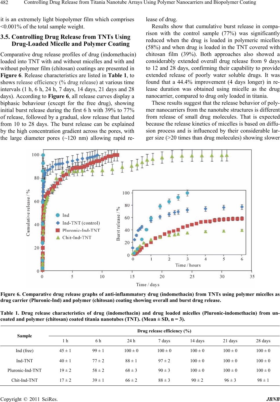 Controlling Drug Release from Titania Nanotube Arrays Using Polymer Nanocarriers and Biopolymer Coating Copyright © 2011 SciRes. JBNB 482 lease of drug. it is an extremely light biopolymer film which comprises <0.001% of the total sample weight. Results show that cumulative burst release in compa- rison with the control sample (77%) was significantly reduced when the drug is loaded in polymeric micelles (58%) and when drug is loaded in the TNT covered with chitosan film (39%). Both approaches also showed a considerably extended overall drug release from 9 days to 12 and 28 days, confirming their capability to provide extended release of poorly water soluble drugs. It was found that a 44.4% improvement (4 days longer) in re- lease duration was obtained using micelle as the drug nanocarrier, compared to drug only loaded in titania. 3.5. Controlling Drug Release from TNTs Using Drug-Loaded Micelle and Polymer Coating Comparative drug release profiles of drug (indomethacin) loaded into TNT with and without micelles and with and without polymer film (chitosan) coatings are presented in Figure 6. Release characteristics are listed in Table 1, to shows release efficiency (% drug release) at various time intervals (1 h, 6 h, 24 h, 7 days, 14 days, 21 days and 28 days). According to Figure 6, all release curves display a biphasic behaviour (except for the free drug), showing initial burst release during the first 6 h with 39% to 77% of release, followed by a gradual, slow release that lasted from 10 to 28 days. The burst release can be explained by the high concentration gradient across the pores, with the large diameter pores (~120 nm) allowing rapid re- These results suggest that the release behavior of poly- mer nanocarriers from the nanotube structures is different from release of small drug molecules. That is expected because the release kinetics of micelles is based on diffu- sion process and is influenced by their considerable lar- ger size (>20 times than drug molecules) showing slower Figure 6. Comparative drug release graphs of anti-inflammatory drug (indomethacin) from TNTs using polymer micelles as drug carrier (Pluronic-Ind) and polymer (c hitosan) c oating showing overall and burst dr ug release. Table 1. Drug release characteristics of drug (indomethacin) and drug loaded micelles (Pluronic-indomethacin) from un- coated and polymer (chitosan) coated titania nanotubes (TNT). (Mean ± SD, n = 3). Drug release efficiency (%) Sample 1 h 6 h 24 h 7 days 14 days 21 days 28 days Ind (free) 45 ± 1 99 ± 1 100 ± 0 100 ± 0 100 ± 0 100 ± 0 100 ± 0 Ind-TNT 40 ± 1 77 ± 2 88 ± 1 97 ± 2 100 ± 0 100 ± 0 100 ± 0 Pluronic-Ind-TNT 19 ± 2 58 ± 2 68 ± 3 90 ± 3 100 ± 0 100 ± 0 100 ± 0 Chit-Ind-TNT 17 ± 2 39 ± 1 66 ± 2 88 ± 3 90 ± 2 96 ± 3 98 ± 1 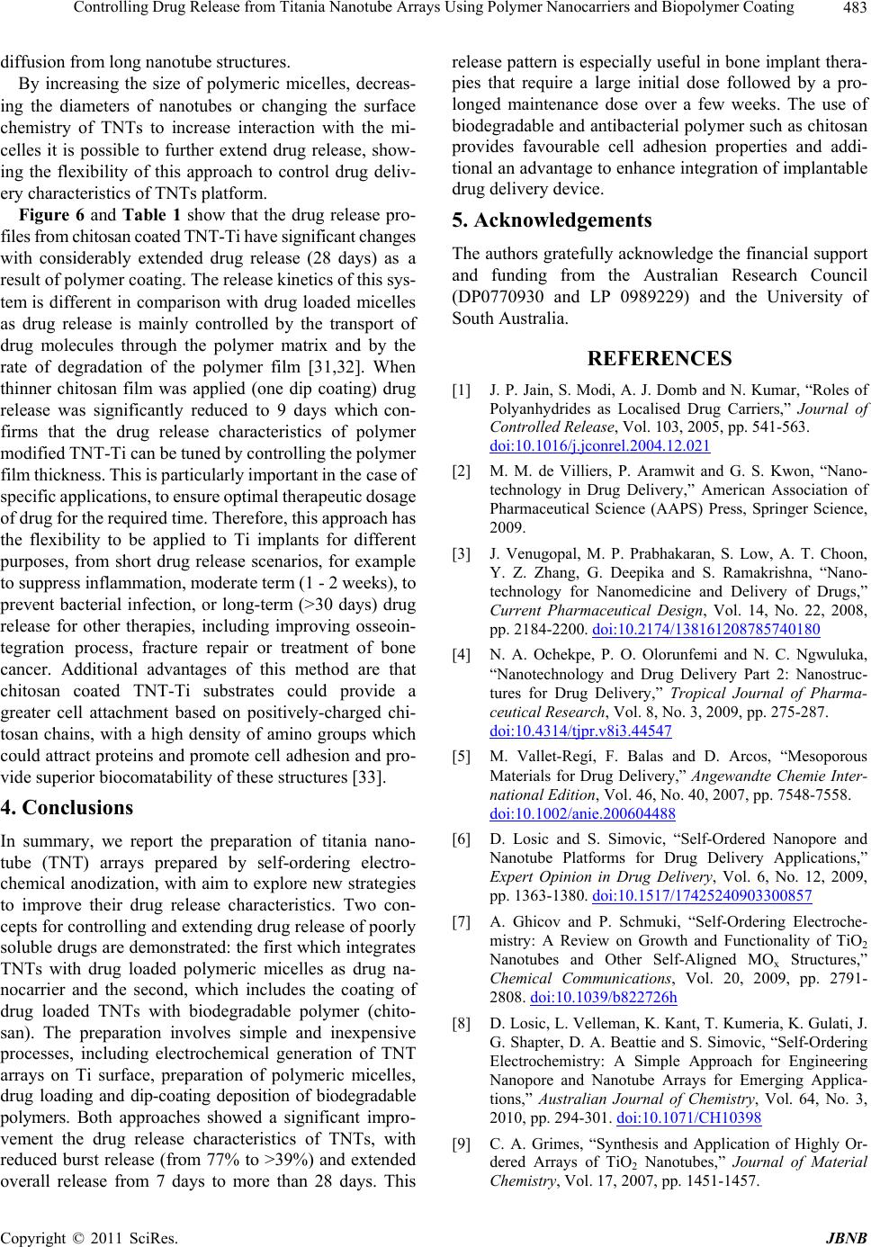 Controlling Drug Release from Titania Nanotube Arrays Using Polymer Nanocarriers and Biopolymer Coating483 diffusion from long nanotube structures. By increasing the size of polymeric micelles, decreas- ing the diameters of nanotubes or changing the surface chemistry of TNTs to increase interaction with the mi- celles it is possible to further extend drug release, show- ing the flexibility of this approach to control drug deliv- ery characteristics of TNTs platform. Figure 6 and Table 1 show that the drug release pro- files from chitosan coated TNT-Ti have significant changes with considerably extended drug release (28 days) as a result of polymer coating. The release kinetics of this sys- tem is different in comparison with drug loaded micelles as drug release is mainly controlled by the transport of drug molecules through the polymer matrix and by the rate of degradation of the polymer film [31,32]. When thinner chitosan film was applied (one dip coating) drug release was significantly reduced to 9 days which con- firms that the drug release characteristics of polymer modified TNT-Ti can be tuned by controlling the polymer film thickness. This is particularly important in the case of specific applications, to ensure optimal therapeutic dosage of drug for the required time. Therefore, this approach has the flexibility to be applied to Ti implants for different purposes, from short drug release scenarios, for example to suppress inflammation, moderate term (1 - 2 weeks), to prevent bacterial infection, or long-term (>30 days) drug release for other therapies, including improving osseoin- tegration process, fracture repair or treatment of bone cancer. Additional advantages of this method are that chitosan coated TNT-Ti substrates could provide a greater cell attachment based on positively-charged chi- tosan chains, with a high density of amino groups which could attract proteins and promote cell adhesion and pro- vide superior biocomatability of these structures [33]. 4. Conclusions In summary, we report the preparation of titania nano- tube (TNT) arrays prepared by self-ordering electro- chemical anodization, with aim to explore new strategies to improve their drug release characteristics. Two con- cepts for controlling and extending drug release of poorly soluble drugs are demonstrated: the first which integrates TNTs with drug loaded polymeric micelles as drug na- nocarrier and the second, which includes the coating of drug loaded TNTs with biodegradable polymer (chito- san). The preparation involves simple and inexpensive processes, including electrochemical generation of TNT arrays on Ti surface, preparation of polymeric micelles, drug loading and dip-coating deposition of biodegradable polymers. Both approaches showed a significant impro- vement the drug release characteristics of TNTs, with reduced burst release (from 77% to >39%) and extended overall release from 7 days to more than 28 days. This release pattern is especially useful in bone implant thera- pies that require a large initial dose followed by a pro- longed maintenance dose over a few weeks. The use of biodegradable and antibacterial polymer such as chitosan provides favourable cell adhesion properties and addi- tional an advantage to enhance integration of implantable drug delivery device. 5. Acknowledgements The authors gratefully acknowledge the financial support and funding from the Australian Research Council (DP0770930 and LP 0989229) and the University of South Australia. REFERENCES [1] J. P. Jain, S. Modi, A. J. Domb and N. Kumar, “Roles of Polyanhydrides as Localised Drug Carriers,” Journal of Controlled Release, Vol. 103, 2005, pp. 541-563. doi:10.1016/j.jconrel.2004.12.021 [2] M. M. de Villiers, P. Aramwit and G. S. Kwon, “Nano- technology in Drug Delivery,” American Association of Pharmaceutical Science (AAPS) Press, Springer Science, 2009. [3] J. Venugopal, M. P. Prabhakaran, S. Low, A. T. Choon, Y. Z. Zhang, G. Deepika and S. Ramakrishna, “Nano- technology for Nanomedicine and Delivery of Drugs,” Current Pharmaceutical Design, Vol. 14, No. 22, 2008, pp. 2184-2200. doi:10.2174/138161208785740180 [4] N. A. Ochekpe, P. O. Olorunfemi and N. C. Ngwuluka, “Nanotechnology and Drug Delivery Part 2: Nanostruc- tures for Drug Delivery,” Tropical Journal of Pharma- ceutical Research, Vol. 8, No. 3, 2009, pp. 275-287. doi:10.4314/tjpr.v8i3.44547 [5] M. Vallet-Regí, F. Balas and D. Arcos, “Mesoporous Materials for Drug Delivery,” Angewandte Chemie Inter- national Edition, Vol. 46, No. 40, 2007, pp. 7548-7558. doi:10.1002/anie.200604488 [6] D. Losic and S. Simovic, “Self-Ordered Nanopore and Nanotube Platforms for Drug Delivery Applications,” Expert Opinion in Drug Delivery, Vol. 6, No. 12, 2009, pp. 1363-1380. doi:10.1517/17425240903300857 [7] A. Ghicov and P. Schmuki, “Self-Ordering Electroche- mistry: A Review on Growth and Functionality of TiO2 Nanotubes and Other Self-Aligned MOx Structures,” Chemical Communications, Vol. 20, 2009, pp. 2791- 2808. doi:10.1039/b822726h [8] D. Losic, L. Velleman, K. Kant, T. Kumeria, K. Gulati, J. G. Shapter, D. A. Beattie and S. Simovic, “Self-Ordering Electrochemistry: A Simple Approach for Engineering Nanopore and Nanotube Arrays for Emerging Applica- tions,” Australian Journal of Chemistry, Vol. 64, No. 3, 2010, pp. 294-301. doi:10.1071/CH10398 [9] C. A. Grimes, “Synthesis and Application of Highly Or- dered Arrays of TiO2 Nanotubes,” Journal of Material Chemistry, Vol. 17, 2007, pp. 1451-1457. Copyright © 2011 SciRes. JBNB  Controlling Drug Release from Titania Nanotube Arrays Using Polymer Nanocarriers and Biopolymer Coating 484 doi:10.1039/b701168g [10] K. C. Popat, L. Leoni, C. A. Grimes and T. A. Desai, “Influence of Engineered Titania Nanotubular Surfaces on Bone Cells,” Biomaterials, Vol. 28, No. 21, 2007, pp. 3188-3197. doi:10.1016/j.biomaterials.2007.03.020 [11] P. A. Tran, L. Sarin, R. H. Hurt and T. J. Webster, “Op- portunities for Nanotechnology-Enabled Bioactive Bone Implants,” Journal of Material Chemistry, Vol. 19, No. 18, 2009, pp. 2653-2659. doi:10.1039/b814334j [12] E. Alpaslan, B. Ercan and T. J. Webster, “Anodized 20 nm Diameter Nanotubular Titanium for Improved Blad- der Stent Applications,” International Journal of Nano- medicine, Vol. 6, 2011, pp. 219-225. [13] J. Park, S. Bauer, K. von der Mark and P. Schmuki, “Nanosize and Vitality: TiO2 Nanotube Diameter Directs Cell Fate,” Nano Letters, Vol. 7, No. 6, 2007, pp. 1686- 1691. doi:10.1021/nl070678d [14] Y. Y. Song, F. Schmidt-Stein, S. Bauer and P. Schmuki, “Amphiphilic TiO2 Nanotube Arrays: An Actively Con- trollable Drug Delivery System,” Journal of American Chemical Society, Vol. 131, No. 12, 2009, pp. 4230-4233. doi:10.1021/ja810130h [15] L. M. Chamberlain, K. S. Brammer, G. W. Johnston, S. Chien and S. Jin, “Microphage Inflammatory Response to TiO2 Nanotube Surface,” Journal of Biomaterials and Nanobiotechnology, Vol. 2, 2011, pp. 293-300. doi:10.4236/jbnb.2011.23036 [16] H. Liu and T. J. Webster, “Nanomedicine for Implants: A Review of Studies and Necessary Experimental Tools,” Biomaterials, Vol. 28, No. 2, 2007, pp. 354-369. doi:10.1016/j.biomaterials.2006.08.049 [17] D. Losic, M. A. Cole, B. Dollmann, K. Vasilev and H. J. Griesser, “Surface Modification of Nanoporous Alumina Membranes by Plasma Polymerization,” Nanotechnology, Vol. 19, No. 24, 2008, pp. 5704-5711. doi:10.1088/0957-4484/19/24/245704 [18] S. Simovic, D. Losic and K. Vasilev, “Controlled Drug Release from Porous Materials by Plasma Polymer Depo- sition,” Chemical Communications, Vol. 46, No. 8, 2009, pp. 1317-1319. doi:10.1039/b919840g [19] K. Vasilev, Z. Poh, K. Kant, J. Chan, A. Michelmore and D. Losic, “Tailoring the Surface Functionalities of Titania Nanotube Arrays,” Biomaterials, Vol. 31, No. 3, 2010, pp. 532-540. doi:10.1016/j.biomaterials.2009.09.074 [20] M. S. Aw, S. Simovic, J. Addai-Mensah and D. Losic, “Polymeric Micelles in Porous and Nanotubular Implants as a New System for Extended Delivery of Poorly Solu- ble Drugs,” Journal of Material Chemistry, Vol. 21, No. 20, 2011, pp. 7082-7089. doi:10.1039/c0jm04307a [21] L. Qiu, C. Zheng, Y. Jin and K. Zhu, “Polymeric Micelles as Nanocarriers for Drug Delivery,” Expert Opinions on Therapeutic Patents, Vol. 17, No. 7, 2007, pp. 819-830. [22] M. L. Adams, A. Lavasanifar and G. S. Kwon, “Amphi- philic Block Copolymers for Drug Delivery,” Journal of Pharmaceutical Sciences, Vol. 92, No. 7, 2003, pp. 1343- 1355. doi:10.1002/jps.10397 [23] A. V. Kabanov, E. V. Batrakova and V. Y. Alakhov, “Pluronic® Block Copolymers as Novel Polymer Thera- peutics for Drug and Gene Delivery,” Journal of Con- trolled Release, Vol. 82, No. 2-3, 2002, pp. 189-212. doi:10.1016/S0168-3659(02)00009-3 [24] A. K. Singla and M. Chawla, “Chitosan: Some Pharma- ceutical and Biological Aspects—An Update,” Journal of Pharmacy and Pharmacology, Vol. 53, No. 8, 2001, pp. 1047-1067. doi:10.1211/0022357011776441 [25] H. Ueno, H. Yamada, I. Tanaka, N. Kaba, M. Matsuura and M. Okumura, “Accelerating Effects of Chitosan for Healing at Early Phase of Experimental Open Wound in Dogs,” Biomaterials, Vol. 20, No. 15, 1999, pp. 1407- 1414. doi:10.1016/S0142-9612(99)00046-0 [26] Y. Hu, J. Wang, Z. Zhi, T. Jiang and S. Wang, “Facile Synthesis of 3D Cubic Mesoporous Silica Microspheres with a Controllable Pore Size and Their Application for Improved Delivery of a Water-Insoluble Drug,” Journal of Colloid and Interface Science, Vol. 363, No. 1, 2011, pp. 410-417. doi:10.1016/j.jcis.2011.07.022 [27] K. Kant and D. Losic, “A Simple Approach for Synthe- sis of TiO2 Nanotubes with Through-Hole Morphology,” Physica Status Solidi (RRL)—Rapid Research Letters, Vol. 3, No. 5, 2009, pp. 139-141. [28] K. Kant and D. Losic, “Self-Ordering Electrochemical Synthesis of TiO2 Nanotube Arrays: Controlling the Nanotube Geometry and the Growth Rate,” International Journal of Nanoscience, Vol. 10, No. 1-2, 2011, pp. 1-6. doi:10.1142/S0219581X11007466 [29] Z. L. Tyrrell, Y. Shen and M. Radosz, “Fabrication of Micellar Nanoparticles for Drug Delivery through the Self-Assembly of Block Copolymers,” Progress in Poly- mer Science, Vol. 35, No. 9, 2010, pp. 1128-1143. doi:10.1016/j.progpolymsci.2010.06.003 [30] G. Bonacucina, M. Cespi, G. Mencarelli, G. Giorgioni and G. F. Palmieri, “Thermosensitive Self-Assembling Block Copolymers as Drug Delivery Systems,” Polymers, Vol. 3, No. 2, 2011, pp. 779-811. doi:10.3390/polym3020779 [31] A. Y. Polishcuck, L. A. Zimina, R. Y. Ksenko, A. L. Iordanskii and G. E. Zaikov, “Diffusion-Activation Laws for Drug Release from Polymer Matrices,” Polymer Deg- radation and Stability, Vol. 31, No. 2, 1991, pp. 247-254. doi:10.1016/0141-3910(91)90079-7 [32] S. A. Agnihotri, N. N. Mallikarjuna and T. M. Aminab- havi, “Recent Advances on Chitosan-Based Micro- and Nanoparticles in Drug Delivery,” Journal of Controlled Release, Vol. 100, No. 1, 2004, pp. 5-28. doi:10.1016/j.jconrel.2004.08.010 [33] J. D. Bumgardner, R. Wiser, P. D. Gerard, P. Bergin, B. Chestnutt, M. Marini, V. Ramsey, S. H. Elder and J. A. Gilbert, “Chitosan: Potential Use as a Bioactive Coating for Orthopaedic and Craniofacial/Dental Implants,” Jour- nal of Biomaterials Science-Polymer Edition, Vol. 14, No. 5, 2003, pp. 423-438. doi:10.1163/156856203766652048 Copyright © 2011 SciRes. JBNB |