

Paper Menu >>
Journal Menu >>
 Chinese Medicine, 2011, 2, 138-142 doi:10.4236/cm.2011.24023 Published Online December 2011 (http://www.SciRP.org/journal/cm) Copyright © 2011 SciRes. CM Comparative Efficacy of Tagetes erecta and Centella asiatica Extracts on Wound Healing in Albino Rats Sandipan Chatterjee1, Tigari Prakash1*, Dupadahalli Kotrsha2, Nadendla Rama Rao3, Divakar Goli1 1Department of Phar macology, Acharya & B M Reddy College of Pharmacy, Bangalore, India 2Department of Parasi t ology, Faculty of Medicine, University of Malaysia, Kuala Lumpur, Malaysia 3Department of Pharmaceutical Chemistry, Chalapathi Institute of Pharmaceutical Science, Guntur, India E-mail: [email protected] Received July 20, 2011; revised August 22, 2011; accepted August 29, 2011 Abstract The study was undertaken to evaluate the comparative efficacy of hydroalcoholic extracts of leaves of Ta- getes erecta (T. erecta) and aerial parts of Centella asiatica (C. asiatica) on Excision, Incision and Dead space wound models in albino rats. Extract of T. erecta and C. asiatica (P < 0.001) showed significant in- crease in rate of wound contraction, epithelization and formation of scar faster in excision wound model compare to control group. T. erecta extract (250 and 500 mg/kg) showed significantly increased the wound breaking strength in incision wound model and wet and dry granulation tissue weights, breaking strength in a dead space wound model compare to control and C. asiatica treated group (P < 0.001). In this investigation the work is conclude that the comparison made between the above two plants, the T. erecta extract showed potent wound healing activity then the reported C. asiatica in different wound parameters. Keywords: Centella asiatica, Tagetes erecta, Excision, Incision, Dead Space Wound Model 1. Introduction Wound healing or wound repair, is the body’s natural pr- ocess. Have regenerating dermal and epidermal tissues. When an individual is wounded, a set of events takes place in a predictable fashion to repair the damages. Wo- unds were probably the first medical problem faced by human race. The field of pharmacologic modulation in wound healing is still in its infancy. Indians were acqu- ainted with a far larger number of medicinal plants than the natives of other country. In fact ancient Indian sur- geon sushruta has enumerated large number of herbal dr- ug in the management of wounds singly or in combina- tion. Some Ayurvedic medicinal plants, namely, Centella asiatica, Curcuma longa, Aloe vera, and Tagetes erecta Linn, were found to be effective on wound healing in ex- perimental models. Tagetes erecta (T. erecta) is commonly known as Ma- rigold. The plant is widely distributed throughout India, Asia, Central Europe, USA and Africa. The leaves are reported to be effective against piles, kidney troubles, muscular pain, ulcers, wounds and earache. The pounded leaves are used as an external application to boils and carbuncles. The steam distillation of fresh leaves offer 0.3% of essential oil with a strong, sweet lasting odor and contains d-limonene, ocimene, l-linalyl acetate, l-li- nalool tagetone, n-nonyl aldehyde, lutein [1]. Centella asiatica (C. asiatica) is commonly known as Indian pennywort. The plant is widely distributed in Asia, Africa, and North and South America [2]. This plant is reportedly useful in wound healing and protecting ag- ainst ulcer formation and is reported to have antimicro- bial properties. Extracts of the plant have been shown to accelerate wound healing through topical and systemic routes [3]. The extracts of C. asiatica plant are reported to have sedative, antidepressant, analgesic, and anticon- vulsive effects [4]. It is also reputed within the Indian system of medicine for its use in treating chronic and ob- stinate eczema, psoriasis, syphilis and leprosy [5]. The titrated extract of C. asiatica contains three principal ingredients like asiaticoside, asiaticacid, and madecassic acid. All these ingredients are known to be clinically effective in the treatment of systemic scleroderma, ab- normal scar formation [6] and wound-healing activity [7, 8]. The present study was planned to evaluate the com- parative efficacy of the hydroalcoholic extract of aerial 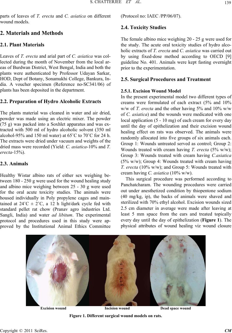 139 S. CHATTERJEE ET AL. parts of leaves of T. erecta and C. asiatica on different wound models. 2. Materials and Methods 2.1. Plant Materials Leaves of T. erecta and arial part of C. asiatica was col- lected during the month of November from the local ar- eas of Burdwan District, West Bengal, India and both the plants were authenticated by Professor Udayan Sarkar, HOD, Dept of Botany, Sonamukhi College, Bankura, In- dia. A voucher specimen (Reference no-SC341/06) of plants has been deposited in the department. 2.2. Preparation of Hydro Alcoholic Extracts The plants material was cleaned in water and air dried, powder was made using an electric mixer. The powder (75 g) was packed into a Soxhlet apparatus and was ex- tracted with 500 ml of hydro alcoholic solvent (350 ml alcohol-95% and 150 ml water) at 65˚C to 70˚C for 24 h. The extracts were dried under vacuum and weights of the dried mass were recorded (Yield: C. asiatica-10% and T. erecta-15%). 2.3. Animals Healthy Wistar albino rats of either sex weighing be- tween 180 - 250 g were used for the wound healing study and albino mice weighing between 25 - 30 g were used for the oral acute toxicity studies. The animals were housed individually in Poly propylene cages and main- tained at 24˚C ± 2˚C, a 12 h light/dark cycle fed with standard pellet rat chow (Pranav agro industries Ltd. Sangli, India) and water ad libitum. The experimental protocol and procedures used in this study were ap- proved by the Institutional Animal Ethics Committee (Protocol no: IAEC /PP/06/07). 2.4. Toxicity Studies The female albino mice weighing 20 - 25 g were used for the study. The acute oral toxicity studies of hydro alco- holic extracts of T. erecta and C. asiatica was carried out by using fixed-dose method according to OECD [9] guideline No. 401. Animals were kept fasting overnight prior to the experimentation. 2.5. Surgical Procedures and Treatment 2.5.1. Excision Wound Model In the present experimental model two different types of creams were formulated of each extract (5% and 10% w/w of T. erecta and the other having 5% and 10% w/w of C. asia tica) and the wounds were medicated with one local application (5 - 10 mg) of each cream for every day until the day of epithelization and their excision wound healing effect on rats was observed. The animals were randomly allocated into five groups of six animals each. Group 1: Wounds untreated served as control; Group 2: Wounds treated with cream having T. erecta (5% w/w); Group 3: Wounds treated with cream having C.asiatica (5% w/w); Group 4: Wounds treated with cream having T. erecta (10% w/w); and Group 5: Wounds treated with cream having C. asiati ca (10% w/w). This surgical procedure was performed according to Panchatcharam. The wounding procedures were carried out under anesthetized condition by thiopentone sodium (40 mg/kg, ip), the backs of animals were shaved and sterilized with 70% ethyl alcohol. Excision wounds sized 2.5 cm diameter in average were made after leaving at least 5 mm space from the ears and treated topically every day until the day of epithelization (Figure 1). The physical attributes of wound healing viz wound closure Excision wound Incision wound Dead space wound Figure 1. Different surgical w ound models on r ats. Copyright © 2011 SciRes. CM 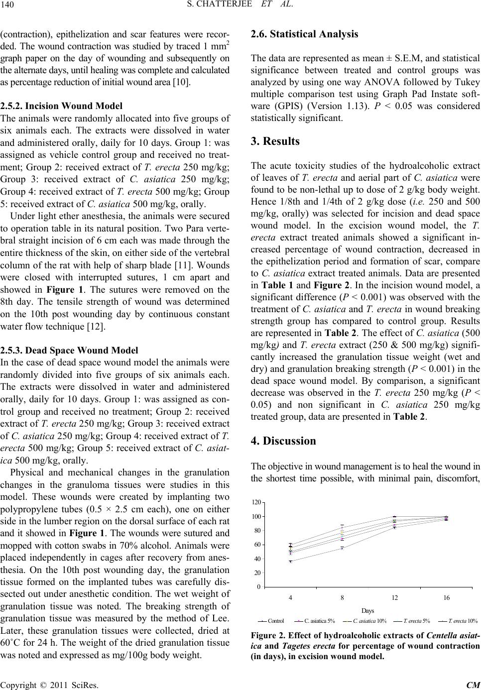 S. CHATTERJEE ET AL. 140 (contraction), epithelization and scar features were recor- ded. The wound contraction was studied by traced 1 mm2 graph paper on the day of wounding and subsequently on the alternate days, until healing was complete and calculated as percentage reduction of initial wound area [10]. 2.5.2. I n cision Wound Model The animals were randomly allocated into five groups of six animals each. The extracts were dissolved in water and administered orally, daily for 10 days. Group 1: was assigned as vehicle control group and received no treat- ment; Group 2: received extract of T. erecta 250 mg/kg; Group 3: received extract of C. asiatica 250 mg/kg; Group 4: received extract of T. erecta 500 mg/kg; Group 5: received extract of C. asiatica 500 mg/kg, orally. Under light ether anesthesia, the animals were secured to operation table in its natural position. Two Para verte- bral straight incision of 6 cm each was made through the entire thickness of the skin, on either side of the vertebral column of the rat with help of sharp blade [11]. Wounds were closed with interrupted sutures, 1 cm apart and showed in Figure 1. The sutures were removed on the 8th day. The tensile strength of wound was determined on the 10th post wounding day by continuous constant water flow technique [12]. 2.5.3. Dead Spac e Wound Model In the case of dead space wound model the animals were randomly divided into five groups of six animals each. The extracts were dissolved in water and administered orally, daily for 10 days. Group 1: was assigned as con- trol group and received no treatment; Group 2: received extract of T. erecta 250 mg/kg; Group 3: received extract of C. asia tica 250 mg/kg; Group 4: received extract of T. erecta 500 mg/kg; Group 5: received extract of C. asiat- ica 500 mg/kg, orally. Physical and mechanical changes in the granulation changes in the granuloma tissues were studies in this model. These wounds were created by implanting two polypropylene tubes (0.5 × 2.5 cm each), one on either side in the lumber region on the dorsal surface of each rat and it showed in Figure 1. The wounds were sutured and mopped with cotton swabs in 70% alcohol. Animals were placed independently in cages after recovery from anes- thesia. On the 10th post wounding day, the granulation tissue formed on the implanted tubes was carefully dis- sected out under anesthetic condition. The wet weight of granulation tissue was noted. The breaking strength of granulation tissue was measured by the method of Lee. Later, these granulation tissues were collected, dried at 60˚C for 24 h. The weight of the dried granulation tissue was noted and expressed as mg/100g body weight. 2.6. Statistical Analysis The data are represented as mean ± S.E.M, and statistical significance between treated and control groups was analyzed by using one way ANOVA followed by Tukey multiple comparison test using Graph Pad Instate soft- ware (GPIS) (Version 1.13). P < 0.05 was considered statistically significant. 3. Results The acute toxicity studies of the hydroalcoholic extract of leaves of T. erecta and aerial part of C. asiatica were found to be non-lethal up to dose of 2 g/kg body weight. Hence 1/8th and 1/4th of 2 g/kg dose (i.e. 250 and 500 mg/kg, orally) was selected for incision and dead space wound model. In the excision wound model, the T. erecta extract treated animals showed a significant in- creased percentage of wound contraction, decreased in the epithelization period and formation of scar, compare to C. asiatica extract treated animals. Data are presented in Table 1 and Figure 2. In the incision wound model, a significant difference (P < 0.001) was observed with the treatment of C. asiatica and T. erecta in wound breaking strength group has compared to control group. Results are represented in Table 2. The effect of C. asiatica (500 mg/k g ) and T. erecta extract (250 & 500 mg/kg) signifi- cantly increased the granulation tissue weight (wet and dry) and granulation breaking strength (P < 0.001) in the dead space wound model. By comparison, a significant decrease was observed in the T. erecta 250 mg/kg (P < 0.05) and non significant in C. asiatica 250 mg/kg treated group, data are presented in Table 2. 4. Discussion The objective in wound management is to heal the wound in the shortest time possible, with minimal pain, discomfort, 0 20 40 60 80 100 120 4 812 16 Da y s Control C. asiatica 5%C . a sia tic a 10 % T. erecta 5% T . erecta 10% Figure 2. Effect of hydroalcoholic extracts of Centella asiat- ica and Tagetes erecta for percentage of wound contraction (in days), in excision wound model. Copyright © 2011 SciRes. CM 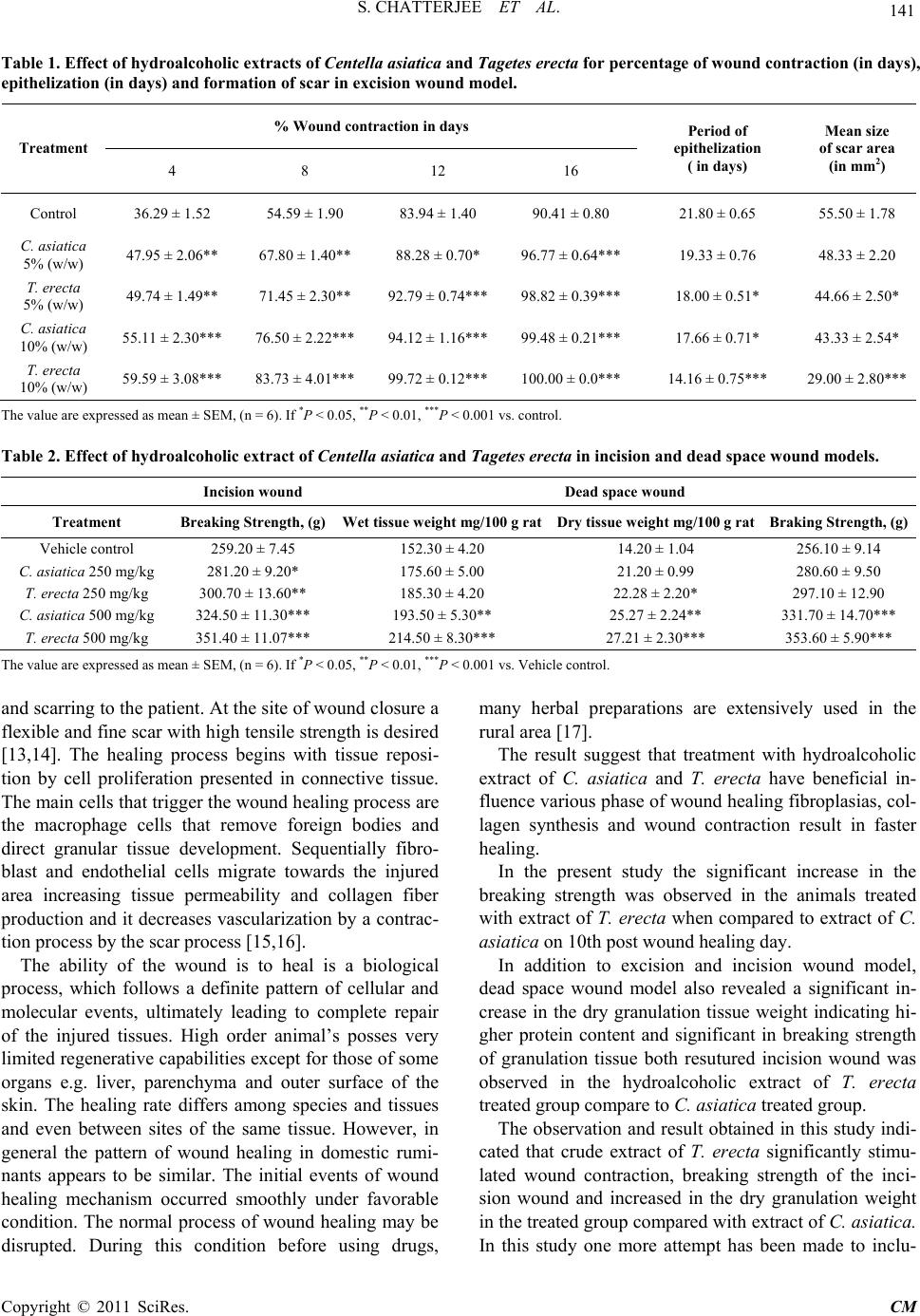 141 S. CHATTERJEE ET AL. Table 1. Effect of hydroalcoholic extracts of Centella asiatica and Tagetes erecta for percentage of wound contract ion (in days), epithelization (in days) and formation of scar in exci sion wound model. % Wound contraction in days Treatment 4 8 12 16 Period of epithelization ( in days) Mean size of scar area (in mm2) Control 36.29 ± 1.52 54.59 ± 1.90 83.94 ± 1.40 90.41 ± 0.80 21.80 ± 0.65 55.50 ± 1.78 C. asiatica 5% (w/w) 47.95 ± 2.06** 67.80 ± 1.40** 88.28 ± 0.70* 96.77 ± 0.64*** 19.33 ± 0.76 48.33 ± 2.20 T. erecta 5% (w/w) 49.74 ± 1.49** 71.45 ± 2.30** 92.79 ± 0.74*** 98.82 ± 0.39*** 18.00 ± 0.51* 44.66 ± 2.50* C. asiatica 10% (w/w) 55.11 ± 2.30*** 76.50 ± 2.22*** 94.12 ± 1.16*** 99.48 ± 0.21*** 17.66 ± 0.71* 43.33 ± 2.54* T. erecta 10% (w/w) 59.59 ± 3.08*** 83.73 ± 4.01*** 99.72 ± 0.12*** 100.00 ± 0.0*** 14.16 ± 0.75*** 29.00 ± 2.80*** The value are expressed as mean ± SEM, (n = 6). If *P < 0.05, **P < 0.01, ***P < 0.001 vs. control. Table 2. Effect of hydroalcoholic ext ract of Centella asiatica and Tagetes erecta in incision and dead space wound models. Incision wound Dead space wound Treatment Breaking Strength, (g) Wet tissue weight mg/100 g ratDry tissue weight mg/100 g rat Braking Strength, (g) Vehicle control 259.20 ± 7.45 152.30 ± 4.20 14.20 ± 1.04 256.10 ± 9.14 C. asiatica 250 mg/kg 281.20 ± 9.20* 175.60 ± 5.00 21.20 ± 0.99 280.60 ± 9.50 T. erecta 250 mg/kg 300.70 ± 13.60** 185.30 ± 4.20 22.28 ± 2.20* 297.10 ± 12.90 C. asiatica 500 mg/kg 324.50 ± 11.30*** 193.50 ± 5.30** 25.27 ± 2.24** 331.70 ± 14.70*** T. erecta 500 mg/kg 351.40 ± 11.07*** 214.50 ± 8.30*** 27.21 ± 2.30*** 353.60 ± 5.90*** The value are expressed as mean ± SEM, (n = 6). If *P < 0.05, **P < 0.01, ***P < 0.001 vs. Vehicle control. and scarring to the patient. At the site of wound closure a flexible and fine scar with high tensile strength is desired [13,14]. The healing process begins with tissue reposi- tion by cell proliferation presented in connective tissue. The main cells that trigger the wound healing process are the macrophage cells that remove foreign bodies and direct granular tissue development. Sequentially fibro- blast and endothelial cells migrate towards the injured area increasing tissue permeability and collagen fiber production and it decreases vascularization by a contrac- tion process by the scar process [15,16]. The ability of the wound is to heal is a biological process, which follows a definite pattern of cellular and molecular events, ultimately leading to complete repair of the injured tissues. High order animal’s posses very limited regenerative capabilities except for those of some organs e.g. liver, parenchyma and outer surface of the skin. The healing rate differs among species and tissues and even between sites of the same tissue. However, in general the pattern of wound healing in domestic rumi- nants appears to be similar. The initial events of wound healing mechanism occurred smoothly under favorable condition. The normal process of wound healing may be disrupted. During this condition before using drugs, many herbal preparations are extensively used in the rural area [17]. The result suggest that treatment with hydroalcoholic extract of C. asiatica and T. erecta have beneficial in- fluence various phase of wound healing fibroplasias, col- lagen synthesis and wound contraction result in faster healing. In the present study the significant increase in the breaking strength was observed in the animals treated with extract of T. erecta when compared to extract of C. asiatica on 10th post wound healing day. In addition to excision and incision wound model, dead space wound model also revealed a significant in- crease in the dry granulation tissue weight indicating hi- gher protein content and significant in breaking strength of granulation tissue both resutured incision wound was observed in the hydroalcoholic extract of T. erecta treated group compare to C. asiatica treated group. The observation and result obtained in this study indi- cated that crude extract of T. erecta significantly stimu- lated wound contraction, breaking strength of the inci- sion wound and increased in the dry granulation weight in the treated group compared with extract of C. asiatica. In this study one more attempt has been made to inclu- Copyright © 2011 SciRes. CM  S. CHATTERJEE ET AL. 142 sion T. erecta plant in the management of wound healing in folk medicine is justifiable. 5. Conclusions In the present work it can be concluded that the com- parison made between the above plants of hydroalcoholic extract of T. erecta showed promising wound healing activity then the reported C. asiatica in different wound parameters. 6. Acknowledgements The authors are grateful to Premnath Reddy, Chairman Acharya Institute, and Principal, Acharya & B. M. Re- ddy College of Pharmacy, Bangalore, India, for the con- stant support and facilities to this study. 7. References [1] T. Ghosh, A. Bose, G. K. Dash and T. K. Maity, “Wound Healing Activity of Tagetes erecta Linn Leaves,” 2004. URL http://www. Pharmainfo.net/exclusive/reviews [2] B. Brinkhaus, “Centella asiatica in Traditional and Mod- ern Phytomedicine―a Pharmacological and Clinical Pro- file, Part I: Botany Chemistry Preparations,” Perfusion, Vol. 11, 1998, pp. 466-474. [3] B. Brinkhaus, “Centella asiatica in Traditional and Mod- ern Phytomedicine―a Pharmacological and Clinical Pro- file, Part II: Botany Chemistry Preparations,” Perfusion, Vol. 11, 1998, pp. 508-520. [4] R. P. Lubadie, “An Ehnopharmacognostic Approach to the Search for Immunomodulators of Plant Origin,” Planta Medica, Vol. 55, 1989, pp. 339-348. doi:10.1055/s-2006-962024 [5] S. Chaudhary, S. Poddar, S. Sarkar and P. K. Das, “New Multidrug Regimen with Indigenous Drugs and Dapsone in the Treatment of Lepromatous Leprosy,” Indian Jour- nal of Dermatology, Vol. 32, No. 3, 1987, pp. 63-67. [6] S. S. Hong, J. K. Kim, H. Li and C. K. Shim, “Advanced Formulation and Pharmacological Activity of Hydrogel of the Titrated Extract of Centella asiatica,” Archives of Pharmacal Research, Vol. 28, No. 4, 2005, pp. 502-508. doi:10.1007/BF02977683 [7] A. Shukla, A. M. Rasik, G. K. Jain, R. Shankar, D. K. Kulashrestha and B. N. Dhawan, “In vitro Wound Heal- ing Activity of Asiaticoside Isolated from Centella asiat- ica,” Journal of Ethnopharmacology, Vol. 65, No. 1, 1999, pp. 1-11. doi:10.1016/S0378-8741(98)00141-X [8] S. Somashekar, S. L. Udupa and A. L. Udupa, “Effect of Centella asiatica Linn (Umbelliferae) on Normal and Dexmetasone Suppressed Wound Healing in Wistar Al- bino Rats,” The International Journal of Lower Extremity Wounds, Vol. 5, 2006, p. 136. [9] Organization for Economic cooperation and development (OECD), “OECD Guidelines for Testing of Chemicals Acute Oral Toxicity,” OECD, Paris, 1993. p. 401. [10] J. J. P. Morton and M. H. Malone, “Evaluation of Vul- nary Activity by an Open Wound Procedure in Rats,” Archives Internationales de Pharmacodynamie, Vol. 196, 1997, pp. 117-126. [11] H. P. Ehrlich and T. K. Hunt, “Effect of Cortisone and Anabolic Steroids on the Tensilestrength of Healing Wounds,” British Journal of Surgery, Vol. 170, 1969, pp. 203-206. [12] K. H. Lee, “Studies on the Mechanism of Action of Sali- cylates II. Retardation of Wound Healing by Aspirin,” Journal of Pharmaceutical Sciences, Vol. 57, No. 6, 1968, pp. 1042-1043. doi:10.1002/jps.2600570633 [13] B. S. Reddy, R. K. K. Reddy, V. G. M. Naidu, K. Mad- husudhana, S. B. Agwane, S. Ramakrishna and P. V. Di- wan, “Evaluation of Antimicrobial, Antioxidant and Wound Healing Potentials of Holoptelea integrifolia,” Journal of Ethnopharmacology, Vol. 115, No. 2, 2008, pp. 249-256. doi:10.1016/j.jep.2007.09.031 [14] I. Süntar, U. Koca, H. Keleş and E. K. Akko, “Wound Healing Activity of Rubus sanctus Schreber (Rosaceae): Preclinical Study in Animal Models,” Evidence-Based Complementary and Alternative Medicine, Vol. 2011, Article ID: 816156. [15] P. J. Michelle, M. Cristiana, T. G. R. Ana Lu´ cia, T. F. Alik, A. F. R. Rodney, M. O. S. Ilza, A. F. Mary and E. C. Jo˜ao, “Evaluation of Wound Healing Properties of Ar- rabidaea chica Verlot Extract,” Journal of Ethnophar- macology, Vol. 118, No. 3, 2008, pp. 361-366. doi:10.1016/j.jep.2008.04.024 [16] S. K. Prasad, R. Kumar, D. K. Patel and S. Hemalatha, “Wound Healing Activity of Withania Coagulans in Str- eptozotocin-Induced Diabetic Rats,” Pharmaceutical Bi- ology, Vol. 48, No, 12, 2010, pp. 1397-1404. doi:10.3109/13880209.2010.486837 [17] M. M. Alam, S. A. Islam, Y. Mohammed, N. S. Juyena and M. A. Hashim, “Comparative Efficacy of Two Me- dicinal Plant Extracts and an Antibiotic on Wound Heal- ing,” Pakistan Journal of Biological Sciences, Vol. 8, No. 5, 2005, pp. 740-743. doi:10.3923/pjbs.2005.740.743 Copyright © 2011 SciRes. CM |