

Paper Menu >>
Journal Menu >>
 Advances in Molecular Imaging, 2012, 2, 1-3 http://dx.doi.org/10.4236/ami.2012.21001 Published Online January 2012 (http://www.SciRP.org/journal/ami) MRI Finding in Delayed Extensive Brain Lesions after Radiation Therapy: Cortical Laminar Necrosis and White Matter Myelinolysis Yeting Zhou1, Guangsheng Wang2, Xiaodong Chen2, Daoming Tong2 1Medical Evaluation Unit, Shuyang People’Hospital, Shuyang, China 2Departments of Neurology, Shuyang People’ Ho sp ital, Shuyang, Chi n a Email: [email protected] Received September 11, 2011; revised October 13, 2011; accepted November 2, 2011 ABSTRACT The focal and diffuse cerebral white matter injury can be caused by treatment with radiation therapy for cranial tumors. However, the literature rarely describes a MRI finding in radiation-induced delayed extensive cerebral injury. Our ob- jective was to report a r are case who had a delayed ex tensive hyperintensity injury in brain on MRI after radiation ther- apy due to nasopharyngeal cancer. A MRI was performed on a 40-year-old patient with extensive brain damage who had the radiation therapy two years ago. MRI finding was evaluated. On MRI, T2-weighted MRI showed an extensive hyperintensity after treated by irradiation. The radiographic pattern of extensive cerebral injury is relatively distinct. It involves the white matter and gray matter in cerebral, cerebellum, medulla oblongata, pons, internal capsule and thala- mus bilaterally. Our observations demonstrate that the extensive hyperintensity lesions in brain on MRI after radiation therapy is a cortical laminar necrosis and white matter myelinolysis. Keywords: Radiation-Induced Brain Damage; Magnetic Resonance Imaging; Encephalopathy; Cortical Laminar Necrosis; Demyelination 1. Introduction Radiation therapy can prolong survival of cancer patients to improve their quality of life, but it occasionally induces the human body, including brain radiation injury. Radia- tion-induced brain damage is considered an uncommon delayed complication among cancer survivors who have undergone whole brain irradiation for brain tumors [1-5]. However, the extensive damages after brain radiotherapy, especially in extensive bilateral brain, cerebellum and brain stem damage, bilateral internal capsule and thala- mus represent a rare late-onset events. In this paper we report a nasopharyngeal carcinoma patient who had a rare delayed complication of a wide range of brain dam- age after radiotherapy and his brain magnetic resonance (MR) imaging finding. 2. Meterial and Methods A 40-year-old male patien t, he h ad su ffered resectio n and radiation therapy because of the nasopharyngeal carci- noma for 2 years. He was symptom-free until 1 month before admission at which time he presented with symp- toms of bilateral progressive weakness to complete dis- ability, slurred speech, and slig htly dysphagia. At the age of 38 years, he had a series of episodes of “nasopharyngeal carcinoma, auricle inflammation” and he was begun treatment with radiation therapy such as 70 Gy nasopharyngeal and gamma knife 8 Gy irradiation in hospital, the patient’s symptoms slowly recovered and he was administered. The temperature was 37.3˚C, the pulse 94, and the respirations 22. The blood pressure was 130/90 mmHg. A general physical examination revealed no im- portant abnormalities. On neurologic examination, the patient was fully alert and oriented; his speech was slu- rred. He had four limbs palsy with grade 3-4 in the upper limbs and grade 3 in the lower limbs, the sensory impair- ment was not demonstrated. The deep tendon reflexes of the extremities were disappeared. The bilateral Hoffm- ann’s sign and Babinski’s sign were present, but decline in cognitive facu lties. Th e cran ia l-nerv e func tions were no- rmal. On admission, a brain CT scan, brain MRI, and the others examination were performed .Brain MRI finding, pathogenesis and prognosis were then analyzed. 3. Result Brain CT scan showed postoperative changes. Brain T2- weighted cranial magnetic resonance imaging (MRI) C opyright © 2012 SciRes. AMI 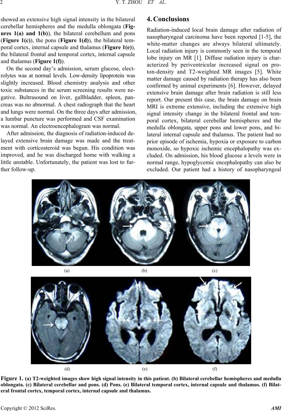 Y. T. ZHOU ET AL. 2 showed an extensive h igh s ign al in tens it y in th e bil ater al cerebellar hemispheres and the medulla oblongata (Fig- ures 1(a) and 1(b)), the bilateral cerebellum and pons (Figure 1(c)), the pons (Figure 1(d)), the bilateral tem- poral cortex, internal capsule and thalamus (Figure 1(e)), the bilateral frontal and temporal cortex, internal capsule and thalamus (Figure 1(f)). On the second day’s admission, serum glucose, elect- rolytes was at normal levels. Low-density lipoprotein was slightly increased. Blood chemistry analysis and other toxic substances in the serum screening results were ne- gative. Bultrasound on liver, gallbladder, spleen, pan- creas was no abnormal. A chest radiograph that the heart and lungs were normal. On the three days af ter admission , a lumbar puncture was performed and CSF examination was normal. An electroencephalogram was normal. After admission, the diagnosis of radiation-induced de- layed extensive brain damage was made and the treat- ment with corticosteroid was begun. His condition was improved, and he was discharged home with walking a little unstable. Unfortunately, the patient was lost to fur- ther follow-up. 4. Conclusions Radiation-induced local brain damage after radiation of nasopharyngeal carcinoma have been reported [1-5], the white-matter changes are always bilateral ultimately. Local radiation injury is commonly seen in the temporal lobe injury on MR [1]. Diffuse radiation injury is char- acterized by periventricular increased signal on pro- ton-density and T2-weighted MR images [5]. White matter damage caused by radiation therapy has also been confirmed by animal experiments [6]. However, delayed extensive brain damage after brain radiation is still less report. Our present this case, the brain damage on brain MRI is extreme extensive, including the extensive high signal intensity change in the bilateral frontal and tem- poral cortex, bilateral cerebellar hemispheres and the medulla oblongata, upper pons and lower pons, and bi- lateral internal capsule and thalamus. The patient had no prior episode of ischemia, hypoxia or exposure to carbon monoxide, so hypoxic ischemic encephalopathy was ex- cluded. On admission, his blood glucose a levels were in normal range, hypoglycemic encephalopathy can also be excluded. Our patient had a history of nasopharyngeal (a) (b) (c) (d) (e) (f) Figure 1. (a) T2-weighted images show high signal intensity in this patient. (b) Bilateral cerebellar hemispheres and medulla oblongata. (c) Bilateral cerebellar and pons. (d) Pons. (e) Bilateral temporal cortex, internal capsule and thalamus. (f) Bilat- eral frontal cortex, temporal cortex, internal capsule and thalamus. Copyright © 2012 SciRes. AMI  Y. T. ZHOU ET AL. 3 cancer and ear infection, and his symptoms was relieved when he was undergone a surgery and radiation therapy of nasopharyngeal carcinoma in two years ago. In add- tion, CT scan of the nasopharyngeal and systemic ex- amination had no evidence of recurrence or metastasis of cancer on admission, the diagnosis of radiation-induced delayed extensive brain damage had been made. Although our patient had an extensive brain damage, especially the extensive high signal change in the pons, the patient is neither the clinical manifestations of the network of nuclei and interconnecting tracts damages in the upper brain stem, nor the clinical signs of brain nuclei damage in the upper pons. Despite patient had a high signal changes in the cortex, he did not diminish the clinical man ifestations of in telligence. This s hows that th e patient’s brain damage is mainly in white matter, while less in gray matter. Prior study had confirmed that it is a radiation leukoencephalopathy [7]. Radiation leukoen- cephalopathy is not very common a complication after radiotherapy. The mechanism of the delayed brain dam- age after radiation therapy has not yet reached a complete consensus. Most studies considered that the radiation therapy-induced delayed brain damage is associated with the demyelination and necrosis [2,7,8]. It has also been confirmed by histopathology [7]. In some instances, the delayed brain damage is associated with ischemia or vascular occlusion [7,9]. Recent, other suggests that these high signals represent neuronal apoptosis [10]. High sig- nals in the white matter on MRI represent a demyelina- tion which has been largely accepted, but we considered that the high signals in the cortex may represent a corti- cal laminar necrosis. Therefore, the radiation therapy- induced delayed extensive brain damage is a relatively rare cortical laminar necrosis and white matter myeli- nolysis. On the other hand, MRI is not only helpful for diagnosis, but also to predict the prognosis of patient. In most cases, delayed leukoencephalopathy after radiation therapy have been improved or relieved [1]. Patient with the formation of cyst may cause a poor functional recov- ery [1]. Rarely, the patient with dementia often develops a poor prognosis, even death [11]. This shows that the prognosis of delayed radioactive brain damage often re- lated with the severity of brain lesions. In our case, MRI showed high sign al although more extensiv e distribution, but not very serious. The outcome of patient has been improved by continuous use of corticosteroid treatment. This suggests that these high signals not only represent a cortical laminar necrosis and white matter myelinolysis, but also confirm that the radiation therapy-induced corti- cal laminar necrosis and white matter myelinolysis is not completely irreversible. REFERENCES [1] Y. X. Wang, A. D. King, H. Zhou, et al., “Evolution of Radiation-Induced Brain Injury: MR Imaging-Based Study,” Radiology, Vol. 254, No. 1, 2010, pp. 210-218. doi:10.1148/radiol.09090428 [2] J. S. Tsuruda, K. E. Kortman, W. G. Bradley, et al., “Ra- diation Effects on Cerebral White Matter: MR Evalua- tion,” American Journal of Roentgenology, Vol. 149, No. 1, 1987, pp. 165-171. [3] Y. I. Chan, S. F. Leung, A. D. King, et al., “Late Radia- tion Injury to the Temporal Lobes: Morphologic Evalua- tion at MR Imaging,” Radiology, Vol. 213, No. 3, 1999, pp. 800-807. [4] A. Asai and K. Kawamoto, “Radiation-Induced Brain Injury,” Brain Nerve, Vol. 60, No. 2, 2008, pp. 123-129. [5] P. L. Khong, D. L. Kwong, G. C. Chan, et al., “Diffu- sion-Tensor Imaging for the Detection and Quantification of Treatment-Induced White Matter Injury in Children with Medulloblastoma: A Pilot Study,” American Journal of Neuroradiology, Vol. 24, No. 4, 2003, pp. 734-740. [6] S. Wang, E. X. Wu, D. Qiu, et al., “Longitudinal Diffu- sion Tensor Magnetic Resonance Imaging Study of Ra- diation-Induced White Matter Damage in a Rat Model,” Cancer Research, Vol. 69, No. 3, 2009, pp. 1190-1198. [7] P. E. Valk and W. P. Dillon, “Radiation Injury of the Brain,” American Journal of Neuroradiology, Vol. 12, No. 1, 1991, pp. 45-62. [8] M. Becker, G. Schroth, P. Zbären, et al., “Long-Term Changes Induced by High-Dose Irradiation of the Head and Neck Region: Imaging Findings,” Radiographics, Vol. 17, No. 1, 1997, pp. 5-26. [9] A. Muthukrishnan, M. Bajoghli and J. M. Mountz, “De- layed Development of Radiation Vasculopathy of the Brain Stem Confirmed by F-18 FDG PET in a Case of Anaplastic Astrocytoma,” Clinical Nuclear Medicine, Vol. 32, No. 7, 2007, pp. 527-531. doi:10.1097/RLU.0b013e31806469ef [10] K. Sano, K. Morii, Sato M, et al., “Radiation-Induced Diffuse Brain Injury in the Neonatal Rat Model-Radia- tion-Induced Apoptosis of Oligodendrocytes,” Neurologia Medico-Chirurgica (Tokyo), Vol. 40, No. 10, 2000, pp. 495-499. doi:10.2176/nmc.40.495 [11] M. C. Vigliani, C. Duyckaerts, J. J. Hauw, et al., “De- mentia Following Treatment of Brain Tumors with Ra- diotherapy Administered Alone or in Combination with Nitrosourea-Based Chemotherapy: A Clinical and Patho- logical Study,” Journal of Neuro-Oncology, Vol. 41, No. 2, 1999, pp. 137-149. doi:10.1023/A:1006183730847 Copyright © 2012 SciRes. AMI |