

Paper Menu >>
Journal Menu >>
 Open Journal of Stomatology, 2011, 1, 207-211 doi:10.4236/ojst.2011.14032 Published Online December 2011 (http://www.SciRP.org/journal/ojst/ OJST ). Published Online December 2011 in SciRes. http://www.scirp.org/journal/OJST Osteoblastoma of the mandible: a case report with immunohistochemical evaluation Evanthia Chrysomali1, Ourania Schoinohoriti2, Nadia Theologie-Lygidakis2, Lambros Goutzanis2, Ioannis Iatrou2 1Department of Oral Pathology and Medicine, School of Dentistry, University of Athens, Athens, Greece; 2Department of Oral and Maxillofacial Surgery, Children’s Hospital “P. and A. Kyriakou”, School of Dentistry, University of Athens, Athens, Greece. Email: [email protected] Received 19 September 2011; revised 23 October 2011; accepted 5 November 2011. ABSTRACT Background: Osteoblastoma, a rare osteoblastic tumor, constitutes approximately 1% of all primary bo ne tu- mors. Aim: The purpose of this report is to present an osteoblastoma of the mandible, with particular emphasis on the differential diagnosis of this rare tu- mor. Methods-Results: the lesion showed osteoblast- like cells, stromal cells, osteoclast-like cells and inter- lacing trabeculae of osteoid. Because of the high cel- lularity of the tumor, immunohistochemical analysis was performed using the cell proliferation marker Ki-67, the p53 and the anti-apoptotic protein Bcl-2. The lesion demonstrated low to moderate prolifera- tive activity and no immunoreactivity was detected for p53. An interesting finding was the Bcl-2 expres- sion by the multinucleated osteoclast-like giant cells, in contrast to the lack of Bcl-2 expression from osteo- blast-like and stromal tumor cells. Conclusions: To our knowledge the Bcl-2 expression has not yet been in- vestigated in benign or malignant osteoblastic tumors. This finding may be related to the molecular mecha- nisms regulating the apoptosis of osteoclast-like tu- mor cells or their function. Keywords: Osteoblastoma; Mandible; Reconstruction; Immunohistochemistry; Ki-67; Bcl-2; p53; Bone Tu- mors 1. INTRODUCTION Osteoblastoma represents about 1% of all primary bone tumors. Approximately 11% of the bone osteoblastomas are localized to the skull and nearly half of these cases involve the mandilble [1,2]. Osteoblastoma usually ap- pears in the second decade of life as a painful expans ion of the bone cortex with non specific radiographic appea- rance. Histologically, it is characterized by osteoblast- like cells proliferation forming trabeculae of osteoid and immature bone in a highly vascularized stroma [2-6]. These cells are usually uniform in the benign or “classi- cal” osteoblastoma, but they may exhibit nuclear hyper- chromatism or/and pleomorphism with features of epi- thelioid appearance in the “atypical type” of osteoblas- toma [5,7]. The most important diagnostic consideration is to dif- ferentiate osteoblastoma from osteoblastic osteosarcoma, especially in case of a borderline osteoblastic lesion in- termediate osteoblastoma and osteosarcoma [2,4,5,7]. In the maxillofacial region the diagnosis of osteoblastoma is often difficult and careful clinicopathological correla- tion may be needed, since many jaw lesions may present similar overlapping demographic, clinical, radiographic and histopat hological features [3 ,4] . The purpose of this article is to present a case of os- teoblastoma involving the anterior mandible with parti- cular emphasis on the differential diagnosis and the im- munohistochemical features of this rare tumor using the cell proliferation marker Ki67, the p53 and the anti- apoptotic protein Bcl-2. 2. CASE REPORT A 14-year-old female with free medical history was re- ferred to our clinic because of a 6-month history of pro- gressive painful discomfort located to the anterior man- dible. Oral examination showed a minimal expansion of the labial cortex at the anterior parasymphysal area of the mandible related to the incisors, canines and right first premolar. Motility of the adjacent teeth was noted, but the teeth were vital. There was no paraesthesia, the overlying mucosa was intact and no regional lymph nodes were palpable. Orthopantomogram revealed an irregularly-shaped radiolucency without sclerotic bor- ders, in the anterior mandible extending from the right first premolar to the left canine and from the alveolar 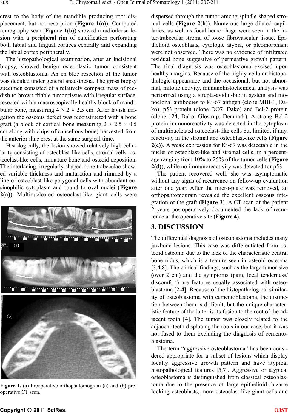 E. Chrysomali et al. / Open Journal of Stomatology 1 (2011) 207-211 208 crest to the body of the mandible producing root dis- placement, but not resorption (Figure 1(a)). Computed tomography scan (Figure 1(b)) showed a radiodense le- sion with a peripheral rim of calcification perforating both labial and lingual cortices centrally and expanding the labial cortex peripherally. The histopathological examination, after an incisional biopsy, showed benign osteoblastic tumor consistent with osteoblastoma. An en bloc resection of the tumor was decided under general anaesthesia. The gross biopsy specimen consisted of a relatively compact mass of red- dish to brown friable tumor tissue with irregular surface, resected with a macroscopically healthy block of mandi- bular bone, measuring 4 × 2 × 2.5 cm. After lavish irri- gation the osseous defect was reconstructed with a bone graft (a block of cortical bone measuring 2 × 2.5 × 0.5 cm along with chips of cancellous bone) harvested from the anterior iliac crest at the same surgical time. Histologically, the lesion showed relatively high cellu- larity consisting of osteoblast-like cells, stromal cells, os- teoclast-like cells, immature bone and osteoid deposition. The interlacing, irregularly-shaped bone trabeculae show- ed variable thickness and maturation and rimmed by a line of osteoblast-like polygonal cells with abundant eo- sinophilic cytoplasm and round to oval nuclei (Figure 2(a)). Multinucleated osteoclast-like giant cells were Figure 1. (a) Preoperative orthopantomogram (a) and (b) pre- operative CT scan. dispersed through the tumor among spindle shaped stro- mal cells (Figure 2(b)). Numerous large dilated capil- laries, as well as focal hemorrhage were seen in the in- ter-trabecular stroma of loose fibrovascular tissue. Epi- thelioid osteoblasts, cytologic atypia, or pleomorphism were not observed. There was no evidence of infiltrated residual bone suggestive of permeative growth pattern. The final diagnosis was osteoblastoma excised upon healthy margins. Because of the highly cellular histopa- thologic appearance and the occasional, but not abnor- mal, mitotic activity, immunohistochemical analysis was performed using a strepta-avidin-biotin system and mo- noclonal antibodies to Ki-67 antigen (clone MIB-1, Da- ko), p53 protein (clone DO7, Dako) and Bcl-2 protein (clone 124, Dako, Glostrup, Denmark). A strong Bcl-2 protein immunoreactivity was detected in the cytoplasm of multinucleated osteoclast-lik e cells but limited, if any, reactivity in the stromal and osteoblast-like cells (Figure 2(c)). A weak expression for Ki-67 was detectable in the nuclei of osteoblast-like and stromal cells, in a percent- age ranging from 10 % to 25% of the tumor cells (Figure 2(d)), while no immunoreact ivity was detected for p53. The patient recovered well; she was asymptomatic without any signs of recurrence on follow-up evaluation after one year. After the micro-plate was removed, an orthopantomogram revealed the excellent osseous inte- gration of the graft (Figure 3). A CT scan of the patient 2 years postoperatively documented the lack of recur- rence at the operative site (Figure 4). 3. DISCUSSION The differential diagnosis of osteoblastoma includes many jawbone lesions. This case was differentiated from os- teoid osteoma due to the lack of the characteristic central bone nidus, which is a feature seen in osteoid osteoma [3,4,8]. The clinical findings, such as the large tumor size (over 2 cm) and the symptoms (pain, local tenderness/ discomfort) are features usually associated with osteo- blastoma [2-4]. Because of the histopathological similar- ity of osteoblastoma with cementoblastoma, the distinc- tion between them is difficult, but the unique character- istic feature of the latter is its fusion to the root of the ad- jacent tooth [4]. The tumor was closely related to the adjacent teeth displacing the roots in our case, but it was not fused to them excluding the diagnosis of cemento- blastoma. (a) (b) The term “aggressive osteoblastoma” has been consi- dered appropriate for a subset of lesions which display locally aggressive growth pattern and have atypical histopathological features [5,7]. Aggressive or atypical osteoblastoma is distinguished from classical osteoblas- toma due to the presence of large epithelioid, bizarre looking osteoblasts, more osteoclast-like giant cells and C opyright © 2011 SciRes. OJST 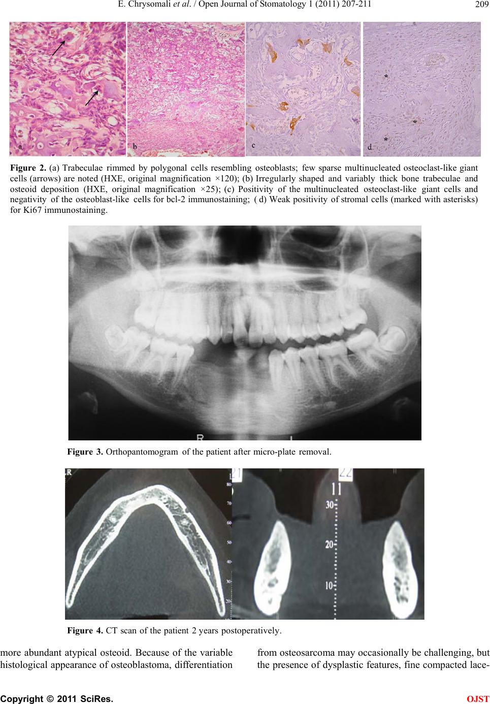 E. Chrysomali et al. / Open Journal of Stomatology 1 (2011) 207-211 Copyright © 2011 SciRes. 209 Figure 2. (a) Trabeculae rimmed by polygonal cells resembling osteoblasts; few sparse multinucleated osteoclast-like giant cells (arrows) are noted (HXE, original magnification ×120); (b) Irregularly shaped and variably thick bone trabeculae and osteoid deposition (HXE, original magnification ×25); ( c) Positivity of the multinucleated osteoclast-like giant cells and negativity of the osteoblast-like cells for bcl-2 immunostaining; (d) Weak positivity of stromal cells (marked with asterisks) for Ki67 immunostaining. Figure 3. Orthopantomogram of the patient after micro-plate remova l. Figure 4. CT scan of the patient 2 years postoperatively. more abundant atypical osteoid. Because of the variable histological appearance of osteoblastoma, differentiation from osteosarcoma may occasionally be challenging, but the presence of dysplastic features, fine compacted lace- OJST  E. Chrysomali et al. / Open Journal of Stomatology 1 (2011) 207-211 210 like osteoid strands, invasion into host bone, high rate of mitotic activity or atypical mitoses aid in the diagnosis of osteosarcoma [1,2,4,5]. Despite the high cellularity in th e case presented here, there was no evidence of epithelioid or dysplastic fea- tures, or growth pattern suggestive of malignancy. The Ki-67 immunostaining disclosed a relatively low to mo- derate cell proliferation index and th e tumor cells did not show any immunoreactivity fo r the p53 protein. Oliveira et al. investigated the cell proliferation marker PCNA, as well as the p53 immunohistochemical expression and p53 gene mutations in classical, atypical osteoblastomas in comparison to osteosarcomas [7]. Atypical osteoblas- tomas, osteosarcomas and tumor recurrence were statis- tically correlated with a high PCNA labelling index and p53 immunoexpression [7]. The histopathological and immunohistochemical find- ings in our case were indicative for the classical osteo- blastoma. A long follow-up period may be needed to draw firm conclusions concerning the benign or potential ag- gressive clinical course of this lesion. Aggressive beha- vior may be within the biologic spectrum of osteoblas- tomas; the histopathology may not appear to be a reli- able predictor of aggressiveness [1]. The predictive val- ue of cell proliferation or other molecular markers in the biologic behavior of osteoblastoma variants remains to be completely determined. The Bcl-2 gene and its protein product promote cellu- lar survival; therefore correlation of the Bcl-2 expression with clinical outcome has been examined in many types of tumors. Recently the Bcl-2 protein expression has been correlated—along with other biomarkers—with the progression and prognosis of osteosarcoma [9]; however its expression has not yet, to our knowledge, been in- vestigated in benign osteoblastic tumors. An interesting finding in the present case was the immunohistochemi- cal detection of the Bcl-2 protein in the multinucleated osteoclast-like giant cells, in contrast to the lack of ex- pression of this protein in the osteoblast-like and stro mal tumor cells. This finding may be related to the molecular mechanisms regulating the apoptosis of osteoclast-like tumor cells or their function. Anti-apoptotic Bcl-2 family members such as Bcl-2 and Bcl-xL seem to play a sig- nificant role, not only in the apoptosis of osteoclasts, but also in the bone resorbing function of these cells [10]. The treatment options of osteoblastoma include con- servative surgical excision, excision with vigorous cu- rettage followed by bur ablation of the margins and co- pious irritation or en block resection [4]. Local recur- rence related to inadequately removed tumors have been reported in a rate of 14% [11]. Lesions treated with en bloc resection or resection yielding tumor-free margins exhibit minimal likelihood of recurrence; we believe that such radical procedures are fully justified in pediatric patients suffering from potentially aggressive lesions like osteoblastomas. Despite the propensity of recurren- ce, primary reconstruction seems to be worthwhile, and should be taken into account when mandibular integrity can be preserved. Long-term follow-up is recommended, of at least 2 years, not only to minimize the risk of un- diagnosed recurrence, but also to guarantee the grafts’ survival and integ ration. REFERENCES [1] Lucas, D., Unni, K., McLeod, R., O’Connor, M. and Sim, F. (1994) Osteoblastoma: Clinicopathologic study of 306 cases. Human Pathology, 25, 117-134. doi:10.1016/0046-8177(94)90267-4 [2] Capodiferro, S., Maiorano, E., Giardina, C., Lacaita, M., Lo Muzio, L. and Favia, G. (2005) Osteoblastoma of the mandible: Clinicopathologic study of four cases and lit- erature review. Head and Neck, 27, 616-621. doi:10.1002/hed.20192 [3] Jones, A., Prihoda, T., Kacher, J., Odingo, N. and Freed- man, P. (2006) Osteoblastoma of the maxilla and mandi- ble: A report of 24 cases, review of the literature and dis- cussion of its relationship to osteoid osteoma of the jaws. Oral Surgery, Oral Medicine, Oral Pathology, Oral Ra- diology & Endodontics, 102, 639-650. doi:10.1016/j.tripleo.2005.09.004 [4] Rawal, Y., Angiero, F., Allen, C., Kalmar, J., Sedghizadeh, P. and Steinhilber, A. (2006) Gnathic osteoblastoma: Cli- nicopathologic review of seven cases with long-term fol- low-up. Oral Oncology, 42, 123-130. doi:10.1016/j.oraloncology. 2005.04.016v [5] Lypka, M., Goos, R., Yamashita, D. and Melrose, R. (2008) Aggressive osteoblastoma of the mandible. Inter- national Journal of Oral and Maxillofacial Surgery, 37, 675-678. doi:10.1016/j.ijom.2008.01.013 [6] Alvares, C.A., Gião, D.M., Casati, A.L., Negrão, F.R. and Sant’Ana, E. (2005) Osteoblastoma of the mandible: Systematic review of the literature and report of a case. Dentomaxillofacial Radiology, 34, 1-8. doi:10.1259/dmfr/24385194 [7] Oliveira De, C., Mendonça, B., Camargo De, O., Pinto, E., Nascimento, S., et al. (2007) Classical osteoblastoma, atypical osteoblastoma, and osteosarcoma. A comparative study based on clinical, histological, and biological pa- rameters. Clinics, 62, 167-174. doi:10.1590/S1807-59322007000200012 [8] Van der Waal, L., Greebe, R. and Elias, E. (1983) Benign osteoblastoma or osteoid osteoma of the maxilla. Inter- national Journal of Oral and Maxillofacial Surgery, 12, 355-358. [9] Wu, X., Cai, Z.D., Lou, L.M. and Zhu, Y.B. (2011) Ex- pressions of p53, c-MYC, BCL-2 and apoptotic index in human osteosarcoma and their correlations with progno- sis of patients. Cancer Epidemiology, Epub Ahead of Print. [10] Iwasawa, M., Miyazaki, T., Nagase, Y., Akiyama, T., Kadono, Y., Nakamura, M. et al. (2009) The antiapop- C opyright © 2011 SciRes. OJST  E. Chrysomali et al. / Open Journal of Stomatology 1 (2011) 207-211 Copyright © 2011 SciRes. 211 OJST totic protein Bcl-xL negatively regulates the bone re- sorbing activity of osteoclasts in mice. Journal of Clini- cal Investigation, 119, 3149-3159. [11] Gordon, S., MacIntosh, R. and Wesley, R. (2001) A re- view of osteoblastoma and case report of metachronous osteoblastoma and unicystic ameloblastoma. Oral Sur- gery, Oral Medicine, Oral Pathology, Oral Radiology & Endodontics, 91, 570-575. doi:10.1067/moe.2001.113349 |