

Paper Menu >>
Journal Menu >>
 Open Journal of Stomatology, 2011, 1, 150-157 doi:10.4236/ojst.2011.14022 Published Online December 2011 (http://www.SciRP.org/journal/ojst/ OJST ). Published Online December 2011 in SciRes. http://www.scirp.org/journal/OJST Immune function in Japanese schoolchildren with sleep-disordered breathing: a preliminary study with analyses of salivary markers Hiroshi Ueda, Atsushi Horihata, Eka Matsumoto, Genki Watanabe, Myongsun Koh, Kotaro Tanimoto, Kazuo Tanne Department of Orthodontics and Craniofacial Developmental Biology, Graduate School of Biomedical Sciences, Hiroshima Univer- sity, Hiroshima, Japan. Email: [email protected] Received 25 August 2011; revised 6 October 2011; accepted 21 October 2011. ABSTRACT The aim of this study was to assess the prevalence of sleep-disordered breathing (SDB) symptoms among primary schoolchildren, and to objectively determine the influence of SDB on the intra-oral environment through the analysis of saliva. A questionnaire survey was conducted among approximately 400 children from a primary school in Hiroshima. Parents were asked to complete the questionnaire and provide their contact information if they allowed the collection of saliva samples from their children. Thirty-eight chil- dren agreed to participate in the saliva study. Habit- ual snoring and cessation of breathing during sleep were found in approximately 8% and 1% of children, respectively. The present results showed significant correlations between snoring and mouth breathing. A significant association between excessive daytime sle- epiness (EDS) and learning problems was found. Fur- thermore, among children between the ages of 7 and 12 years, those with EDS and learning problems tended to be older. SDB symptom scores were statisti- cally significant only in relation to EDS. The present study also demonstrated significantly higher levels of salivary IgA and cortisol in children with sleep-re- lated disorders. The present study determined the pre- valence and characteristics of SDB among Japanese primary schoolchildren and their effects on the oral environment. Approximately 8% of primary school- children with habitual snoring might need to be care- fully monitored for SDB symptoms and immune sta- tus to ensure proper psychological and physical deve- lopment. Keywords: Schoolchildren; Sleep-Disordered Breathing Symptoms; Immune Function; Salivary Markers 1. INTRODUCTION Sleep-disordered breathing (SDB), including sleep apnea syndrome (SAS), is an important cause of morbidity for both adults and children [1-5]. In children in particular, SDB is of great concern because it can cause neurobe- havioral and growth-related problems, and affect aca- demic performance [6-9]. The estimated prevalence of SAS in children ranges from 1% to 3% and the incidence of habitual snoring has been reported to be 7% - 12% in previous studies [10-13]. Therefore, the prevalence of SDB and its influence on daily life are significant and cannot be neglected in growing children. In the fields of dentistry and orthodontics, mouth breathing patients are known to have narrow upper den- tition and long facial height resulting from the backward and downward displacement of the mandible. This asso- ciation has been explained by the fact that mouth brea- thing causes reduced activity of the facial and mastica- tory muscles, leading to the appearance of skeletal defor- mities represented by an open bite with a small and dis- tally located mandible. In addition, the descriptive term “adenoid face”, consisting of narrow dentition, protrude- ing teeth, and lip incompetence at rest has been docu- mented in the literature for at least a century [14-16]. Over the last decades, the effect of SDB on the immu- ne system has been reported frequently [17]. The meas- urement of secretory immunoglobulin A (IgA) levels in the saliva is a convenient and frequently used indicator of immune status. SDB during childhood differs signify- cantly from that in adults in terms of various parameters such as symptoms and pathogenesis. Therefore, an appro- priate differential study to discriminate SDB symptoms in children is necessary. The aim of this study was to assess the prevalence of SDB symptoms in primary schoolchildren and to object- tively determine its influence on the intra-oral environ- ment by means of saliva analysis. 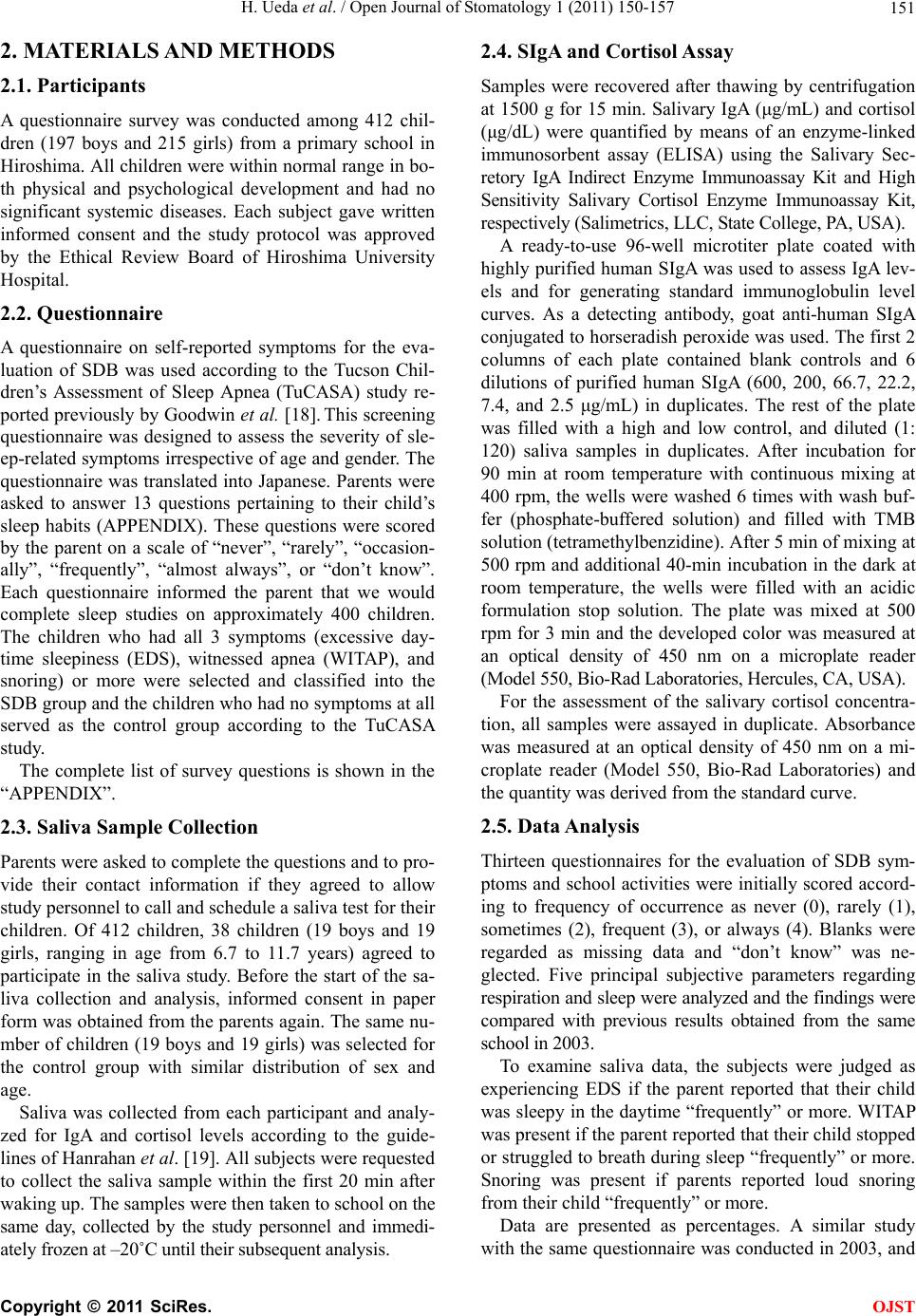 H. Ueda et al. / Open Journal of Stomatology 1 (2011) 150-157 151 2. MATERIALS AND METHODS 2.1. Participants A questionnaire survey was conducted among 412 chil- dren (197 boys and 215 girls) from a primary school in Hiroshima. All children were within normal range in bo- th physical and psychological development and had no significant systemic diseases. Each subject gave written informed consent and the study protocol was approved by the Ethical Review Board of Hiroshima University Hospital. 2.2. Questionnaire A questionnaire on self-reported symptoms for the eva- luation of SDB was used according to the Tucson Chil- dren’s Assessment of Sleep Apnea (TuCASA) study re- ported previously by Goodwin et al. [18]. This screening questionnaire was designed to assess the severity of sle- ep-related symptoms irrespective of age and gender. The questionnaire was translated into Japanese. Parents were asked to answer 13 questions pertaining to their child’s sleep habits (APPENDIX). These questions were scored by the parent on a scale of “never”, “rarely”, “occasion- ally”, “frequently”, “almost always”, or “don’t know”. Each questionnaire informed the parent that we would complete sleep studies on approximately 400 children. The children who had all 3 symptoms (excessive day- time sleepiness (EDS), witnessed apnea (WITAP), and snoring) or more were selected and classified into the SDB group and the children who had no symptoms at all served as the control group according to the TuCASA study. The complete list of survey questions is shown in the “APPENDIX”. 2.3. Saliva Sample Collection Parents were asked to complete the questions and to pro- vide their contact information if they agreed to allow study personnel to call and schedule a saliva test for their children. Of 412 children, 38 children (19 boys and 19 girls, ranging in age from 6.7 to 11.7 years) agreed to participate in the saliva study. Before the start of the sa- liva collection and analysis, informed consent in paper form was obtained from the parents again. The same nu- mber of children (19 boys and 19 girls) was selected for the control group with similar distribution of sex and age. Saliva was collected from each participant and analy- zed for IgA and cortisol levels according to the guide- lines of Hanrahan et al. [19]. All subjects were requested to collect the saliva sample within the first 20 min after waking up. The samples were then taken to school on the same day, collected by the study personnel and immedi- ately frozen at –20˚C until their subsequent analysis. 2.4. SIgA and Cortisol Assay Samples were recovered after thawing by centrifugation at 1500 g for 15 min. Salivary IgA (μg/mL) and cortisol (μg/dL) were quantified by means of an enzyme-linked immunosorbent assay (ELISA) using the Salivary Sec- retory IgA Indirect Enzyme Immunoassay Kit and High Sensitivity Salivary Cortisol Enzyme Immunoassay Kit, respectively (Salimetrics, LLC, State College, PA, USA). A ready-to-use 96-well microtiter plate coated with highly purified human SIgA was used to assess IgA lev- els and for generating standard immunoglobulin level curves. As a detecting antibody, goat anti-human SIgA conjugated to horseradish peroxide was used. The first 2 columns of each plate contained blank controls and 6 dilutions of purified human SIgA (600, 200, 66.7, 22.2, 7.4, and 2.5 μg/mL) in duplicates. The rest of the plate was filled with a high and low control, and diluted (1: 120) saliva samples in duplicates. After incubation for 90 min at room temperature with continuous mixing at 400 rpm, the wells were washed 6 times with wash buf- fer (phosphate-buffered solution) and filled with TMB solution (tetramethylbenzidine). After 5 min of mixing at 500 rpm and additional 40-min incubation in the dark at room temperature, the wells were filled with an acidic formulation stop solution. The plate was mixed at 500 rpm for 3 min and the developed color was measured at an optical density of 450 nm on a microplate reader (Model 550, Bio-Rad Laboratories, Hercules, CA, USA). For the assessment of the salivary cortisol concentra- tion, all samples were assayed in duplicate. Absorbance was measured at an optical density of 450 nm on a mi- croplate reader (Model 550, Bio-Rad Laboratories) and the quantity was derived from the standard curve. 2.5. Data Analysis Thirteen questionnaires for the evaluation of SDB sym- ptoms and school activities were initially scored accord- ing to frequency of occurrence as never (0), rarely (1), sometimes (2), frequent (3), or always (4). Blanks were regarded as missing data and “don’t know” was ne- glected. Five principal subjective parameters regarding respiration and sleep were analyzed and the findings were compared with previous results obtained from the same school in 2003. To examine saliva data, the subjects were judged as experiencing EDS if the parent reported that their child was sleepy in the daytime “frequently” or more. WITAP was present if the parent reported that their child stopped or struggled to breath during sleep “frequently” or more. Snoring was present if parents reported loud snoring from their child “frequently” or more. Data are presented as percentages. A similar study with the same questionnaire was conducted in 2003, and C opyright © 2011 SciRes. OJST 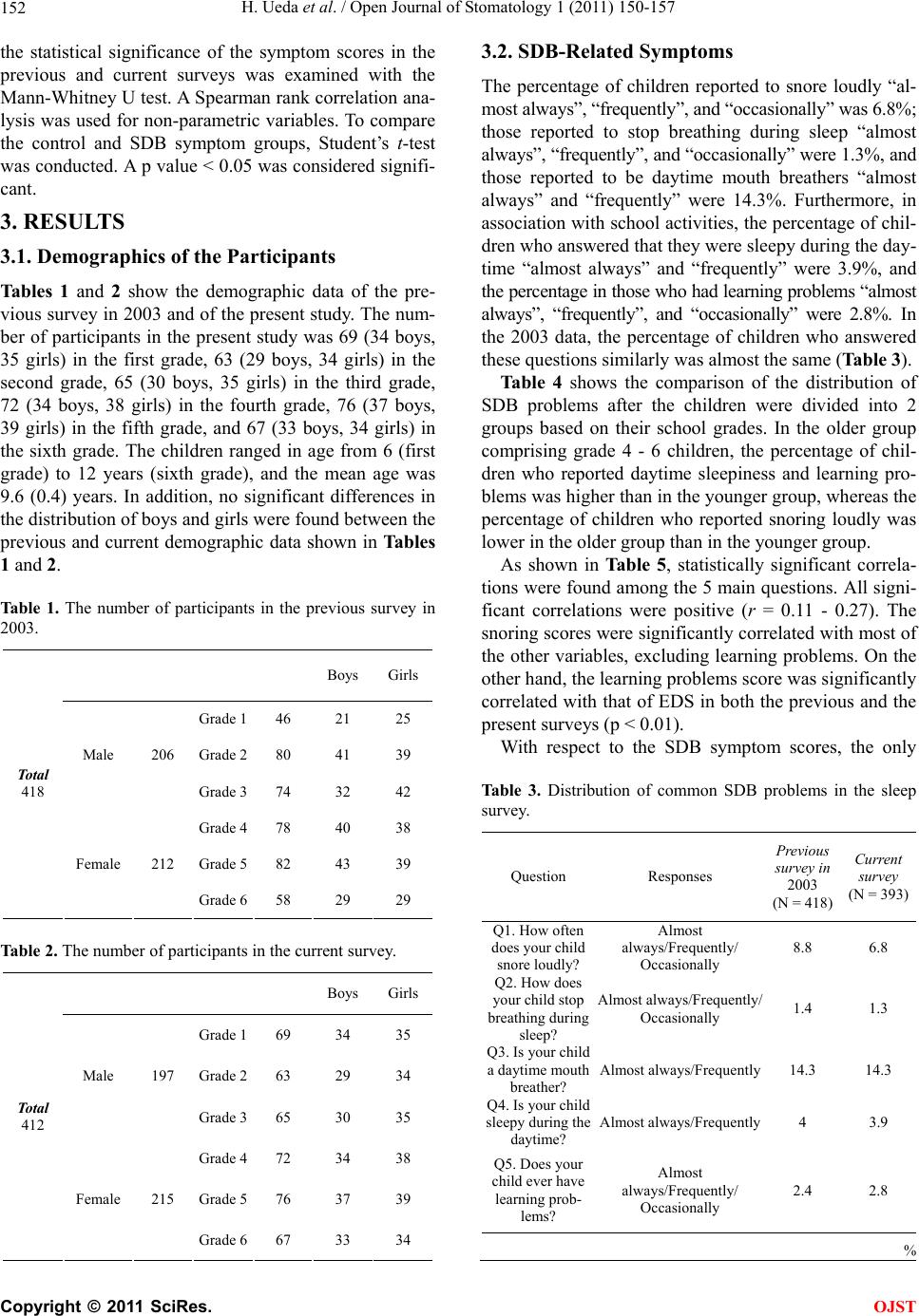 H. Ueda et al. / Open Journal of Stomatology 1 (2011) 150-157 152 the statistical significance of the symptom scores in the previous and current surveys was examined with the Mann-Whitney U test. A Spearman rank correlation ana- lysis was used for non-parametric variables. To compare the control and SDB symptom groups, Student’s t-test was conducted. A p value < 0.05 was considered signifi- cant. 3. RESULTS 3.1. Demographics of the Participants Tables 1 and 2 show the demographic data of the pre- vious survey in 2003 and of the present study. The num- ber of participants in the present study was 69 (34 boys, 35 girls) in the first grade, 63 (29 boys, 34 girls) in the second grade, 65 (30 boys, 35 girls) in the third grade, 72 (34 boys, 38 girls) in the fourth grade, 76 (37 boys, 39 girls) in the fifth grade, and 67 (33 boys, 34 girls) in the sixth grade. The children ranged in age from 6 (first grade) to 12 years (sixth grade), and the mean age was 9.6 (0.4) years. In addition, no significant differences in the distribution of boys and girls were found between the previous and current demographic data shown in Tables 1 and 2. Table 1. The number of participants in the previous survey in 2003. Boys Girls Grade 1 46 21 25 Grade 2 80 41 39 Male 206 Grade 3 74 32 42 Grade 4 78 40 38 Grade 5 82 43 39 Total 418 Female 212 Grade 6 58 29 29 Table 2. The number of participants in the current survey. Boys Girls Grade 1 69 34 35 Grade 2 63 29 34 Male 197 Grade 3 65 30 35 Grade 4 72 34 38 Grade 5 76 37 39 Total 412 Female 215 Grade 6 67 33 34 3.2. SDB-Related Symptoms The percentage of children reported to snore loudly “al- most always”, “frequently”, and “occasionally” was 6.8%; those reported to stop breathing during sleep “almost always”, “frequently”, and “occasionally” were 1.3%, and those reported to be daytime mouth breathers “almost always” and “frequently” were 14.3%. Furthermore, in association with school activities, the percentage of chil- dren who answered that they were sleepy during the day- time “almost always” and “frequently” were 3.9%, and the percentage in those who had learning problems “almost always”, “frequently”, and “occasionally” were 2.8%. In the 2003 data, the percentage of children who answered these questions similarly was almost the same (Table 3). Table 4 shows the comparison of the distribution of SDB problems after the children were divided into 2 groups based on their school grades. In the older group comprising grade 4 - 6 children, the percentage of chil- dren who reported daytime sleepiness and learning pro- blems was higher than in the younger group, whereas the percentage of children who reported snoring loudly was lower in the older group than in the younger group. As shown in Tabl e 5 , statistically significant correla- tions were found among the 5 main questions. All signi- ficant correlations were positive (r = 0.11 - 0.27). The snoring scores were significantly correlated with most of the other variables, excluding learning problems. On the other hand, the learning problems score was significantly correlated with that of EDS in both the previous and the present surveys (p < 0.01). With respect to the SDB symptom scores, the only Table 3. Distribution of common SDB problems in the sleep survey. Question Responses Previous survey in 2003 (N = 418) Current survey (N = 393) Q1. How often does your child snore loudly? Almost always/Frequently/ Occasionally 8.8 6.8 Q2. How does your child stop breathing during sleep? Almost always/Frequently/ Occasionally 1.4 1.3 Q3. Is your child a daytime mouth breather? Almost always/Frequently 14.3 14.3 Q4. Is your child sleepy during the daytime? Almost always/Frequently 4 3.9 Q5. Does your child ever have learning prob- lems? Almost always/Frequently/ Occasionally 2.4 2.8 % C opyright © 2011 SciRes. OJST 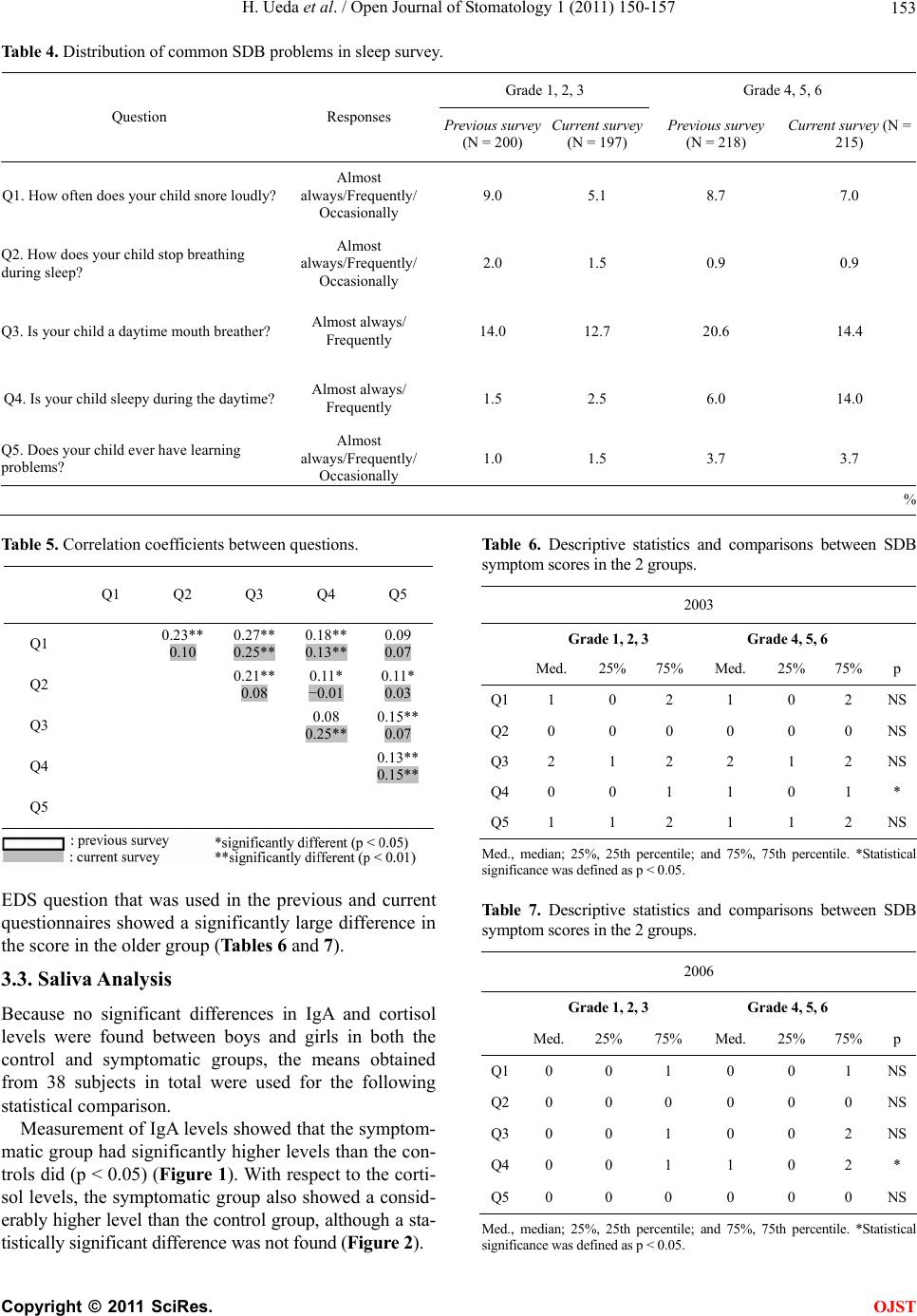 H. Ueda et al. / Open Journal of Stomatology 1 (2011) 150-157 Copyright © 2011 SciRes. 153 Table 4. Distribution of common SDB problems in sleep survey. Grade 1, 2, 3 Grade 4, 5, 6 Question Responses Previous survey (N = 200) Current survey (N = 197) Previous survey (N = 218) Current survey (N = 215) Q1. How often does your child snore loudly? Almost always/Frequently/ Occasionally 9.0 5.1 8.7 7.0 Q2. How does your child stop breathing during sleep? Almost always/Frequently/ Occasionally 2.0 1.5 0.9 0.9 Q3. Is your child a daytime mouth breather? Almost always/ Frequently 14.0 12.7 20.6 14.4 Q4. Is your child sleepy during the daytime? Almost always/ Frequently 1.5 2.5 6.0 14.0 Q5. Does your child ever have learning problems? Almost always/Frequently/ Occasionally 1.0 1.5 3.7 3.7 % Table 5. Correlation coefficients between questions. Table 6. Descriptive statistics and comparisons between SDB symptom scores in the 2 groups. Q1 Q2 Q3 Q4 Q5 Q1 0.23** 0.10 0.27** 0.25** 0.18** 0.13** 0.09 0.07 Q2 0.21** 0.08 0.11* −0.01 0.11* 0.03 Q3 0.08 0.25** 0.15** 0.07 Q4 0.13** 0.15** Q5 2003 Grade 1, 2, 3 Grade 4, 5, 6 Med. 25% 75% Med. 25% 75% p Q11 0 2 1 0 2 NS Q20 0 0 0 0 0 NS Q32 1 2 2 1 2 NS Q40 0 1 1 0 1 * Q51 1 2 1 1 2 NS Med., median; 25%, 25th percentile; and 75%, 75th percentile. *Statistical significance was defined as p < 0.05. EDS question that was used in the previous and current questionnaires showed a significantly large difference in the score in the older group (Tables 6 and 7). Table 7. Descriptive statistics and comparisons between SDB symptom scores in the 2 groups. 2006 Grade 1, 2, 3 Grade 4, 5, 6 Med. 25% 75% Med. 25% 75% p Q10 0 1 0 0 1 NS Q20 0 0 0 0 0 NS Q30 0 1 0 0 2 NS Q40 0 1 1 0 2 * Q50 0 0 0 0 0 NS 3.3. Saliva Analysis Because no significant differences in IgA and cortisol levels were found between boys and girls in both the control and symptomatic groups, the means obtained from 38 subjects in total were used for the following statistical comparison. Measurement of IgA levels showed that the symptom- matic group had significantly higher levels than the con- trols did (p < 0.05) (Figure 1). With respect to the corti- sol levels, the symptomatic group also showed a consid- erably higher level than the control group, although a sta- tistically significant difference was not found (Figur e 2). Med., median; 25%, 25th percentile; and 75%, 75th percentile. *Statistical significance was defined as p < 0.05. OJST  H. Ueda et al. / Open Journal of Stomatology 1 (2011) 150-157 154 0 200 400 600 800 Control groupSymptom group * IgA (μg/mL) * :significantly different (p < 0.05) 0 200 400 600 800 Control groupSymptom group * IgA (μg/mL) * :significantly different (p < 0.05) Figure 1. Comparison of the IgA levels between control and SDB symptom groups. 0. 5.0 10.0 15.0 20.0 25.0 Control groupSymptom group Cortisol (μg/dL) 0. 5.0 10.0 15.0 20.0 25.0 Control groupSymptom group Cortisol (μg/dL) Figure 2. Comparison of the cortisol levels between control nd SDB symptom groups. a 4. DISCUSSION In the present study, a consciousness survey was con- ducted for the assessment of SDB symptoms and prob- lem behaviors among primary schoolchildren. The influ- ence of these factors on salivary secretory immuno- globulin A and cortisol levels was also examined. Sleep apnea syndrome (SAS) has been of increasing concern among the middle-aged population in Japan in recent years, and the physical and psychological development of children is considered to be greatly influenced by sle- ep quality [2-4]. Mental retardation from chronic hypo- xia, low stature from hyposecretion of growth hormones, and slow learning due to daytime sleepiness are listed as examples of the association between SDB and physical and psychological development; hence, children in pri- mary school were defined as the target of the present study. 4.1. Prevalence of SDB Symptoms In this study, habitual snoring and cessation of breathing during sleep were found in approximately 8% and 1% of children, respectively. Several studies from other coun- tries have reported the prevalence of sleep problems in a population-based sample of children. Gozal and Pope re- ported that the mean prevalence of snoring among 1588 children was 9% in the United States [7]. A similar pre- valence of snoring among children was reported in Fr- ance (10%) [20] and in Italy (7.3%) [12]. The overall snoring prevalence in the previous and current surveys from our group was at the upper limit of the range re- ported by others. With respect to cessation of breathing, the present re- sults were similar to those of previous studies reporting that SAS affects approximately 1% - 2% children [2, 10,13]. Cessation of breathing during sleep is not the same as diagnostic SAS. However, apneic symptoms such as cessation of breathing, difficulty in periodic breathing, and nocturnal choking might be the indicators of the presence of SAS. The present results showed significant correlations between snoring and mouth breathing. Mouth breathing is one of the characteristic SAS symptoms [13]. Fur- thermore, nasal influences on mouth breathing, snoring, and sleep apnea have been reported [21]. Therefore, it is reasonable to assume that mouth breathing caused by nasal obstruction leads to loud snoring, apneic breathing, and disturbed sleep. In this study, a strong association between EDS and learning problems was detected. This finding suggests that poor sleep quality such as sleep fragmentation and nocturnal hypoxemia might induce EDS, which ulti- mately influences academic performance and ability. In agreement with our results, EDS, as well as various be- havioral problems such as inattention, hyperactivity, and aggression in school were reported in association with sleep-related disorders in previous studies [7-9]. Inter- estingly, Gozal demonstrated that improvements in scho- ol performance could be induced by remediation of SAS in first-grade children with substantial academic impair- ment in school-related activities [6]. Furthermore, among children between 7 and 12 years of age, those with EDS and learning problems were more likely to be older than those without. Our data regarding SDB symptom scores were statistically significant only with respect to EDS. This may be due to the difference in mean sleep times between older and younger children, which were 9 h 18 min (32 min) (first grade) and 7 h 38 min (56 min) (sixth grade), respectively (data not sho- wn). The current lifestyle of Japanese children, which includes longer study periods after school and the resul- ting delay in bedtime, might be a significant factor affec- ting children’s sleep. The Tucson Children’s Assessment of Sleep Apnea (TuCASA) study is a prospective cohort study that was designed to determine the prevalence of objectively do- cumented SDB in preadolescent children and to investi- gate its relationship to symptoms, performance on neuro- behavioral measures, and physiologic and anatomic risk factors [18]. Although screening questionnaires for chil- dren can easily show bias, most TuCASA components C opyright © 2011 SciRes. OJST  H. Ueda et al. / Open Journal of Stomatology 1 (2011) 150-157 155 are regarded as standard questions that are found in sleep-habit questionnaires pertaining to SDB in children. 4.2. Association of SDB Symptoms with Salivary Immune Factors The results of the present study showed significantly hi- gher levels of salivary IgA and cortisol in children with sleep-related disorders. Secretory IgA, which is meas- ured in the saliva, is the predominant antibody detected in bodily secretions. IgA levels are believed to indicate the functional status of the entire mucosal immune sys- tem [22]. As a source of biological fluid for physical examination, saliva has several advantages. Collection of saliva is less intrusive and easier than that of urine or blood, especially for children. The most likely explanation for the significantly high concentration of salivary IgA in the symptom group is that sleep-related disorders negatively affect immune functions and increase IgA levels for protection against certain infections. The release of salivary IgA could be an indicator of intra-oral and upper airway inflammation. Therefore, salivary IgA provides a major defense against potential pathogens by preventing colonization and rep- lication on the mucosal surfaces of the oral cavity and the upper respiratory tract. Previous studies demonstra- ted that recurrent upper respiratory infection was associ- ated with increased prevalence of SDB symptoms in children [12,23,24]. The use of self-reported stress as a measurement me- thod can be associated with bias and variation in study results [25]. The psychological status of patients can be reflected in their stress level. Cortisol is a stress hormone that indicates hypothalamic-pituitary-adrenal (HPA) axis activity and is a reliable biomarker of stress that is easily measured in saliva [26]. The cortisol awakening re- sponse, which is measured during the first 30 - 45 min after waking up in the morning, is an especially reliable marker of HPA axis activity [27]. Yehuda et al. demon- strated that a single salivary cortisol sample taken im- mediately after waking up can provide an accurate esti- mate of urinary cortisol and mean salivary cortisol thr- oughout the day [28]. The present results indicated that SDB symptoms can constitute a stressor capable of ele- vating cortisol levels, and suggested that children in pri- mary school with greater adrenocortical reactivity might have a considerable level of psychological stress. In the field of dentistry, especially orthodontics, many studies have shown the influence of breathing disorders caused by respiratory obstruction with the large soft pal- ate, tonsils, tongue, and nasal inflammation on craniofa- cial morphology. Animal studies demonstrated that spe- cific skeletal and dental changes, including an increased lower facial height, anterior downward tipping of the occlusal plane, and dual bites, crossbite, and open bites occur within 24 months after nasal obstruction in mon- keys [29,30]. Bresolin et al. investigated the relation- ship between mouth breathing in allergic children and dento-facial development [29], and demonstrated that mouth breathers had longer faces with narrower maxilla and retrognathic mandibles [31]. These studies show that breathing disorders are strongly correlated with cranio- facial morphology. Moreover, a small oral cavity with narrow dentition associated with retrognathism due to undergrowth of the mandible could induce SDB. There- fore, anatomical disorders carry a certain risk in terms of predisposition to the development of respiratory disor- ders such as SAS. In conclusion, the present study determined the pre- valence and characteristics of SDB among Japanese primary schoolchildren and its influence on the oral en- vironment. Approximately 8% of primary schoolchildren with habitual snoring might need to be carefully moni- tored for SDB symptoms and immune status to ensure appropriate psychological and physical development. REFERENCES [1] American Academy of Pediatrics (2002) Clinical practice guideline: Diagnosis and management of childhood sleep apnea syndrome. Pediatrics, 109, 704-712. doi:10.1542/peds.109.4.704 [2] Ali, N.J., Pitson, D.J. and Stradling, J.R. (1993) Snoring, sleep disturbance and behaviour in 4 - 5 year olds. Ar- chives of Disease in Childhood, 68, 360-366. doi:10.1136/adc.68.3.360 [3] Gottlieb, D.J., Vezina, R.M., Chase, C., Lesko, S.M., Hee- ren, T.C., Weese-Mayer, D.E., Auerbach, S.H. and Corwin, M.J. (2003) Symptoms of sleep-disordered breathing in 5-year-old children are associated with sleepiness and problem behaviors. Pediatrics, 112, 870-877. doi:10.1542/peds.112.4.870 [4] Schechter, M.S. (2001) Section on pediatrics pulmonol- ogy subcommittee obstructive sleep apnea syndrome. Technical report: Diagnosis and management of child- hood obstructive sleep apnea syndrome. Pediatrics, 109, e69. doi:10.1542/peds.109.4.e69 [5] Matsumoto, E., Tanaka, E., Tabe, H., Wakisaka, N., Na- kata, Y., Ueda, H., Hori, T., Abe, T. and Tanne K. (2007) Sleep architecture and the apnea-hypopnea index in chil- dren with obstructive-sleep apnea syndrome. Journal of Oral Rehabilitation, 34, 112-120. doi:10.1111/j.1365-2842.2006.01676.x [6] Gozal, D. (1998) Sleep-disordered breathing and school performance in children. Pediatrics, 102, 616-620. doi:10.1542/peds.102.3.616 [7] Gozal, D. and Pope, D.W. (2001) Snoring during early childhood and academic performance at ages thirteen to fourteen years. Pediatrics, 107, 1394-1399. doi:10.1542/peds.107.6.1394 [8] Owens, J., Opipari, L., Nobile, C. and Spirito, A. (1998) Sleep and daytime behavior in children with obstructive C opyright © 2011 SciRes. OJST  H. Ueda et al. / Open Journal of Stomatology 1 (2011) 150-157 Copyright © 2011 SciRes. 156 OJST sleep apnea and behavioral sleep disorders. Pediatrics, 102, 1178-1184. doi:10.1542/peds.102.5.1178 [9] Downey, R., Perkin, R.M. and MacQuarrie, J. (1993) Upper airway resistance syndrome: Sick, symptomatic but unrecognized. Sleep, 16, 620-623. doi:10.1378/chest.107.4.963 [10] Gislason, T. and Benediktsdottir, B. (1995) Snoring, ap- neic episodes, and nocturnal hypoxemia among children 6 months to 6 years old: An epidemiologic study of lower limit of prevalence. Chest, 107, 963-966. [11] Brunetti, L., Rana, S., Lospalluti, M.L., Pietrafesa, A., Francavilla, R., Fanelli, M. and Armenio, L. (2001) Prevalence of obstructive sleep apnea syndrome in a co- hort of 1207 children of southern Italy. Chest, 120, 1930-1935. doi:10.1378/chest.120.6.1930 [12] Corbo, G.M., Fuciarelli, F. and Foresi, A. (1989) Snoring in children: Association with respiratory symptoms and passive smoking. British Medical Journal, 299, 1491- 1494. doi:10.1136/bmj.299.6714.1491 [13] Carrol, J.L., McColley, S.A., Marcus, C.L., Curtis, S. and Loughlin, G.M. (1995) Inability of clinical history to dis- tinguish primary snoring from obstructive sleep apnea syndrome in children. Chest, 108, 610-618. doi:10.1378/chest.108.3.610 [14] Linder-Aronson, S. (1970) Adenoids: Their effect on mode of breathing and nasal airflow and their relation- ship to characteristic of the facial skeleton and the denti- tion. Acta Otolaryngologica Supplementum, 265, 1-132. [15] Harvold, E.P., Vargervik, K. and Chierici, G. (1973) Pri- mate experiments on oral sensation and dental malocclu- sions. American Journal of Orthodontics, 63, 494-508. doi:10.1016/0002-9416(73)90162-0 [16] Proffit, W.R. and Fields, H.W. (1993) The etiology of orthodontic problem. Contemporary Orthodontics, 2nd Edition, Mosby, St. Louis, 105-138. [17] O’Leary, A. (1990) Stress, emotion, and human immune function. Psychological Bulletin, 108, 363-383. doi:10.1037/0033-2909.108.3.363 [18] Goodwin, J.L., Babar, S.I., Kaemingk, K.L., Rosen, G.M., Morgan, W.J., Sherrill, D.L. and Quan, S.F. (2003) Symp- toms related to sleep-disordered breathing in White and Hispanic children. The Tucson Children’s Assessment of Sleep Apnea Study Chest, 124, 196-203. [19] Hanrahan, K., McCarthy, A.M., Kleiber, C., Lutgendorf, S. and Tsalikian, E. (2006) Strategies for salivary cortisol collection and analysis in research with children. Applied Nursing Research, 19, 95-101. doi:10.1016/j.apnr.2006.02.001 [20] Teculescu, D.B., Caillier, I. and Perrin, P. (1992) Snoring in French preschool children. Pediatric Pulmonology, 13, 239-244. doi:10.1002/ppul.1950130412 [21] Olsen, K. and Kern, E.B. (1995) Nasal influences on snoring and obstructive sleep apnea. Mayo Clinic Pro- ceedings, 65, 1095-1105. [22] Mestecky, J. (1993) Saliva as a manifestation of the common mucosal immune system. Annals of the New York Academy of Sciences, 694, 184-194. doi:10.1111/j.1749-6632.1993.tb18352.x [23] Frank, Y., Kravath, R., Pollak, C.P. and Weitzman, E.D. (1983) Obstructive sleep apnea and its therapy: Clinical and polysomnographic manifestations. Pediatrics, 71, 737-742. [24] Brouillette, R.T., Fernbach, S.K. and Hunt, C.E. (1982) Obstructive sleep apnea in infants and children. Journal of Pediatric, 100, 31-40. doi:10.1016/S0022-3476(82)80231-X [25] Suter, P.M., Maire, R., Holtz, D. and Vetter, W. (1997) Relationship between self-perceived stress and blood pres- sure. Journal of Human Hypertension, 11, 171-176. doi:10.1038/sj.jhh.1000409 [26] Kirschbaum, C. and Hellhammer, D.H. (1994) Salivary cortisol in psychoneuroendocrine research: Recent de- velopments and applications. Psychoneuroendocrinology, 19, 313-333. doi:10.1016/0306-4530(94)90013-2 [27] Prussner, J.C., Wolf, O.T., Hellhammer, D.H., Buske- Kirschbaum, A., Von Auer, K., Jobst, S., Kaspers, D. and Kirschbaum, C. (1997) Free cortisol levels after awak- ening: A reliable biological marker for the assessment of adreno-cortical activity. Life Sciences, 61, 2539-2549. doi:10.1016/S0024-3205(97)01008-4 [28] Yehuda, R., Halligan, S.L., Yang, R.K., Guo, L.S., Ma- kotkine, I., Singh, B. and Pickholtz, D. (2003) Relation- ship between 24-hour urinary-free cortisol excretion and salivary cortisol levels sampled from awakening to bed- time in healthy subjects. Life Sciences, 73, 349-358. doi:10.1016/S0024-3205(03)00286-8 [29] Yamada, T., Tanne, K., Miyamoto, K. and Yamauchi, K. (1997) Influences of nasal respiratory obstruction on cra- niofacial growth in young Macaca fuscata monkeys. American Journal of Orthodontics and Dentofacial Or- thopedics, 111, 38-43. doi:10.1016/S0889-5406(97)70300-7 [30] Miller, A.J., Vargervik, K. and Chierici, G. (1984) Ex- perimentally induced neuromuscular changes during and after nasal airway obstruction. American Journal of Or- thodontics, 85, 385-392. doi:10.1016/0002-9416(84)90159-3 [31] Bresolin, D., Sharpiro, P.A., Sharpiro, G.G., Chapko, M.K. and Dassel, S. (1983) Mouth breathing in allergic chil- dren: Its relationship to dentofacial development. Ameri- can Journal of Orthodontics, 83, 334-343. doi:10.1016/0002-9416(83)90229-4  H. Ueda et al. / Open Journal of Stomatology 1 (2011) 150-157 157 APPENDIX Screeni n g Questionnaire 1) Does your child stop breathing during sleep? 2) Does your child struggle to breathe during sleep? 3) Do you ever shake your child during sleep to make him/her breathe again? 4) Do your child’s lips ever turn blue or purple while he/she is sleeping? 5) Are you ever concerned about your child’s breath- ing during sleep? 6) How often does your child snore loud? 7) How often does your child have a sore throat? 8) Does your child complain of morning headaches? 9) Is your child a daytime mouth breather? 10) Is your child sleepy during the daytime? 11) Does your child fall asleep at school? 12) Does your child fall asleep while watching televi- sion? 13) Does your child have learning problems? Possible Responses 1) Almost always 2) Frequently 3) Occasionally 4) Rarely 5) Never 6) Don’t know C opyright © 2011 SciRes. OJST |