

Paper Menu >>
Journal Menu >>
 Open Journal of Stomatology, 2011, 1, 195-201 doi:10.4236/ojst.2011.14030 Published Online December 2011 (http://www.SciRP.org/journal/ojst/ OJST ). Published Online December 2011 in SciRes. http://www.scirp.org/journal/OJST Radiation-inducible promoter mediated PUMA gene in the the treatment of Tca8113 Dong-Sheng Yu1, We i Zhao1, Jia-Xian Luo1, Bing Liu1, Xiao-Lin Wu2 1Institute of Stomatological Research, Sun Yat-sen University, Guangzhou, China; 2Department of Stomatology, The Conde S. Januário Hospital Centre, Macao, China. Email: [email protected] Received 12 August 2011; revised 20 September 2011; accepted 3 October 2011. ABSTRACT Objective: The aim of this study is to explore the therapeutic effect of the PUMA gene mediated by ra- diation-inducible promoters in the treatment of ton- gue squamous cell carcinoma. Methods: Recombi- nant pcDNA3.1 (+)/E-PUMA was constructed, in which the PUMA gene was mediated by a synthetic radiation inducible promoter. The recombined plas- mids were transfected into the Tca8113 cell and xeno- grafts of human tongue squamous carcinoma in na- ked mice respectively. After 24 h, the tumors were tr- eated with 3 Gy of irradiation to upregulate the PU- MA gene expression. PUMA mRNA was detected by RT-PCR. Proliferating cell nuclear antigen (PCNA) and apoptosis were detected by immunohistochemi- cal method and in situ end-labeling (ISEL) respecti- vely. The data were analyzed using the SPSS11.0 soft- ware package for chi-square test. Results: Compared with the control group, the comparative survival rate of Tca8113 cells in the PUMA/IR group was markedly decreased and the xenografts were signifycantly sup- pressed. Up-regulation of PUMA gene expression was observed in the Tca8113 cells and in the xenografts after irradiation. The apoptosis indices of the Tca- 8113 cells and xenograft with irradiation were mar- kedly higher than those without irradiation. At the same time, the proliferation indices of the Tca8113 cells and xenografts with irradiation were markedly lower than those without irradiation. Conclusions: radiation-inducible promoters can serve as molecular switches to improve the expression of PUMA gene in tongue squamous cell carcinoma both in vivo and in vitro. Low-dose induction radiation can significantly improve therapeutic efficiency. Keywords: Blast; Graph Theory; Redundant Sequences; CD-HIT 1. INTRODUCTION The p53 up-regulated modulator of apoptosis (PUMA) was newly discovered in 2001, which possessed power- ful pro-apoptotic effect as a member of the Bcl-2 family and the key protein during the apoptosis caused by ra- diation [1-3]. Although its specific mechanism for in- ducing apoptosis needs further explanation, the pro- apoptotic function has achieved rather good results, be- coming a promising new target in gene therapy [4-6]. Radiation-inducible promoters can regulate and control the downstream therapeutic gene expression, a new stra- tegy for radiation-gene therapy founded in recent years [7,8]. It combines traditional radiation treatment with gene therapy, which may reduce radiation dosage and in- creases efficacy of gene therapy [9,10]. Human tongue squamous cell carcinoma is the most common type of neoplasm in the oral cavity [11,12]. Little is known about the role of PUMA in the targeted treatment of hu- man tongue squamous cell carcinoma. This research adopt- ed radiation-inducible promoters to mediate PUMA gene for treating human tongue squamous cell carcinoma in vivo or in vitro, to observe the synergistic effect of radia- tion and to explore a new policy for the targeted therapy of tongue squamous cell carcinoma. 2. MATERIALS AND METHODS 2.1. Cells and Cell Culture Tongue squamous carcinoma cells (China Center for Ty- pe Culture Collection Wuhan University Hubei China) were cultured in RPMI 1640 (GIBCO, Carlsbad, CA, USA) culture medium supplemented with 10% fetal calf serum (GIBCO), 50 U/ml penicillin G, and 50 µg/ml streptomycin. Subculture occurred weekly (seeded at 5 × 10⁵ cells/ml), with feeding with complete medium twi- ce a week. All cells were cultured in a humidified 5% CO2 environment at 37˚C. 2.2. Animals BALB/c naked mice were provided by the Experimen-  D.-S. Yu et al. / Open Journal of Stomatology 1 (2011) 195-201 196 tal Animal Center of Sun Yat-sen University. Animal ex- periments were performed with the permission from the Animal Ethical Commission of Sun Yat-sen University, China. 2.3. pcDNA (+)–3.1/E-PUMA Construction All restriction and modifying enzymes were supplied by Promega or Roche and were used according to the ma- nufacturer’s instructions. DNA isolation and purification were carried out using the appropriate kit from Qiagen (Crawley, UK). Nhe I and Hind III were used for conduct- ing double digestion of pMD18-T-E, producing DNA frag- ments containing radiation inducible promoter sequences. The six-CArG elements enhancer (E6) was cloned using the complementary single-strand-edoligodeoxyribo-nu- cleotides pair: 5’-AGATCTGC TAGC(CCATATAA-GG)6- TCGCGAA-3’ and 5’-T CGCGA(CCTTATATGG)6GC- TAGCAGATCTA-3’, which were synthesized by the Son- gon Company, Shanghai, China. Linkers were produced by mixing 0.05 nmol of each oligonucleotide in 5 µl vo- lumes, heating them at 55˚C for 5 min and then cooling them to room temperature. They were inserted into PU- MA upstream of the plasmid pcDNA (+)3.1-PUMA, ob- taining the eukaryotic expression plasmid pcDNA (+)3.1- E-PUMA,which carries the radiation inducible promo- ter mediated PUMA gene. 2.4. The Killing Effect of PUMA Gene Mediated by Radiation-Inducible Promoter in Vitro Grouping. Tca8113 cells were divided into four groups as follows: Control group: no intervention; PUMA group: PUMA gene transfection only; IR group: irradiation only; PU- MA/IR group: PUMA gene transfection and irradiation. Transfection of DNA. Tca8113 cells were transfected with Lipofectamine (GIBCO) by modifying the manu- facturer’s procedure based on our previous study [7,8]. Tca8113 cells (2 × 10⁵) were seeded into a six-well plate at 2 × 10⁵ cells per well, and conventionally maintained for 24 h. A total of 24 wells of Tca8113 cells were pre- pared to be transfected. Two micrograms of plasmid DNA (10 µL) was mixed with 90 µL of serum-free me- dium. This mixture was then incubated with a mixture of 15 µL lipofectamine (2 mg/mL) and 85 µL serum-free medium for 45 min at room temperature. Subsequently, the DNA-Lipofectamine complex (200 µL) was added to each well. Radiation procedure. After transfection for 24 h, the transfected cells were irradiated by linear accelerator (Primvs, Siemens, Germany) at a dose of 3 Gy. We cho- se the radiation dose of 3 Gy based on our previous re- search [8]. The monolayers of the transfected cells in the control group were also mock-irradiated and subse qu- ently treated identically as with the irradiated monolay- ers of transfected cells. Microculture tetrazolium assay. A micro-culture te- trazolium (MTT) was used to assess cell viability. All ex- periments were performed in triplicate. The results were expressed as percentages of the control, which was con- sidered 100%. RNA preparation and RT-PCR. After IR for 48 h, total mRNA from transfected Tca8113 cells was ex- tracted using an RNeasy Mini Kit (Qiagen, USA) ac- cording to the manufacturer’s in structions. RT-PCR was carried out using a reverse transcription kit (Promega, USA) and human β-actin gene was used as the internal control. The PUMA gene primers were as follows: 5’-T- GTCGAATAAACGCTTTACAAAC-3’ (forward) and 5’- AACGTTTGTAATG ATGG CTTCTG-3’ (reverse). The β-actin primers were as follows: 5’-GGTCGGAGTCA- ACGGATTTGGTCG-3’ (forward) and 5’-CCTCCGAC- GCCTGCTTCAC CAC-3’ (reverse). The transcript lev- els were normalized according to transcription of the β- actin internal control, and the products were resolved by agarose gel electrophoresis. The intensity was quantified by image-analysis software (NIH Image; Apple, Cuper- tino, CA, USA). Detection of apoptosis by flow cytometry. After trans- fection for 48 h, both adherent and floating cells were harvested and centrifuged. The cell pellets were washed with phosphate-buffered saline (PBS) and then fixed with overnight with 70% ethanol. After incubation in phosphate-citric acid, the cells were resuspended in PBS containing propidium iodide (10 µg/mL) and RNase (10 µg/mL) .The cells were quantified with a FACScan flow cytometer (Becton Dickinson). Salmon sperm DNA (ss- DNA) was used as the DNA toxicity control. 2.5. Tumor Inhibition Effect in Vivo Establishment of transplantable tumor model. The freeze-stored Tca8113 cells were revived in an incubator with 5% CO2 at 37˚C, and subcultured in RPMI-1640 containing 100 mL/L fetal bovine serum. The trypsin method was utilized to collect the cancer cells. Naked mice 6 to 8 weeks old were given subcutaneous inject- tions at 6 × 10⁶ inoculated tumor cells for each mouse. When the diameters of transplanted tumor reached 8 mm, the treatments were begun. Plasmid transfection and radiation therapy. The mice with transplanted tumors were randomly divided into four groups with 10 mice for each group: (A) con- trol group, (B) transfection group, (C) radiation group, and (D) transfection and radiation group. Then, 2 μg of the plasmid pcDNA3.1 (+)/E-PUMA was dissolved into 100 μL of serum-free RPMI-1640 culture medium. At the same time, 80 μL serumfree RPMI-1640 culture me- C opyright © 2011 SciRes. OJST  D.-S. Yu et al. / Open Journal of Stomatology 1 (2011) 195-201 197 dium was used to dilute 20 μL Lipofectamine. Both of them were mixed and shaken well, and another 800 μL of serum-free RPMI-1640 culture medium was added into the mixture. This prepared miscible liquid was used for intratumoral injection (100 μL/mouse). After 24 h transfection, groups C and D were exposed to 3 Gy of radiation via a linear accelerator. Description of tumor growth curve. Vernier caliper was used to measure maximum of the tumors and maximum transverse diameter of tumors (b) every four days, and the maximum tumor size of multiple tumors were re- corded. Tumor size was calculated using the formula V = 1/2(a × b2). On the 20th day of transfection, the animals were euthanized and tumor growth curves were con- structed. RNA preparation and RT-PCR. After transfection for 96 h, the naked mice were sacrificed and the tumor tissues were subsequently collected. RT-PCR was em- ployed as in the in vitro experiment described above. Detect PCNA expression. The proliferating cell nu- clear antigen (PCNA) was detected with immunohisto- chemical kit (Boster, Wuhan, China) according to the manufacturer’s protocol: Briefly, the tumor specimens were conventionally fixed, embedded, sectioned, depar- affinized, incubated for 15 min at RT in 3% H2O2 diluted in methanol; add diluted primary antibody and incubate overnight at 4˚C; The sections were then washed three times with buffer solution, secondary antibodies (rabbit anti-rat monoclonal antibodies 1/75 dilution) were added, and the specimens were incubated for 30 min at room temperature. ABC reagent was added and the sections were incubated for 30 min and immersed in hematoxylin for 90 s for counterstaining. Primary antibodies were replaced with PBS for the blank control whereas oral mucosal cells were used for the positive control. Proliferation indices (PIs) were calculated by observ- ing the proportion of positively staining cells. Staining was considered positive when the cell nuclei had been stained brown. The number of positive cells and total observed cells was counted under light microscopy (×400 magnification) with a 10 × 10 grid. The observers randomly selected distinct fields during three separate evaluations in each slide. Ten sections were used to evaluate every group. PIs were calculated using the fol- lowing formula: PI = (positive ly staining cells/total ob- served cells) × 100%. In situ end-labeling (ISEL) detects apoptosis. Apop- tosis was detected by in situ end-labeling (ISEL) with an apoptosis labeling kit (Boster, Wuhan, China) according to the manufacturer’s protocol. Tumor specimens were conventionally fixed, embedded, sectioned, and deparaf- finized. Endogenous peroxidases were then blocked by immersing the slides in 3% H2O2 for 30 min at room temperature. The slides were incubated in ISEL solution in an 18˚C ice bath for 2 h. ABC solution was then ap- plied and the slides were incubated at room temperature for 30 min. DAB was subsequently applied for 5 min at room temperature. Counterstaining was done by immer- sion in hematoxylin for 60 s. Finally, the slides were rehydrated and topped with coverslips. For the blank control, the DIG-dUTP was replaced with TBS solution, whereas oral mucosal cells were used in the positive control.The calculation of AI is the same as that of PI. 2.6. Statistical Analysis All experiments, as well as the measurements on each experiment, were run in triplicate. The experimental data are presented as means ±s.d. Statistical analyses were performed by a one-way ANOVA and chi-square test us- ing SPSS software 11.0 (SPSS Inc., Chicago, IL, USA). A P value of 0.05 was considered statistically signify- cant. 3. RESULTS 3.1. Inhibition of Tca8113 Growth Cells in Vitro The variability of Tca8113 cells was detected by MTT assay. Figure 1 shows that Tca8113 cells grew rapidly in the blank control group. The Tca8113 cells in the PUMA group and the IR group were significantly inhibited com- pared with the control group. Furthermore, the growth of Tca8113 cells in the PUMA/IR group was significantly more inhibited than in any other group. 3.2. Expression of PUMA mRNA in Transfected Tca8113 Cells After IR for 48 h, the PUMA expression was detected by RT-PCR. Figure 2 shows that the mRNA levels in the PUMA/IR group (0.782 ± 0.035) were significantly Figure 1. Tca8113 cell growth in vitro. The growth of Tca8113 cells in the PUMA/IR group was significantly inhibited compared with any other group. There was significant difference between the PUMA group and the PUMA/IR group. C opyright © 2011 SciRes. OJST  D.-S. Yu et al. / Open Journal of Stomatology 1 (2011) 195-201 198 1900 bp 750 bp Figure 2. PUMA transcription after trans- fection into Tca8113 cells. Lines M, DNA maker; Line 1, product of β-actin (750 bp); Line 2, product of PUMA gene in the PUMA/IR group; Line 3, product of PUMA gene in the PUMA group. higher than those in the PUMA group (0.527 ± 0.013, P < 0.01). This result suggests that the synthetic radiation inducible promoters combined with IR up-regulated PUMA gene expression in the transfected Tca8113 cells. 3.3. FACScan Analysis of Cells Apoptosis After IR for 48 h, the percentage of cell apoptosis in the PUMA/IR group was about (26.32 ± 2.32)%, which is significantly higher than the (16.65 ± 1.44)% in the PUMA group (P < 0.05) and the (9.76 ± 0.85)% in the IR group (P < 0.01) (Figure 3). These results indicate that IR marked enhanced the anticancer ability of PUMA ge- ne therapy in vitro. 3.4. Tumor Gr owth Curve The transplanted tumor growth curves of the naked mice from different groups are shown in Figure 4. PUMA gene transfection and 3Gy irradiation exposure have cer- tain inhibitory effects on tumor growth, but are obvi- ously lower than that of the radiation gene therapy. 0 2 4 6 8 10 12 14 16 18 ContrPUMA IRPUMA/IR Apoptosis rate (% ) Figure 3. Cells apoptosis in different group were analyzed by flow cytometry 48 h after irradiation. The apoptosis index was highest (26.32 ± 2.32)% in the PUMA⁄IR group, which was significantly higher than (16.65 ± 1.44) in the PUMA group (P < 0.05) and (9.76 ± 0.85)% in the IR group (P < 0.01). Figure 4. The growth curves of tumors in naked mice. Tumor growth was fast in the blank con- trol group, but significantly depressed in the IR, PUMA and PUMA⁄IR groups. Furthermore, there was a significant difference between the PUMA group and PUMA/IR group. 3.5. PUMA mRNA Expression in the Xenografts PUMA gene mRNA expression is merely detected in the control group and the IR group. The PUMA gene frag- ment was detected in both the PUMA group and the PUMA/IR group (Figure 5), but the band intensity of the former is significantly higher than that of the latter. Computer image analyzer scans of the electrophoretic band grey values were compared with the β-actin grey value and their ratios are shown in Ta b l e 1 . The differ- ences between the two groups are significant (P < 0.05). Radiation treatment increases PUMA expression level. 3.6. PCNA Staining and ISEL Labeling Results The PCNA staining and ISEL labeling results are shown in Figures 6(a)-(d) and Figure 6(A)-(D), whereas the PI and AI results are in Table 2. 4. DISCUSSION Apoptosis is a gene-determined active cell death. It plays a key role in the normal development of multi-cellular organisms. The occurrence of tumor is closely linked with 2000 bp 750 bp 1000 bp 500 bp 250 bp M 1 2 3 4 5 Figure 5. Agarose gel electrophoresis of RT- PCR products M: DNA marker; 1: control group; 2: radiation group; 3: tranfection and radiation group; 4: tranfection group. C opyright © 2011 SciRes. OJST 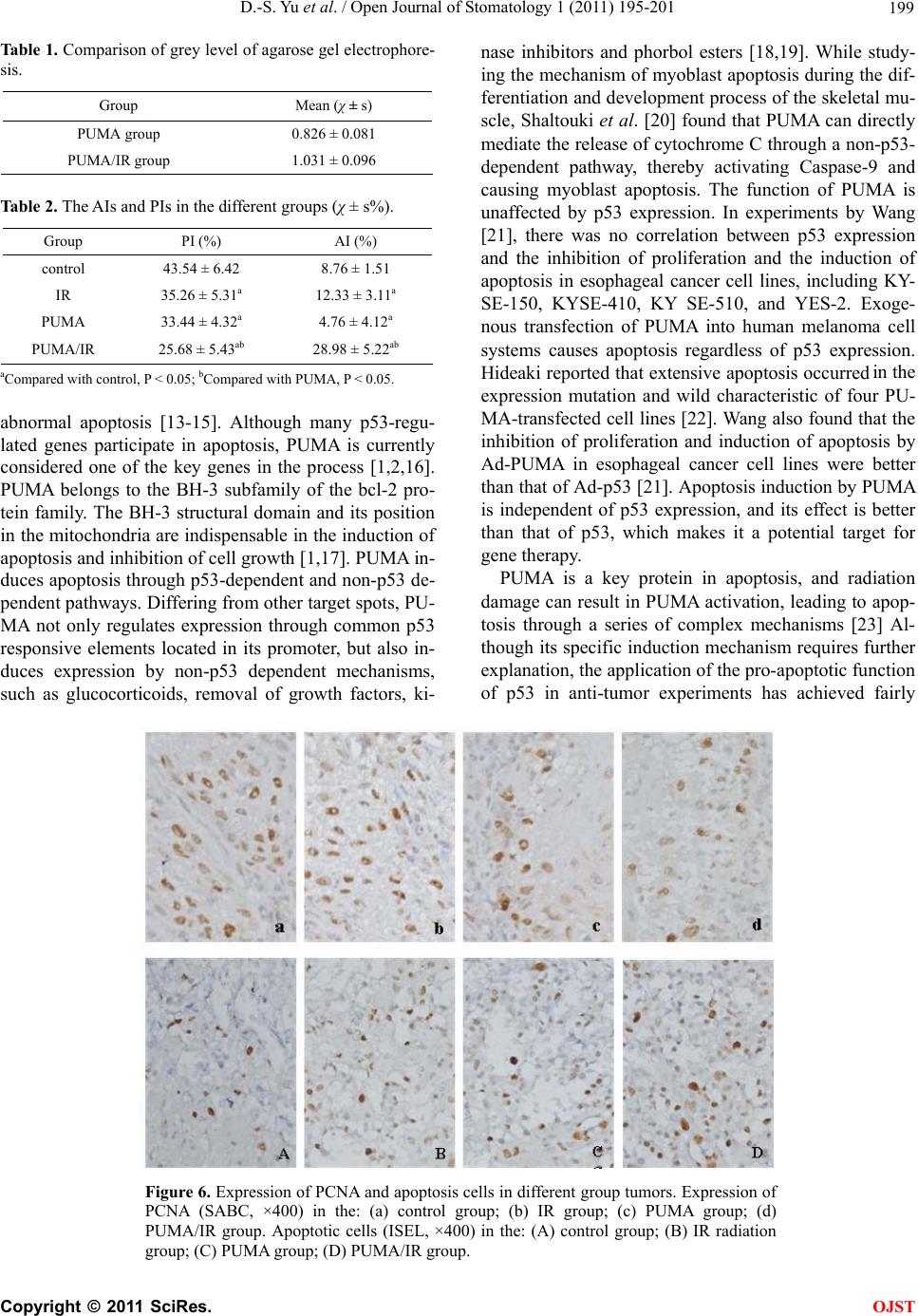 D.-S. Yu et al. / Open Journal of Stomatology 1 (2011) 195-201 Copyright © 2011 SciRes. 199 Table 1. Comparison of grey level of agarose gel electrophore- sis. Group Mean (χ ± s) PUMA group 0.826 ± 0.081 PUMA/IR group 1.031 ± 0.096 Table 2. The AIs and PIs in the different groups (χ ± s%). Group PI (%) AI (%) control 43.54 ± 6.42 8.76 ± 1.51 IR 35.26 ± 5.31a 12.33 ± 3.11a PUMA 33.44 ± 4.32a 4.76 ± 4.12a PUMA/IR 25.68 ± 5.43ab 28.98 ± 5.22ab aCompared with control, P < 0.05; bCompared with PUMA, P < 0.05. abnormal apoptosis [13-15]. Although many p53-regu- lated genes participate in apoptosis, PUMA is currently considered one of the key genes in the process [1,2,16]. PUMA belongs to the BH-3 subfamily of the bcl-2 pro- tein family. The BH-3 structural domain and its position in the mitochondria are indispensable in the induction of apoptosis and inhibition of cell growth [1,17]. PUMA in- duces apoptosis through p53-dependent and non-p53 de- pendent pathways. Differing from other target spots, PU- MA not only regulates expression through common p53 responsive elements located in its promoter, but also in- duces expression by non-p53 dependent mechanisms, such as glucocorticoids, removal of growth factors, ki- nase inhibitors and phorbol esters [18,19]. While study- ing the mechanism of myoblast apoptosis during the dif- ferentiation and development process of the skeletal mu- scle, Shaltouki et al. [20] found that PUMA can directly mediate the release of cytochrome C through a non-p53- dependent pathway, thereby activating Caspase-9 and causing myoblast apoptosis. The function of PUMA is unaffected by p53 expression. In experiments by Wang [21], there was no correlation between p53 expression and the inhibition of proliferation and the induction of apoptosis in esophageal cancer cell lines, including KY- SE-150, KYSE-410, KY SE-510, and YES-2. Exoge- nous transfection of PUMA into human melanoma cell systems causes apoptosis regardless of p53 expression. Hideaki reported that extensive apoptosis occurred in the expression mutation and wild characteristic of four PU- MA-transfected cell lines [22]. Wang also found that the inhibition of proliferation and induction of apoptosis by Ad-PUMA in esophageal cancer cell lines were better than that of Ad-p53 [21]. Apoptosis induction by PUMA is independent of p53 expression, and its effect is better than that of p53, which makes it a potential target for gene therapy. PUMA is a key protein in apoptosis, and radiation damage can result in PUMA activation, leading to apop- tosis through a series of complex mechanisms [23] Al- though its specific induction mechanism requires further explanation, the application of the pro-apoptotic function of p53 in anti-tumor experiments has achieved fairly Figure 6. Expression of PCNA and apoptosis cells in different group tumors. Expression of PCNA (SABC, ×400) in the: (a) control group; (b) IR group; (c) PUMA group; (d) PUMA/IR group. Apoptotic cells (ISEL, ×400) in the: (A) control group; (B) IR radiation group; (C) PUMA group; (D) PUMA/IR group. OJST 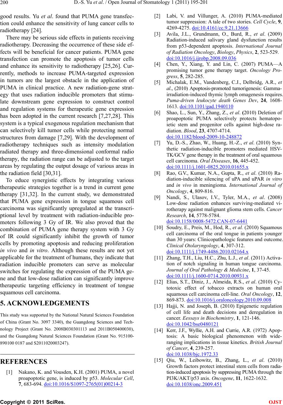 D.-S. Yu et al. / Open Journal of Stomatology 1 (2011) 195-201 200 good results. Yu et al. found that PUMA gene transfec- tion could enhance the sensitivity of lung cancer cells to radiotherapy [24]. There may be serious side effects in patients receiving radiotherapy. Decreasing the occurrence of these side ef- fects will be beneficial for cancer patients. PUMA gene transfection can promote the apoptosis of tumor cells and enhance its sensitivity to radiotherapy [25,26]. Cur- rently, methods to increase PUMA-targeted expression in tumors are the largest obstacle in the application of PUMA in clinical practice. A new radiation-gene strat- egy that uses radiation inducible promoters that stimu- late downstream gene expression to construct control and regulation systems for therapeutic gene expression has been adopted in the current research [7,27,28]. This system is a typical exogenous regulation mechanism that can selectively kill tumor cells while protecting normal structures from damage [7,29]. With the development of radiotherapy techniques such as intensity modulation radiated therapy and three-dimensional conformal radio therapy, the radiation range can be adjusted to the target areas by regulating the output dosage of various areas in the radiation field [30,31]. To educe synergistic effects by integrating various therapeutic strategies together is a trend in current gene therapy [31,32]. In the current study, we demonstrated that PUMA gene expression in tongue squamous cell carcinoma was significantly upregulated at the transcri- ptional level by treatment with radiation-inducible pro- moters following 3 Gy of IR. We also proved that the combination of PUMA gene therapy system with 3 Gy of IR could significantly inhibit the growth of tumor cells by promoting apoptosis and reducing proliferation in vivo and in vitro. Although these results are not yet applicable for the treatment of humans, they indicate that radiation inducible promoters can serve as molecular switches for regulating the expression of the PUMA ge- ne and that low-dose radiation can significantly improve therapeutic targeting efficiency in treatment of tongue squamous cell carcinoma. 5. ACKNOWLEDGEMENTS This study was supported by the National Natural Sciences Foundation of China (Grant No. 3097 3340), the Guangdong Sciences and Tech- nology Project (Grant No. 2008B030301113 and 2011B050400030), and the Guangdong Natural Sciences Foundation (Grant No. 915100- 890100 0187 and S2011020003247). REFERENCES [1] Nakano, K. and Vousden, K.H. (2001) PUMA, a novel proapoptotic gene, is induced by p53. Molecular Cell, 7, 683-694. doi:10.1016/S1097-2765(01)00214-3 [2] Labi, V. and Villunger, A. (2010) PUMA-mediated tumor suppression: A tale of two stories. Cell Cycle, 9, 4269-4275. doi:10.4161/cc.9.21.13666 [3] Avila, J.L., Grundmann, O., Burd, R., et al. (2009) Radiation-induced salivary gland dysfunction results from p53-dependent apoptosis. International Journal of Radiation Oncology, Biology, Physics, 2, 523-529. doi:10.1016/j.ijrobp.2008.09.036 [4] Chen, Y., Xiang, Y. and Lin, C. (2007) PUMA—A promising tumor gene therapy target. Oncology Pro- gress, 5, 282-285. [5] Michalak, E.M., Vandenberg, C.J., Delbridg, A.R., et al., (2010) Apoptosis-promoted tumorigenesis: Gamma- irradiation-induced thymic lymph omagenesis requires. Puma-driven leukocyte death Genes Dev, 24, 1608- 1613. doi:10.1101/gad.1940110 [6] Shao, L., Sun, Y., Zhang, Z., et al. (2010) Deletion of proapoptotic PUMA selectively protects hematopo- ietic stem and progenitor cells against high-dose ra- diation. Blood, 23, 4707-4714. doi:10.1182/blood-2009-10-248872 [7] Yu, D.-S., Zhao, W., Huang, H.-Z., et al. (2010) Syn- thetic radiation-inducible promoters mediated HSV- TK/GCV gene therapy in the treatment of oral squamous cell carcinoma. Oral Diseases, 16, 445-452. doi:10.1111/j.1601-0825.2010.01655.x [8] Rao, G.V., Kumar, N.A., Gupta, R., et al. (2010) Ra- diation-inducible silencing of uPA and uPAR in vitro and in vivo in meningioma. International Journal of Oncology, 4, 809-816. [9] Nandi, S., Ulasov, I.V., Tyler, M.A., et al. (2008) Low-dose radiation enhances surviving-mediated vi- rotherapy against malignant glioma stem cells. Cancer Research, 14, 5778-5784. doi:10.1158/0008-5472.CAN-07-6441 [10] Soudry, E., Preis, M., Hod, R., et al. (2010) Squamous cell carcinoma of the oral tongue in patients younger than 30 years: Clinicopathologic features and outcome. Clinical Otolaryngology, 4, 307-312. doi:10.1111/j.1749-4486.2010.02164.x [11] Zhang, T.H., Liu, H.C., Zhu, L.J., et al. (2011) Activa- tion of notch signaling in human tongue carcinoma. Journal of Oral Pathology & Medicine, 1, 37-45. doi:10.1111/j.1600-0714.2010.00931.x [12] Elias, S.T., Diniz, J., Almeida, R.S., et al. (2010) Cy- totoxic effect of tobacco extracts on human oral squamous cell carcinoma cell-line. Oral Oncology, 12, 869-873. doi:10.1016/j.oraloncology.2010.09.008 [13] Hajji, N. and Joseph, B. (2010) Epigenetic regulation of cell life and death decisions and deregulation in cancer. Eessays in Biochemistry, 1, 121-146. doi:10.1042/bse0480121 [14] Kerr, J.F., Wyllie, A.H. and Currie, A.R. (1972) Apop- tosis: A basic biological phenomenon with wide- ranging implications in tissue kinetics. British Journal of Cancer, 4, 239-257. doi:10.1038/bjc.1972.33 [15] Qiu, W., Leibowitz, B., Zhang, L., et al. (2010) Growth factors protect intestinal stem cells from radia- tion-induced apoptosis by suppressing PUMA through the PI3K/AKT/p53 axis. Oncogene, 11, 1622-1632. doi:10.1038/onc.2009.451 C opyright © 2011 SciRes. OJST  D.-S. Yu et al. / Open Journal of Stomatology 1 (2011) 195-201 201 [16] Chipuk, J.E. and Green, D.R. (2009) PUMA cooper- ates with direct activator proteins to promote mito- chondrial outer membrane permeabilization and apop- tosis. Cell Cycle, 17, 2692-2696. doi:10.4161/cc.8.17.9412 [17] Yu, J. and Zhang, L. (2008) PUMA, a potent killer with or without p53. Oncogene, 27, S71-S83. doi:10.1038/onc.2009.45 [18] Hong, L.Z., Zhao, X.Y. and Zhang, H.L. (2010) p53-mediated neuronal cell death in ischemic brain injury. Neuroscience Bulletin, 3, 232-240. doi:10.1007/s12264-010-1111-0 [19] Shaltouki, A., Freer, M., Mei, Y., et al. (2007) In- creased expression of the pro-apoptotic Bcl2 family member PUMA is required for mitochondrial release of cytochrome C and the apoptosis associated with skeletal myoblast differentiation. Apoptosis, 12, 2143- 2154. doi:10.1007/s10495-007-0135-z [20] Wang, H., Qian, H., Yu, J., et al. (2006) Administra- tion of PUMAadenovirus increases the sensitivity of esophageal cancer cellsto anticancer drugs. Cancer Biology & Therapy, 4, 380-389. doi:10.4161/cbt.5.4.2477 [21] Hideaki, I., Takao, K. and Toru, M. (2005) Efficacy of PUMA for malignant glioma cells regard less of the p53 status. Human Gene Therapy, 6, 685-692. [22] Vousden, K.H. (2005) Apotosis. p53 and PUMA: A deadly duo. Science, 5741, 1685-1686. doi:10.1126/science.1118232 [23] Yu, J., Yue, W., Wu, B., et al. (2006) PUMA sensitizes lung cancer cells to chemotherapeutic agents and irra- diation. Clinical Cancer Research, 9, 2928-2935. doi:10.1158/1078-0432.CCR-05-2429 [24] Qiu, W., Carson-Walter, E.B., Liu, H., et al. (2008) PUMA regulates intestinal progenitor cell radio-sen- sitivity and gastrointestinal syndrome. Cell Stem Cell, 6, 576-583. doi:10.1016/j.stem.2008.03.009 [25] Sasano, N., Enomoto, A., Hosoi, Y., et al. (2010) Eda- ravone, a known free radical scavenger, enhances X-ray-induced apoptosis at low concentrations. Can- cer Letters, 1, 52-57. doi:10.1016/j.canlet.2009.12.020 [26] Tsurushima, H., Yuan, X., Dillehay, L.E., et al. (2008) Radiation-inducible caspase-8 gene therapy for ma- lignant brain tumors. International Journal of Radia- tion Oncology, Biology, Physics, 2, 517-525. doi:10.1016/j.ijrobp.2008.02.002 [27] Marignol, L., Coffey, M., Hollywood, D., et al. (2007) Radiation to control transgene expression in tumors. Cancer Biology & Therapy, 7, 1005-1012. doi:10.4161/cbt.6.7.4477 [28] Vilaboa, N. and Voellmy, R. (2006) Regulatable gene expression systems for gene therapy. Current Gene Therapy, 4, 421-38. doi:10.2174/156652306777934829 [29] Bhide, S.A. and Nutting, C.M. (2010) Recent ad- vances in radiotherapy. BMC Medicine, 8, 25-28. doi:10.1186/1741-7015-8-25 [30] Brahme, A. and Lind, B.K. (2010) A systems biology approach to radiation therapy optimization. Radiation and Environmental Biophysics, 2, 111-124. doi:10.1007/s00411-010-0268-2 [31] John-Aryankalayil, M., Palayoor, S.T., Cerna, D., et al. (2010) Fractionated radiation therapy can induce a molecular profile for therapeutic targeting. Radiation Research, 4, 446-458. doi:10.1667/RR2105.1 [32] Marignol, L., Robson, T., McCarthy, H.O., et al. (2008) The tissue plasminogen activator gene promoter: A novel tool for radiogenic gene therapy of the prostate? Journal of Gene Medicine, 9, 1032-1038. doi:10.1002/jgm.1221 C opyright © 2011 SciRes. OJST |