

Paper Menu >>
Journal Menu >>
 Open Journal of Stomatology, 2011, 1, 185-188 doi:10.4236/ojst.2011.14028 Published Online December 2011 (http://www.SciRP.org/journal/ojst/ OJST ). Published Online December 2011 in SciRes. http://www.scirp.org/journal/OJST Upper mediastinal and paratracheal node dissection in total (pharyngo) laryngectomy, it is really indicated? Didier Dequanter, M. Shahla, P. Paulus, P. Lothaire Department of Head and Neck Surgery, CHU Charleroi, Montigny le Tilleul, Belgium. Email: [email protected] Received 25 July 2011; revised 29 August, 2011; accepted 14 September 2011. ABSTRACT Introduction: Advanced laryngeal and hypopharyn- geal cancers are aggressive tumors with a poor prog- nosis. Multiple lymph node metastases often occur in the neck as well as in the upper mediastinum and thus upper mediastinal dissection is crucial to im- proving the cure rate. However, excessive mediastinal dissection can increase postoperative morbidity and mortality making it important to employ the proper technique and appropriate extent of dissection. In the present study, we aimed to determine the need and the prognostic importance of mediastinal dissection in patients with advanced carcinoma of the upper aerodigestive tract. Methods: A retrospective review of the records of 30 patients who underwent (phar- ynxgo) laryngectomy for advanced squamous cell carcinomas was done. 17 patients had laryngeal car- cinomas, 13 had hypopharyngeal carcinomas. The mediastinal dissection was designed to remove mainly the paratracheal and retrooesophageal lymph nodes. Results: 60 neck dissections and 30 mediastinal dis- section were performed in 30 patients and yielded positive nodes were found in 20/30 patients. Neck nodes were positive in 9/17 of the patients with la- ryngeal cancer and 11/13 of the patients with hypo- pharyngeal cancers respectively. Positive nodes were detected in the neck regardless of T stage. The medi- astinal nodes were positive in 0% of the patients with laryngeal cancer. Upper mediastinal metastases were detected positive in 6/13 of the hypopharyngeal pa- tients. In these patients, mediastinal metastases were associated with tumors greater than 35 mm. The ma- jority of positive paratracheal nodes were less than 1 cm in diameter and appeared negative preoperatively. 0% of the patients had positive paratracheal nodes alone in a histologically negative cervical neck dissec- tion Regarding the appropriate extent of dissection, no major complications were observed. Conclusions: There is little controversy about neck dissection in advanced tumors of the (pharyngo) larynx. Laryn- geal carcinomas showed no positive mediastinal no- des in this series. The study highlighted the propen- sity of advanced hypopharyngeal cancers to involve the paratracheal nodes. Keywords: Advanced Head and Neck Cancer; Medi- astinal Dissection; Hypopharynx 1. INTRODUCTION (Pharyngo) laryngeal squamous cell carcinoma is a very aggressive cancer that is generally diagnosed at advan- ced stages and, consequently, has a poor prognosis and low survival. More than 75% of patients with (pharyn- go) laryngeal tumors have stage III or IV disease at presentation [1]. Indeed, cervical metastases are present in 60% - 80% [2,3] and, often, associated with multiple lymph nodes metastases that occur in the upper medi- astinum as well as in the paratracheal area. Although neck dissection is the surgical standard for the treatment of cervical lymph spread the upper mediastinal paratra- cheal nodal group is not routinely included in the dissec- tion. However, metastases to paratracheal lymph nodes occur in 30% of patients, mostly with hypopharyngeal tumors [4]. A 20% frequency of occult metastases to ipsilateral paratracheal lymph nodes has been reported in laryngeal cancer patients with postcricoid lesions and in patients with tumors that involved the pyriform fossa [5]. Moreover, laryngeal/hypopharygeal metastatic disease involving the paratracheal nodal group has been impli- cated in recurrent disease, particulary in patients with peristomal recurrence; this suggest the prognostic sig- nificance of positive paratracheal metastatic spread [6,7]. The purpose of this study was to analyse the paratra- cheal nodal yield and frequency of metastases and de- termine the indications of the upper mediastinal paratra- cheal dissection. 2. METHODS The study group consisted of 30 patients that were not previously treated for advanced squamous cell carcinoma 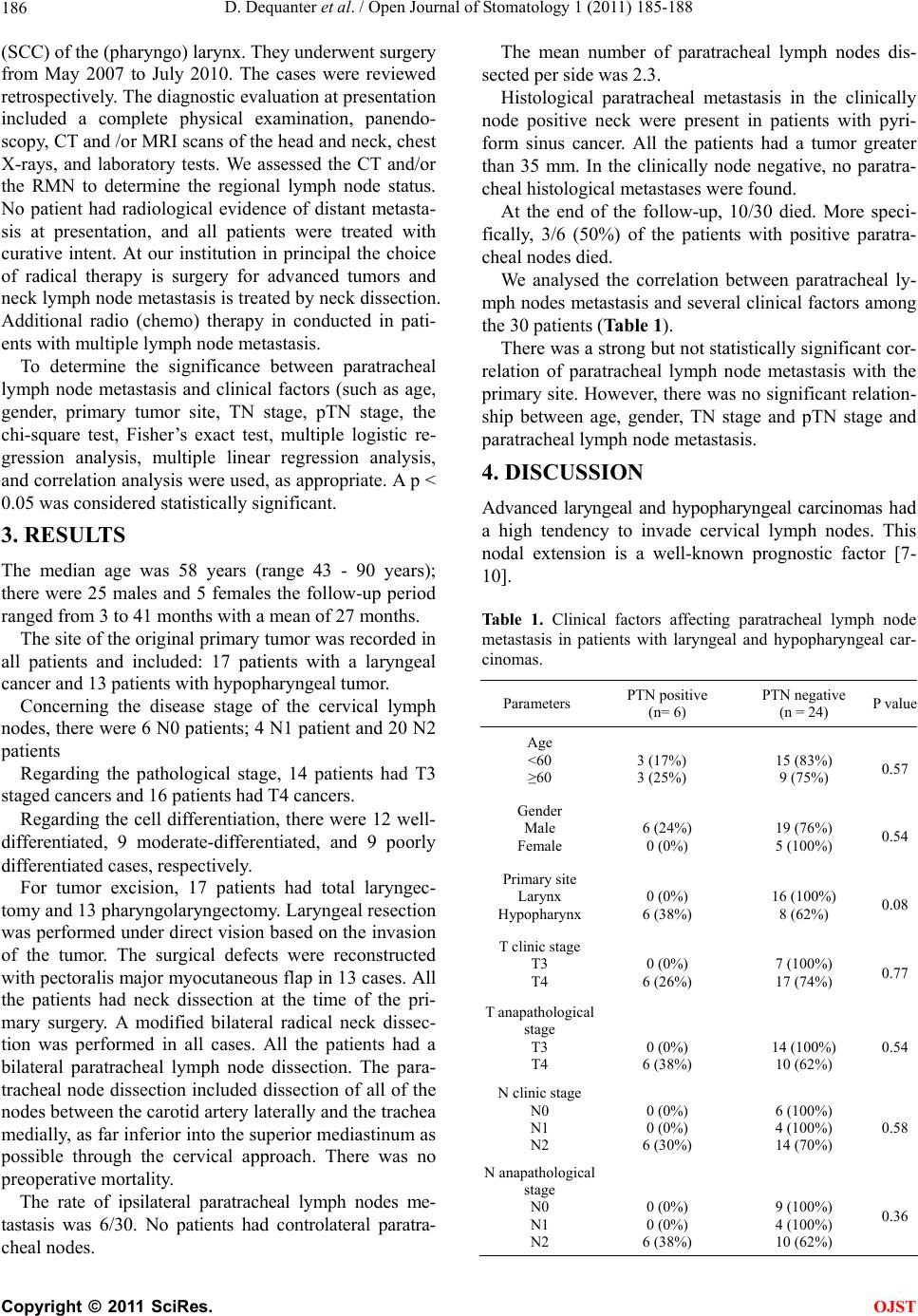 D. Dequanter et al. / Open Journal of Stomatology 1 (2011) 185-188 186 (SCC) of the (pharyngo) larynx. They underwent surgery from May 2007 to July 2010. The cases were reviewed retrospectively. The diagnostic evaluation at presentation included a complete physical examination, panendo- scopy, CT and /or MRI scans of the head and neck, chest X-rays, and laboratory tests. We assessed the CT and/or the RMN to determine the regional lymph node status. No patient had radiological evidence of distant metasta- sis at presentation, and all patients were treated with curative intent. At our institution in principal the choice of radical therapy is surgery for advanced tumors and neck lymph node metastasis is treated by neck dissection. Additional radio (chemo) therapy in conducted in pati- ents with multiple lymph node metastasis. To determine the significance between paratracheal lymph node metastasis and clinical factors (such as age, gender, primary tumor site, TN stage, pTN stage, the chi-square test, Fisher’s exact test, multiple logistic re- gression analysis, multiple linear regression analysis, and correlation analysis were used, as appropriate. A p < 0.05 was considered statistically significant. 3. RESULTS The median age was 58 years (range 43 - 90 years); there were 25 males and 5 females the follow-up period ranged from 3 to 41 months with a mean of 27 months. The site of the original primary tumor was recorded in all patients and included: 17 patients with a laryngeal cancer and 13 patients with hypopharyngeal tumor. Concerning the disease stage of the cervical lymph nodes, there were 6 N0 patients; 4 N1 patient and 20 N2 patients Regarding the pathological stage, 14 patients had T3 staged cancers and 16 patients had T4 cancers. Regarding the cell differentiation, there were 12 well- differentiated, 9 moderate-differentiated, and 9 poorly differentiated cases, respectively. For tumor excision, 17 patients had total laryngec- tomy and 13 pharyngolaryngectomy. Laryngeal resection was performed under direct vision based on the invasion of the tumor. The surgical defects were reconstructed with pectoralis major myocutaneous flap in 13 cases. All the patients had neck dissection at the time of the pri- mary surgery. A modified bilateral radical neck dissec- tion was performed in all cases. All the patients had a bilateral paratracheal lymph node dissection. The para- tracheal node dissection included dissection of all of the nodes between the carotid artery laterally and the trachea medially, as far inferior into the superior mediastinum as possible through the cervical approach. There was no preoperative mortality. The rate of ipsilateral paratracheal lymph nodes me- tastasis was 6/30. No patients had controlateral paratra- cheal nodes. The mean number of paratracheal lymph nodes dis- sected per side was 2.3. Histological paratracheal metastasis in the clinically node positive neck were present in patients with pyri- form sinus cancer. All the patients had a tumor greater than 35 mm. In the clinically node negative, no paratra- cheal histological metastases were found. At the end of the follow-up, 10/30 died. More speci- fically, 3/6 (50%) of the patients with positive paratra- cheal nodes died. We analysed the correlation between paratracheal ly- mph nodes metastasis and several clinical factors among the 30 patients (Table 1). There was a strong but not statistically significant cor- relation of paratracheal lymph node metastasis with the primary site. However, there was no significant relation- ship between age, gender, TN stage and pTN stage and paratracheal lymph node metastasis. 4. DISCUSSION Advanced laryngeal and hypopharyngeal carcinomas had a high tendency to invade cervical lymph nodes. This nodal extension is a well-known prognostic factor [7- 10]. Table 1. Clinical factors affecting paratracheal lymph node metastasis in patients with laryngeal and hypopharyngeal car- cinomas. Parameters PTN positive (n= 6) PTN negative (n = 24) P value Age <60 ≥60 3 (17%) 3 (25%) 15 (83%) 9 (75%) 0.57 Gender Male Female 6 (24%) 0 (0%) 19 (76%) 5 (100%) 0.54 Primary site Larynx Hypopharynx 0 (0%) 6 (38%) 16 (100%) 8 (62%) 0.08 T clinic stage T3 T4 0 (0%) 6 (26%) 7 (100%) 17 (74%) 0.77 T anapathological stage T3 T4 0 (0%) 6 (38%) 14 (100%) 10 (62%) 0.54 N clinic stage N0 N1 N2 0 (0%) 0 (0%) 6 (30%) 6 (100%) 4 (100%) 14 (70%) 0.58 N anapathological stage N0 N1 N2 0 (0%) 0 (0%) 6 (38%) 9 (100%) 4 (100%) 10 (62%) 0.36 C opyright © 2011 SciRes. OJST  D. Dequanter et al. / Open Journal of Stomatology 1 (2011) 185-188 187 Although neck dissection is the surgical gold standard for the treatment of cervical lymphatic spread, the upper mediastinal and paratracheal nodal groups are not rou- tinely included in the dissection. However, Welsh et al. [11] initially reported the im- portance of the paratracheal nodes in lymphatic drainage of the larynx and hypopharynx. Harrison et al. con- firmed that carcinomas of the larynx (especially from the subglottic region), the trachea, and the cervical esopha- gus are at risk for paratracheal and tracheobronchial lymph node metastasis and recommended resection of the manubrium to remove theses lymph nodes and re- duce the risk for parastomal disease recurrence [5]. We- ber et al. [7] reported that paratracheal lymph node me- tastasis were found in 17.6% of the patients with tumors of the larynx, in 8.3% of the patients with pharyngeal tumors and, at least, in 71.4% of the patients with tumors of the esophagus. Some investigators have suggested that ipsilateral paratracheal node dissection should be included as part of a selective neck dissection in all patients with tumors invading the subglottis, pyriform fossa apex and postcri- coid region [12]. In our study, all the patients with advanced laryngeal and hypopharyngeal SCC had a neck dissection extend- ed to the upper mediastinum. Most patients with hypo- pharyngeal SCC had positive paratracheal lymph nodes but only to the ipsilateral side of the primary. All the patients with hypopharyngeal SCC greater than 35 mm had paratracheal lymph node metastasis. Furthemore, paratracheal lymph node metastasis was always associ- ated with the presence of cervical lymph node metastasis. In addition, a strong correlation but, unfortunately, not statistically significant was found between the presence of paratracheal lymph node metastasis and hypoharyn- geal tumors. We recommend that ipsilateral paratracheal node dis- section should be included as part of a selective neck dissection in all patients with tumors invading the hypo- pharynx, certainly in tumors greater than 35 mm. In terms of survival rates, in a prospective study of 50 patients with carcinoma of the larynx, hypopharynx, and cervical esophagus, Timon et al. [13] reported that the rate of paratracheal nodal metastases was 26%. More- over, they reported that the survival in patients with po- sitive paratracheal nodes demonstrated a trend towards poorer survival compared to patients without paratra- cheal nodal involvement, and concomitant involvement of both cervical and paratracheal nodal groups was asso- ciated with the poorest survival propability. In their study, Weber et al. [7] confirmed that survival was significantly reduced by the presence of paratra- cheal lymph nodes. In our study, 50% of the patients with positive paratracheal nodes died. In conclusion, there is little controversy about neck dis- sections in advanced tumors of the larynx and hypo- pahrynx. A similar situation applies to mediadtinal dis- section for hypopharyngeal carcinomas, certainly in tu- mors greater than 35 mm. REFERENCES [1] Hoffman, H.T., Karnell, L.H., Funk, G.F., Robinson, R.A. and Menck, H.R. (1998) The National Cancer Data Base Report on cancer of the head and neck. Archives of Oto- laryngology-Head & Neck Surgery, 124, 951-962. [2] Ho, C.M., Lam, K.H., Wei, W.I., Yuen, P.W., Lam, L.K. (1993) Squamous cell carcinoma of the hypopharynx— Analysis of treatment results. Head Neck, 15, 405-412. doi:10.1002/hed.2880150507 [3] Lefebvre, J.L., Castelain, B., DeLatorre, J.C., Delobelle- Deroide, A. and Vankemmel, B. (1987) Lymph node in- vasion in hypopharynx and lateral epilarynx: A prognos- tic factor. Head & Neck Surgery, 10, 14-18. doi:10.1002/hed.2890100104 [4] Wei, W.I. (2002) The dilemma of treating hypopharyn- geal carcinoma: More or less. Archives of Otolaryngol- ogy-Head & Neck Surgery, 128, 229-232. [5] Buckley, J.G. and Lemman, K. (2000) Cervical node metastases in laryngeal and hypopharyngeal cancer: A prospective analysis of prevalence and distribution. Head Neck, 22, 280-385. doi:10.1002/1097-0347(200007)22:4<380::AID-HED11 >3.0.CO;2-E [6] Harris, H.H. and Butler, E. (1968) Surgical limits in can- cer of the subglottic larynx. Archives of Otolaryngology, 87, 64-67. [7] Weber, R.S., Marvel, J., Smith, P., Hankins, P., Wolf, P. and Goepfert, H. (1993) Paratracheal lymph node dissec- tion for carcinoma of the larynx, hypopharynx and cer- vical esophagus. Archives of Otolaryngology-Head & Neck Surgery, 108, 11-17. [8] Driscoll, W.G., Nagorsky, M.J., Cantrell, R.W. and Johns, M.E. (1983) Carcinoma of the pyriform sinus: Analysis of 102 cases. Laryngoscope, 93, 556-560. [9] Bataini, J.P., Bernier, J., Brugere, J., Jaulerry, C., Picco, C. and Brunin, F. (1985) Natural history of neck diseases in patients with squamous cell carcinoma of oropharynx and pharyngolarynx. Radiotherapy & Oncology, 21, 245- 255. doi:10.1016/S0167-8140(85)80033-5 [10] Lindberg, R. (1972) Distribution of cervical lymph nodes metastases from squamous cell carcinoma of the upper respiratory and digestive tracts. Cancer, 29, 1446-1449. doi:10.1002/1097-0142(197206)29:6<1446::AID-CNCR 2820290604>3.0.CO;2-C [11] Welsh, L.W. (1964) The normal human laryngeal lym- phatics. Annals of Otology, Rhinology, and Laryngology, 73, 569-582. [12] Harrison, D.F.N. (1975) Laryngectomy for subglottic lesions. Laryngoscope, 85, 1208-1210. C opyright © 2011 SciRes. OJST  D. Dequanter et al. / Open Journal of Stomatology 1 (2011) 185-188 Copyright © 2011 SciRes. 188 OJST doi:10.1288/00005537-197507000-00012 [13] Timon, C.V., Toner, M. and Conlon, B.J. (2003) Paratra- cheal lymph node involvement in advanced cancer of the larynx, hypopharynx and cervical esophagus. Laryngo- scope, 113, 1595-1599. doi:10.1097/00005537-200309000-00035 |