

Paper Menu >>
Journal Menu >>
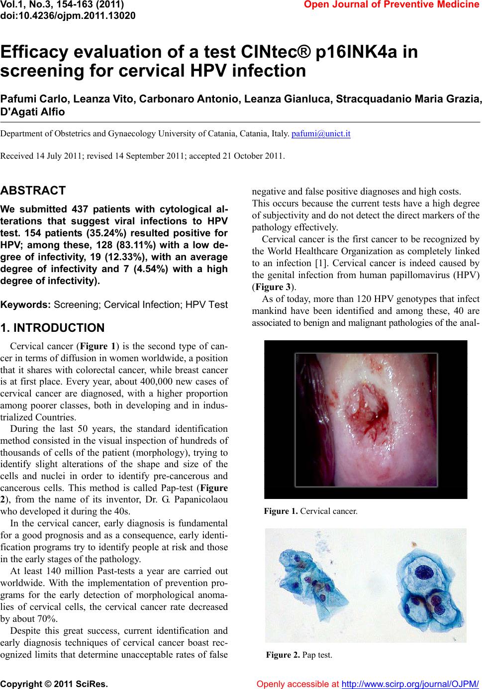 Vol.1, No.3, 154-163 (2011) doi:10.4236/ojpm.2011.13020 C opyright © 2011 SciRes. Openly accessible at http://www.scirp.org/journal/OJPM/ Open Journal of Preventive Medicine Efficacy evaluation of a test CINtec® p16INK4a in screening for cervical HPV infection Pafumi Carlo, Leanza V ito, Carbonaro A ntonio, Leanza Gianluca, S tracquadani o Maria Grazia, D'Agati Alfio Department of Obstetrics and Gynaecology University of Catania, Catania, Italy. [email protected] Received 14 July 2011; revised 14 September 2011; accepted 21 October 2011. ABSTRACT We submitted 437 patients with cytological al- terations that suggest viral infections to HPV test. 154 patients (35.24%) resulted positive for HPV; among these, 128 (83.11%) with a low de- gree of infectivity, 19 (12.33%), with an average degree of infectivity and 7 (4.54%) with a high degree of infectivity). Keywords: Screening; Cervical Infection; HPV Test 1. INTRODUCTION Cervical cancer (Figure 1) is the second type of can- cer in terms of diffusion in women worldwide, a position that it shares with colorectal cancer, while breast cancer is at first place. Every year, about 400,000 new cases of cervical cancer are diagnosed, with a higher proportion among poorer classes, both in developing and in indus- trialized Countries. During the last 50 years, the standard identification method consisted in the visual inspection of hundreds of thousands of cells of the patient (morphology), trying to identify slight alterations of the shape and size of the cells and nuclei in order to identify pre-cancerous and cancerous cells. This method is called Pap-test (Figure 2), from the name of its inventor, Dr. G. Papanicolaou who developed it during the 40s. In the cervical cancer, early diagnosis is fundamental for a good prognosis and as a consequence, early identi- fication programs try to identify people at risk and those in the early stages of the pathology. At least 140 million Past-tests a year are carried out worldwide. With the implementation of prevention pro- grams for the early detection of morphological anoma- lies of cervical cells, the cervical cancer rate decreased by about 70%. Despite this great success, current identification and early diagnosis techniques of cervical cancer boast rec- ognized limits that determine unacceptable rates of false negative and false positive diagnoses and high co sts. This occurs because the current tests have a high degree of subjectivity and do not detect the direct markers of the pathology effectively. Cervical cancer is the first cancer to be recognized by the World Healthcare Organization as completely linked to an infection [1]. Cervical cancer is indeed caused by the genital infection from human papillomavirus (HPV) (Figure 3). As of today, mor e than 120 HPV genotypes that infect mankind have been identified and among these, 40 are associated to benign and malignant pathologies of the a nal - Figure 1. Cervical cancer. Figure 2. Pap test. 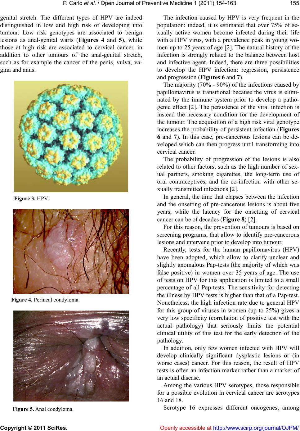 P. Carlo et al. / Open Journal of Preventive Medicine 1 (2011) 154-163 Copyright © 2011 SciRes. Openly accessible at http://www.scirp.org/journal/OJPM/ 155 genital stretch. The different types of HPV are indeed distinguished in low and high risk of developing into tumour. Low risk genotypes are associated to benign lesions as anal-genital warts (Figures 4 and 5), while those at high risk are associated to cervical cancer, in addition to other tumours of the anal-genital stretch, such as for example the cancer of the penis, vulva, va- gina and anus. Figure 3. HPV. Figure 4. Perineal condyloma. Figure 5. Anal condyloma. The infection caused by HPV is very frequent in the population: indeed, it is estimated that over 75% of se- xually active women become infected during their life with a HPV virus, with a prevalence peak in young wo- men up to 25 years of age [2]. The natural history of the infection is strongly related to the balance between host and infective agent. Indeed, there are three possibilities to develop the HPV infection: regression, persistence and progression (Figures 6 and 7). The majority (70% - 90%) of the infections caused by papillomavirus is transitional because the virus is elimi- nated by the immune system prior to develop a patho- genic effect [2]. The persistence of the viral infection is instead the necessary condition for the development of the tumour. The acq uisition of a high risk viral genotype increases the probability of persistent infection (Figures 6 and 7). In this case, pre-cancerous lesions can be de- veloped which can then progress until transforming into cervical cancer. The probability of progression of the lesions is also related to other factors, such as the high number of sex- ual partners, smoking cigarettes, the long-term use of oral contraceptives, and the co-infection with other se- xually transmitted infections [2 ]. In general, the time that elapses between the infection and the onsetting of pre-cancerous lesions is about five years, while the latency for the onsetting of cervical cancer can be of decades (Figure 8) [2]. For this reason, the preven tion of tumours is based on screening programs, that allow to identify pre-cancerous lesions and intervene prior to develop into tumour. Recently, tests for the human papillomavirus (HPV) have been adopted, which allow to clarify unclear and slightly anomalous Pap-tests (the majority of which was false positive) in women over 35 years of age. The use of tests on HPV for this application is limited to a small percentage of all Pap-tests. The sensitivity for detecting the illness by HPV tests is higher than that of a Pap-test. Nonetheless, the high infection rate due to general HPV for this group of viruses in women (up to 25%) gives a very low specificity (correlation of positive test with the actual pathology) that seriously limits the potential clinical utility of this test for the early detection of the pathology. In addition, only few women infected with HPV will develop clinically significant dysplastic lesions or (in worse cases) cancer. For this reason, the result of HPV tests is often an infection marker ra ther than a mar ker of an actual disease. Among the various HPV serotypes, those responsible for a possible evolution in cervical cancer are serotypes 16 and 18. Serotype 16 expresses diferent oncogenes, among f 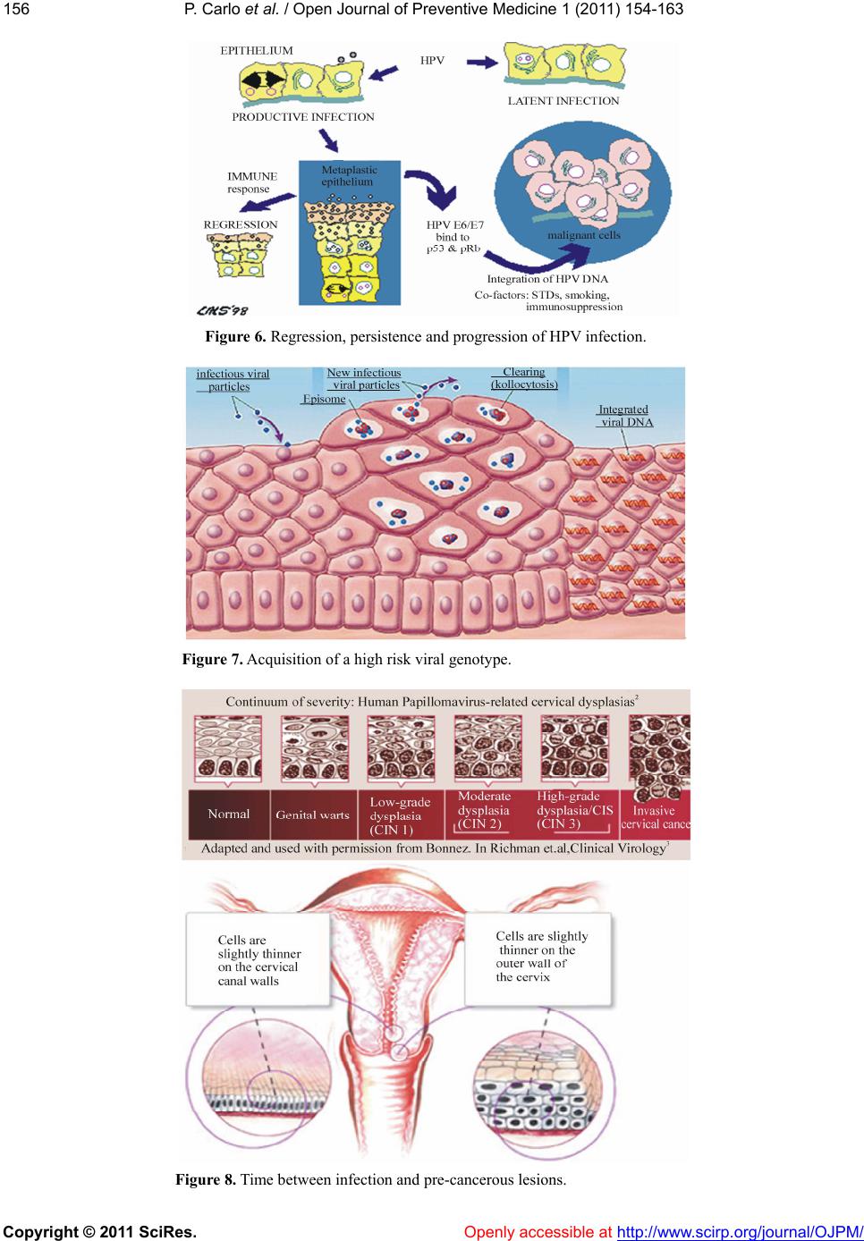 P. Carlo et al. / Open Journal of Preventive Medicine 1 (2011) 154-163 Copyright © 2011 SciRes. http://www.scirp.org/journal/OJPM/ 156 Figure 6. Regression, persistence and progression of HPV infection. Figure 7. Acquisition of a high risk viral genotype. Figure 8. T ime between infection and pre-cancerous lesions. Openly accessible at  P. Carlo et al. / Open Journal of Preventive Medicine 1 (2011) 154-163 Copyright © 2011 SciRes. Openly accessible at http://www.scirp.org/journal/OJPM/ 157 which it seems that E6 and E7 (Figure 9) are of high risk of tumour especially if expressed in basal and para- basal cells subject to replicatio n. (Figure 10) In order for this to take place, specific changes of the host cell are needed, that alter the transcriptional control of the viral genome [3,4]. The expression of viral onco- genes in proliferating cells interferes with the regulation of the cellular cycle and, after numerous bio-chemical interactions, it changes the expression profiles of many genes and/or proteins (Figures 11 and 12) [5]. These changes in the cells of basal and parabasal lay- ers infected from HPV (Figure 13) take place only ra- rely and therefore the tran sformation and carcinogenesis process is fortunately only a very rare consequence of a very common infection. Due to this high incidence of HPV infections in the healthy population, the ideal screening test for cervical cancer should identify the modifications of the basal and parabasal cells induced by the deregulated expression pattern of viral genes; this test should at least in theory, combine the high sensitivity of HPV tests with the high specificity of the cytology, and therefore overcome the Figure 9. Different oncogeneses of HPV-16. Figure 10. HPV infection in basal and parabasal cells. 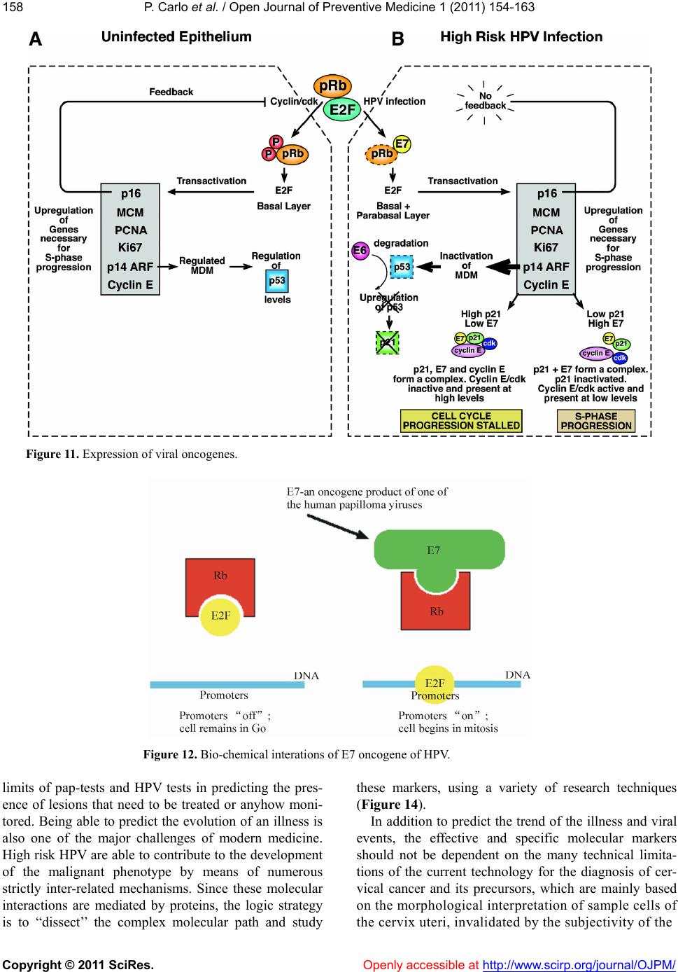 P. Carlo et al. / Open Journal of Preventive Medicine 1 (2011) 154-163 Copyright © 2011 SciRes. Openly accessible at http://www.scirp.org/journal/OJPM/ 158 Figure 11. Expression of viral oncogenes. Figure 12. Bio-chemical interations of E7 oncogene of HPV. limits of pap-tests and HPV tests in predicting the pres- ence of lesions that need to be treated or anyhow moni- tored. Being able to predict the ev olution of an illness is also one of the major challenges of modern medicine. High risk HPV are able to contribute to the development of the malignant phenotype by means of numerous strictly inter-related mechanisms. Since these molecular interactions are mediated by proteins, the logic strategy is to “dissect’’ the complex molecular path and study these markers, using a variety of research techniques (Figure 14). In addition to predict the trend of the illness and viral events, the effective and specific molecular markers should not be dependent on the many technical limita- tions of the current technology for the diagnosis of cer- vical cancer and its precursors, which are mainly based on the morphological interpretation of sample cells of th cervix uteri, invalidated by the subjectivity of the e 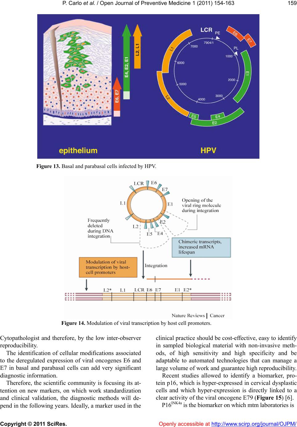 P. Carlo et al. / Open Journal of Preventive Medicine 1 (2011) 154-163 Copyright © 2011 SciRes. Openly accessible at http://www.scirp.org/journal/OJPM/ 159 Figure 13. Basal and parabasal cells in fected by HPV. Figure 14. Modulation of viral transcription by host cell promoters. Cytopathologist and therefore, by the low inter-observer reproducibility. The identification of cellular modifications associated to the deregulated expression of viral oncogenes E6 and E7 in basal and parabasal cells can add very significant diagnostic information. Therefore, the scientific community is focusing its at- tention on new markers, on which work standardization and clinical validation, the diagnostic methods will de- pend in the following years. Ideally, a marker used in the clinical practice should be cost-effective, easy to iden tify in sampled biological material with non-invasive meth- ods, of high sensitivity and high specificity and be adaptable to automated technologies that can manage a large volume of work and guarantee high reproducibility. Recent studies allowed to identify a biomarker, pro- tein p16, which is hyper-expressed in cervical dysplastic cells and which hyper-expression is directly linked to a clear activity of the viral oncogen e E79 (Figure 15) [6]. P16INK4a is the biomarker on which mtm laboratories is 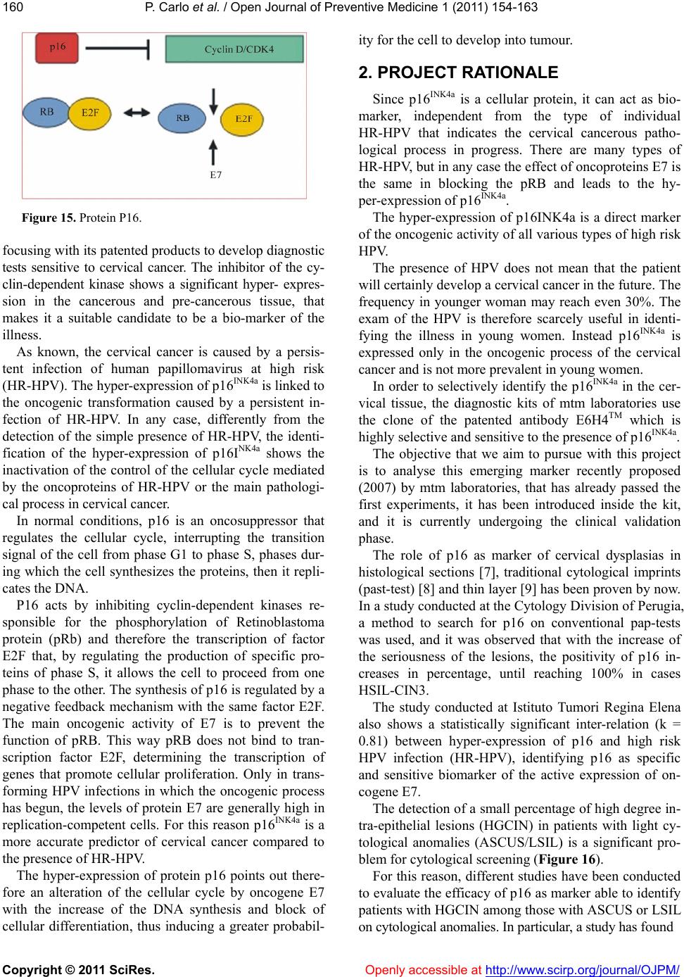 P. Carlo et al. / Open Journal of Preventive Medicine 1 (2011) 154-163 Copyright © 2011 SciRes. Openly accessible at http://www.scirp.org/journal/OJPM/ 160 Figure 15. Protein P16. focusing with its patented products to develop diagnostic tests sensitive to cervical cancer. The inhibitor of the cy- clin-dependent kinase shows a significant hyper- expres- sion in the cancerous and pre-cancerous tissue, that makes it a suitable candidate to be a bio-marker of the illness. As known, the cervical cancer is caused by a persis- tent infection of human papillomavirus at high risk (HR-HPV). The hyper-expression of p16INK4a is linked to the oncogenic transformation caused by a persistent in- fection of HR-HPV. In any case, differently from the detection of the simple presence of HR-HPV, the identi- fication of the hyper-expression of p16INK4a shows the inactivation of the control of the cellular cycle mediated by the oncoproteins of HR-HPV or the main pathologi- cal process in cervical cancer. In normal conditions, p16 is an oncosuppressor that regulates the cellular cycle, interrupting the transition signal of the cell from phase G1 to phase S, phases dur- ing which the cell synthesizes the proteins, then it repli- cates the DNA. P16 acts by inhibiting cyclin-dependent kinases re- sponsible for the phosphorylation of Retinoblastoma protein (pRb) and therefore the transcription of factor E2F that, by regulating the production of specific pro- teins of phase S, it allows the cell to proceed from one phase to the other. The synthesis of p16 is regulated by a negative feedback mechanism with the same factor E2F. The main oncogenic activity of E7 is to prevent the function of pRB. This way pRB does not bind to tran- scription factor E2F, determining the transcription of genes that promote cellular proliferation. Only in trans- forming HPV infections in which the oncogenic process has begun, the levels of protein E7 are generally high in replication-competent cells. For this reason p16INK4a is a more accurate predictor of cervical cancer compared to the presence of HR-HPV. The hyper-expression of protein p16 points out there- fore an alteration of the cellular cycle by oncogene E7 with the increase of the DNA synthesis and block of cellular differentiation, thus inducing a greater probabil- ity for the cell to develop into tumour. 2. PROJECT RATIONALE Since p16INK4a is a cellular protein, it can act as bio- marker, independent from the type of individual HR-HPV that indicates the cervical cancerous patho- logical process in progress. There are many types of HR-HPV, but in any case the effect of oncoproteins E7 is the same in blocking the pRB and leads to the hy- per -e xp ression of p16INK4a. The hyper-expression of p16INK4a is a direct marker of the oncogenic activity of all various types of high risk HPV. The presence of HPV does not mean that the patient will certainly develop a cervical cancer in the future. The frequency in younger woman may reach even 30%. The exam of the HPV is therefore scarcely useful in identi- fying the illness in young women. Instead p16INK4a is expressed only in the oncogenic process of the cervical cancer and is not more prevalent in young women. In order to selectively identify the p16INK4a in the cer- vical tissue, the diagnostic kits of mtm laboratories use the clone of the patented antibody E6H4TM which is highly selective and sens itive to the presence of p16INK4a. The objective that we aim to pursue with this project is to analyse this emerging marker recently proposed (2007) by mtm laboratories, that has already passed the first experiments, it has been introduced inside the kit, and it is currently undergoing the clinical validation phase. The role of p16 as marker of cervical dysplasias in histological sections [7], traditional cytological imprints (past-test) [8] and th in layer [9] has been proven by now. In a study conducted at the Cytology Division of Perugia, a method to search for p16 on conventional pap-tests was used, and it was observed that with the increase of the seriousness of the lesions, the positivity of p16 in- creases in percentage, until reaching 100% in cases HSIL-CIN3. The study conducted at Istituto Tumori Regina Elena also shows a statistically significant inter-relation (k = 0.81) between hyper-expression of p16 and high risk HPV infection (HR-HPV), identifying p16 as specific and sensitive biomarker of the active expression of on- cogene E7. The detection of a small percentage of high degree in- tra-epithelial lesions (HGCIN) in patients with light cy- tological anomalies (ASCUS/LSIL) is a significant pro- blem for cytological screening (Figure 16). For this reason, different studies have been conducted to evaluate the efficacy of p16 as marker able to identify patients with HGCIN among those with ASCUS or LSIL on cytological anomali es. In particular, a study has found 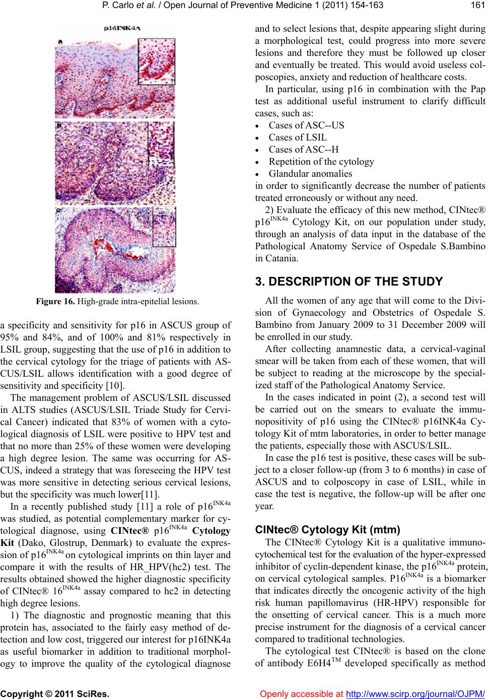 P. Carlo et al. / Open Journal of Preventive Medicine 1 (2011) 154-163 Copyright © 2011 SciRes. Openly accessible at http://www.scirp.org/journal/OJPM/ 161 Figure 16. High-grade intra-epitelial lesions. a specificity and sensitivity for p16 in ASCUS group of 95% and 84%, and of 100% and 81% respectively in LSIL group, suggesting that the use of p16 in addition to the cervical cytology for the triage of patients with AS- CUS/LSIL allows identification with a good degree of sensitivity and specificity [10]. The management problem of ASCUS/LSIL discussed in ALTS studies (ASCUS/LSIL Triade Study for Cervi- cal Cancer) indicated that 83% of women with a cyto- logical diagnosis of LSIL were positive to HPV test and that no more than 25% of these women were developing a high degree lesion. The same was occurring for AS- CUS, indeed a strategy that was foreseeing the HPV test was more sensitive in detecting serious cervical lesions, but the specificity was much lower[11]. In a recently published study [11] a role of p16INK4a was studied, as potential complementary marker for cy- tological diagnose, using CINtec® p16INK4a Cytology Kit (Dako, Glostrup, Denmark) to evaluate the expres- sion of p16INK4a on cytological imprints on thin layer and compare it with the results of HR_HPV(hc2) test. The results obtained showed the higher diagnostic specificity of CINtec® 16INK4a assay compared to hc2 in detecting high degree lesions. 1) The diagnostic and prognostic meaning that this protein has, associated to the fairly easy method of de- tection and low cost, triggered our interest for p16INK4a as useful biomarker in addition to traditional morphol- ogy to improve the quality of the cytological diagnose and to select lesions that, despite appearing slight during a morphological test, could progress into more severe lesions and therefore they must be followed up closer and eventually be treated. This would avoid useless col- poscopies, anxiety and reduction of healthcare costs. In particular, using p16 in combination with the Pap test as additional useful instrument to clarify difficult cases, such as: Cases of ASC--US Cases of LSIL Cases of ASC--H Repetition of the cytology Glandular anoma lies in order to significantly decrease the number of patients treated erroneously or without any need. 2) Evaluate the efficacy of this new method, CINtec® p16INK4a Cytology Kit, on our population under study, through an analysis of data input in the database of the Pathological Anatomy Service of Ospedale S.Bambino in Catania. 3. DESCRIPTION OF THE STUDY All the women of any age that will come to the Divi- sion of Gynaecology and Obstetrics of Ospedale S. Bambino from January 2009 to 31 December 2009 will be enrolled in our study. After collecting anamnestic data, a cervical-vaginal smear will be taken from each of these women, that will be subject to reading at the microscope by the special- ized staff of the Pathological Anatomy Service. In the cases indicated in point (2), a second test will be carried out on the smears to evaluate the immu- nopositivity of p16 using the CINtec® p16INK4a Cy- tology Kit of mtm laboratories, in order to better manage the patients, especially those with ASCUS/LSIL. In case the p16 test is positive, these cases will be sub- ject to a closer follow-up (from 3 to 6 months) in case of ASCUS and to colposcopy in case of LSIL, while in case the test is negative, the follow-up will be after one year. CINtec® Cytology Kit (mtm) The CINtec® Cytology Kit is a qualitative immuno- cytochemical test for the evaluation of the hyper- ex pressed inhibitor of cyclin-dependent kinase, the p16INK4a protein, on cervical cytological samples. P16INK4a is a biomarker that indicates directly the oncogenic activity of the high risk human papillomavirus (HR-HPV) responsible for the onsetting of cervical cancer. This is a much more precise instrument for the diagnosis of a cervical cancer compared to traditional technologies. The cytological test CINtec® is based on the clone f antibody E6H4TM developed specifically as method o 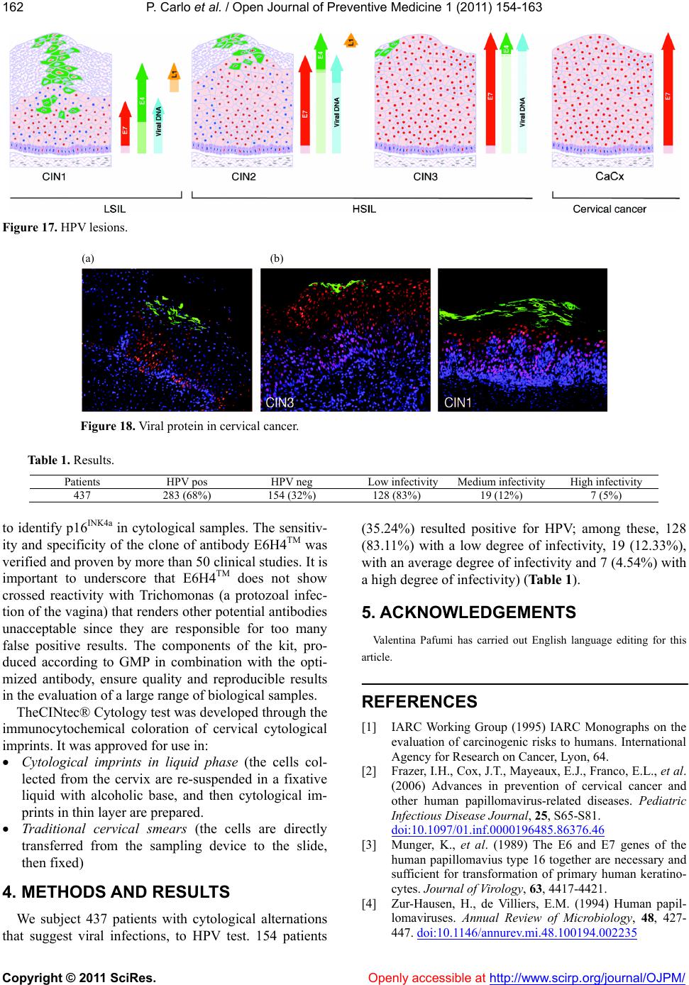 P. Carlo et al. / Open Journal of Preventive Medicine 1 (2011) 154-163 Copyright © 2011 SciRes. http://www.scirp.org/journal/OJPM/ 162 Figure 17. HPV lesions. (a) (b) Figure 18. Viral protein in cervic al cancer. Table 1. Results. Patients HPV pos HPV neg Low infectivityMedium infectivity High infectivity 437 283 (68%) 154 (32%) 128 (83%) 19 (12%) 7 (5%) to identify p16INK4a in cytological samples. The sensitiv- ity and specificity of the clone of antibody E6H4TM was verified and proven by more than 50 clinical studies. It is important to underscore that E6H4TM does not show crossed reactivity with Trichomonas (a protozoal infec- tion of the vagina) that renders other potential antibodies unacceptable since they are responsible for too many false positive results. The components of the kit, pro- duced according to GMP in combination with the opti- mized antibody, ensure quality and reproducible results in the evaluation of a large range of biolog ical samples. Openly accessible at TheCINtec® Cytology test was developed th rough th e immunocytochemical coloration of cervical cytological imprints. It was approved for use in: Cytological imprints in liquid phase (the cells col- lected from the cervix are re-suspended in a fixative liquid with alcoholic base, and then cytological im- prints in thin layer are prepared. Traditional cervical smears (the cells are directly transferred from the sampling device to the slide, then fixed ) 4. METHODS AND RESULTS We subject 437 patients with cytological alternations that suggest viral infections, to HPV test. 154 patients (35.24%) resulted positive for HPV; among these, 128 (83.11%) with a low degree of infectivity, 19 (12.33%), with an average degree of infectivity and 7 (4.54%) with a high degree of infectivity) (Table 1). 5. ACKNOWLEDGEMENTS Valentina Pafumi has carried out English language editing for this article. REFERENCES [1] IARC Working Group (1995) IARC Monographs on the evaluation of carcinogenic risks to humans. International Agency for Research on Cancer, Lyon, 64. [2] Frazer, I.H., Cox, J.T., Mayeaux, E.J., Franco, E.L., et al. (2006) Advances in prevention of cervical cancer and other human papillomavirus-related diseases. Pediatric Infectious Disease Journal, 25, S65-S81. doi:10.1097/01.inf.0000196485.86376.46 [3] Munger, K., et al. (1989) The E6 and E7 genes of the human papillomavius type 16 together are necessary and sufficient for transformation of primary human keratino- cytes. Journal of Virology, 63, 4417-4421. [4] Zur-Hausen, H., de Villiers, E.M. (1994) Human papil- lomaviruses. Annual Review of Microbiology, 48, 427- 447. doi:10.1146/annurev.mi.48.100194.002235  P. Carlo et al. / Open Journal of Preventive Medicine 1 (2011) 154-163 Copyright © 2011 SciRes. Openly accessible at http://www.scirp.org/journal/OJPM/ 163 [5] Zur-Hausen H., (2000) Papillomaviruses causing cancer: Evasion from host-cell control in early events in car- cinogenesis. Journal of the National Cancer Institute, 92, 690-698. doi:10.1093/jnci/92.9.690 [6] Von Knebel-Doeberitz, M. (2002) New markers for cer- vical dysplasia to visualize the genomic chaos created by aberrant oncogenic papillomavirus infections. European Journal of Cancer, 38, 2229-2242. doi:10.1016/S0959-8049(02)00462-8 [7] Benevolo, M., et al. (2006) Immunohistochemical ex- pression of p16INK4a is predictive of HR-HPV infection in cervical low-grade lesion. Modern Pathology, 19, 384- 391. doi:10.1038/modpathol.3800551 [8] Passamonti, B., et al. (2006) P16: A method for directed research on abnormal convenzional pap-test. Pathologica, 98, 417. [9] Bibbo, M., et al. (2002) Procedure for immunocyto- chemical detection of p16INK4a antigen in thin-layer, liq- uid-based specimens. Acta Cytologica, 46, 25-29. doi:10.1159/000326711 [10] Nicolas, W., et al. (2009) Triage of women with ASCUS and LSIL cytology. Cancer Epidemiol Biomarkers Pre- vention, 18, 1341-1349. [11] Meyer, J.L., et al. (2007) Evalution of p16INK4 expression in thin-prep cervical specimens with the CINtec® 16INK4a assay. Cancer Cytopathology, 11, 83-92. |