

Paper Menu >>
Journal Menu >>
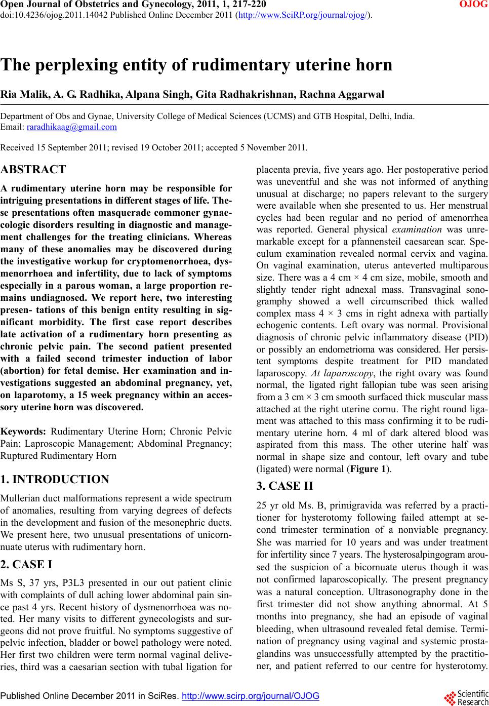 Open Journal of Obstetrics and Gynecology, 2011, 1, 217-220 doi:10.4236/ojog.2011.14042 Published Online December 2011 (http://www.SciRP.org/journal/ojog/ OJOG ). Published Online December 2011 in SciRes. http://www.scirp.org/journal/OJOG The perplexing entity of rudimentary uterine horn Ria Malik, A. G. Radhika, Alpana Singh, Gita Radhakrishnan, Rachna Aggarwal Department of Obs and Gynae, University College of Medical Sciences (UCMS) and GTB Hospital, Delhi, India. Email: [email protected] Received 15 September 2011; revised 19 October 2011; accepted 5 November 2011. ABSTRACT A rudimentary uterine horn may be responsible for intriguing presentations in different stages of life. The- se presentations often masquerade commoner gynae- cologic disorders resulting in diagnostic and manage- ment challenges for the treating clinicians. Whereas many of these anomalies may be discovered during the investigative workup for cryptomenorrhoea, dys- menorrhoea and infertility, due to lack of symptoms especially in a parous woman, a large proportion re- mains undiagnosed. We report here, two interesting presen- tations of this benign entity resulting in sig- nificant morbidity. The first case report describes late activation of a rudimentary horn presenting as chronic pelvic pain. The second patient presented with a failed second trimester induction of labor (abortion) for fetal demise. Her examination and in- vestigations suggested an abdominal pregnancy, yet, on laparotomy, a 15 week pregnancy within an acces- sory uterine horn was discovered. Keywords: Rudimentary Uterine Horn; Chronic Pelvic Pain; Laproscopic Management; Abdominal Pregnancy; Ruptured Rudimentary Horn 1. INTRODUCTION Mullerian duct malformations repr esent a wide spectrum of anomalies, resulting from varying degrees of defects in the development and fusion of the mesonephric ducts. We present here, two unusual presentations of unicorn- nuate uterus wi t h ru di mentary horn. 2. CASE I Ms S, 37 yrs, P3L3 presented in our out patient clinic with complaints of dull achin g lower abdominal pain sin- ce past 4 yrs. Recent history of dysmenorrhoea was no- ted. Her many visits to different gynecologists and sur- geons did not prove fruitful. No symptoms suggestive of pelvic infection, bladder or bowel pathology were noted. Her first two children were term normal vaginal delive- ries, third was a caesarian section with tubal ligation for placenta previa, five years ago. Her postoperative period was uneventful and she was not informed of anything unusual at discharge; no papers relevant to the surgery were available when she presented to us. Her menstrual cycles had been regular and no period of amenorrhea was reported. General physical examination was unre- markable except for a pfannensteil caesarean scar. Spe- culum examination revealed normal cervix and vagina. On vaginal examination, uterus anteverted multiparous size. There was a 4 cm × 4 cm size, mobile, smooth and slightly tender right adnexal mass. Transvaginal sono- gramphy showed a well circumscribed thick walled complex mass 4 × 3 cms in right adnexa with partially echogenic contents. Left ovary was normal. Provisional diagnosis of chronic pelvic inflammatory disease (PID) or possibly an endometrioma was considered. Her persis- tent symptoms despite treatment for PID mandated laparoscopy. At laparoscopy, the right ovary was found normal, the ligated right fallopian tube was seen arising from a 3 c m × 3 cm smooth surfaced thick muscular mass attached at the right uterine cornu. The right round liga- ment was attached to this mass confirming it to be rudi- mentary uterine horn. 4 ml of dark altered blood was aspirated from this mass. The other uterine half was normal in shape size and contour, left ovary and tube (ligated) were normal (Figure 1). 3. CASE II 25 yr old Ms. B, primigravida was referred by a practi- tioner for hysterotomy following failed attempt at se- cond trimester termination of a nonviable pregnancy. She was married for 10 years and was under treatment for infertility since 7 years. The hysterosalpingogram arou- sed the suspicion of a bicornuate uterus though it was not confirmed laparoscopically. The present pregnancy was a natural conception. Ultrasonography done in the first trimester did not show anything abnormal. At 5 months into pregnancy, she had an episode of vaginal bleeding, when ultrasound revealed fetal demise. Termi- nation of pregnancy using vaginal and systemic prosta- glandins was unsuccessfully attempted by the practitio- ner, and patient referred to our centre for hysterotomy. 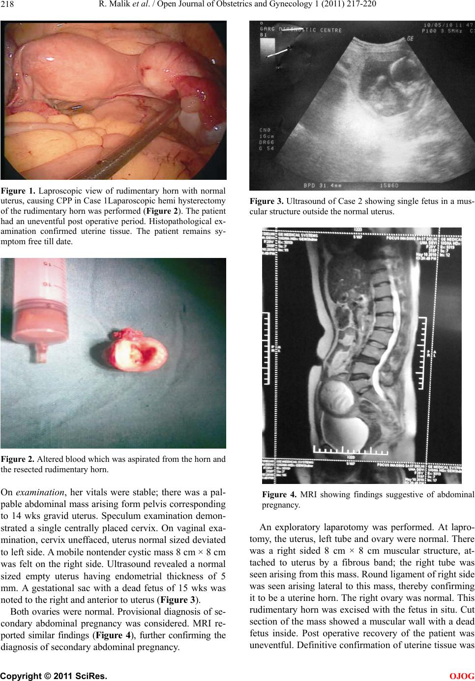 R. Malik et al. / Open Journal of Obstetrics and Gynecology 1 (2011) 217-220 218 Figure 1. Laproscopic view of rudimentary horn with normal uterus, causing CPP in Case 1Laparoscopic hemi hysterectomy of the rudimentary horn was performed (Figure 2). The patient had an uneventful post operative period. Histopathological ex- amination confirmed uterine tissue. The patient remains sy- mptom free till date. Figure 2. Altered blood which was aspirated from the horn and the resected rudimentary horn. On examination, her vitals were stable; there was a pal- pable abdominal mass arising form pelvis corresponding to 14 wks gravid uterus. Speculum examination demon- strated a single centrally placed cervix. On vaginal exa- mination, cervix uneffaced, uterus normal sized deviated to left side. A mobile nontender cystic mass 8 cm × 8 cm was felt on the right side. Ultrasound revealed a normal sized empty uterus having endometrial thickness of 5 mm. A gestational sac with a dead fetus of 15 wks was noted to the right and anterior to uterus (Figure 3). Both ovaries were normal. Provisional diagnosis of se- condary abdominal pregnancy was considered. MRI re- ported similar findings (Figure 4), further confirming the diagnosis of secondary abdominal pregnancy. Figure 3. Ultrasound of Case 2 showing single fetus in a mus- cular structure outside the normal uterus. Figure 4. MRI showing findings suggestive of abdominal pregnancy. An exploratory laparotomy was performed. At lapro- tomy, the uterus, left tube and ovar y were normal. There was a right sided 8 cm × 8 cm muscular structure, at- tached to uterus by a fibrous band; the right tube was seen arising from this mass. Round ligament of right side was seen arising lateral to this mass, thereby confirming it to be a uterine horn. The right ovary was normal. This rudimentary horn was excised with the fetus in situ. Cut section of the mass showed a muscular wall with a dead fetus inside. Post operative recovery of the patient was uneventful. Definitive con firmation of uterine tissue was C opyright © 2011 SciRes. OJOG 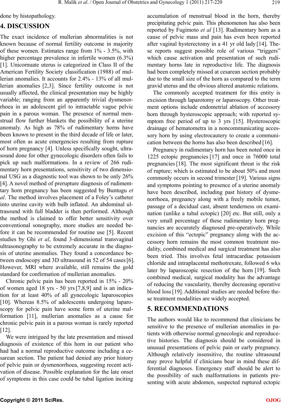 R. Malik et al. / Open Journal of Obstetrics and Gynecology 1 (2011) 217-220 219 done by histo p at hology. 4. DISCUSSION The exact incidence of mullerian abnormalities is not known because of normal fertility outcome in majority of these women. Estimates range from 1% - 3.5%, with higher percentage prevalence in infertile women (6.3%) [1]. Unicornuate uterus is categorized in Class II of the American Fertility Society classification (1988) of mul- lerian anomalies. It accounts for 2.4% - 13% of all mul- lerian anomalies [2,3]. Since fertility outcome is not usually affected, the clinical presentation may be highly variable; ranging from an apparently trivial dysmenor- rhoea in an adolescent girl to intractable vague pelvic pain in a parous woman. The presence of normal men- strual flow further blankets the possibility of a uterine anomaly. As high as 78% of rudimentary horns have been known to presen t in the third decade of life o r later, most often as acute emergencies resulting from rupture of horn pregnancy [4]. Unless specifically sought, ultra- sound done for other gynecologic disorders often fails to pick up such malformations. In a review of 266 rudi- mentary horn presen tations, sensitivity of two dimensio- nal USG as a diagnostic tool was shown to be only 26% [4]. A novel method of prerup ture diag nosis of rudiment- tary horn pregnancy has been suggested by Buntugu et al. The method involves placement of a Foley’s catheter into uterine cavity with bulb inflated. An abdominal ul- trasound with full bladder is then performed. Although the method is claimed to offer better sensitivity over conventional sonography, more studies are needed be- fore it can be recommended for routine use [5]. Recent studies by Ghi et al, found 3-dimensional transvaginal ultrasonography to be extremely accurate in the diagno- sis of uterine anomalies. They found a concordance be- tween endoscopy and 3D ultrasound in 52 of 54 cases [6]. However, MRI where available, still remains the gold standard for confirmation of mullerian anomalies. Chronic pelvic pain has been reported in 15% - 20% of women aged 18 yrs - 50 yrs [7,8,9] and is an indica- tion for at least 40% of all gynecologic laparoscopies [10]. Whereas 8.5% of adolescents undergoing laparo- scopy for pelvic pain have some form of uterine mal- formation [11], mullerian anomalies as a cause for chronic pelvic pain in a parous woman is rarely reported [12]. We were intrigued by the late p resentation and missed diagnosis of existence of this horn in our patient who had had a normal reproductive outcome including a ce- sarean section. The patient had denied any prior history of pelvic pain or dysmenorrhoea, suggesting recent acti- vation of disease. Possible explanation for the late onset of symptoms in this case could be tubal ligation inciting accumulation of menstrual blood in the horn, thereby precipitating pelvic pain. This phenomenon has also been reported by Fugimoto et al [13]. Rudimentary horn as a cause of pelvic mass and pain has even been reported after vaginal hysterectomy in a 41 yr old lady [14]. The- se reports suggest possible role of various “triggers” which cause activation and presentation of such rudi- mentary horns late in reproductive life. The diagnosis had been completely missed at cesarean section probably due to the small size of the horn as compared to the term gravid uterus and the obvious altered anatomic relations. The commonly accepted treatment for this entity is excision through laparotomy or laparoscopy. Other treat- ment options include endometrial ablation of accessory horn through hysteroscopic approach; with reported sy- mptom free period of up to 3 yrs [15]. Hysteroscopic drainage of hematometra in a noncommunicating acces- sory horn by using electrocautery to create a communi- cation between the horns has al so been descr i bed [16]. Pregnancy in rudimentary horn has been noted once in 1225 ectopic pregnancies [17] and once in 76000 total pregnancies [18]. The most significant threat is the risk of rupture; which is estimated to be about 50% and most commonly occurs in second trimester [19]. Various signs and symptoms pointing to presence of a uterine anomaly have been described, including past history of dysme- norrhoea, pregnancy along with a freely mobile tumor, passage of a decidual cast, absent tenderness on exami- nation (unlike a tubal ectopic) [20] etc. But still, only a very small percentage of these rudimentary horn preg- nancies are accurately diagnosed pre-operatively. While excision of this “ectopic” pregnancy along with the ac- cessory horn remains the most common treatment mo- dality, combined medical and surgical treatment has also been tried. This involves fetal intracardiac potassium chloride and intraplacental methotrexate, followed 6 wks later by laparoscopic resection of the horn [19]. Such combined medical, surgical modality has the advantage of reducing the vascularity, thereby decreasing operative blood loss [19]. Additional studies are needed before the- se treatment modalities are widely accepted. 5. RECOMMENDATIONS The authors would like to recommend that clinicians be sensitive to the presence of mullerian anomalies in pa- tients with otherwise normal gynecologic and reproduce- tive histories. The diagnosis should be considered in unusual presentations of pelvic pain or early pregnancy. Although relatively insensitive, the routine ultrasound may prove helpful if clinicians bear in mind these dif- ferential diagnoses. Emergency staff should be alert to the possibility of such malformations in patients pre- senting with acute abdomen, suspected ruptured ectopic C opyright © 2011 SciRes. OJOG  R. Malik et al. / Open Journal of Obstetrics and Gynecology 1 (2011) 217-220 Copyright © 2011 SciRes. 220 or even failed induction with adequate doses of prosta- glandins. A thorough inspection of the pelvis should be performed at the time of any operative procedure. Pro- phylactic resection of a noncommunicating uterine horn should be considered in patients with incidental disco- very on laprotomy to prevent endometriosis and possible future horn pre gnancy. [10] Howard, F.M. (1993) The role of laparoscopy in chronic pelvic pain: Promise and pitfalls. Obstetrical and Gyne- cological Survey, 48, 357-387. doi:10.1097/00006254-199306000-00001 [11] Vercellini, P., Fedele, L., Arcaini, L., et al. (1989) Laparoscopy in the diagnosis of chronic pelvic pain in adolescent women. Journal of Reproductive Medicine, 34, 827. [12] Howard, F.M. (2003) The role of laparoscopy in chronic pelvic pain patient. Clinical Obstetrics and Gynecology, 46, 749-766. doi:10.1097/00003081-200312000-00004 6. CONFLICT OF INTEREST We declare that we have no conflict of interests. [13] Fugimoto, V.Y., Klein, N.A. and Miller, P.B. (1998) Late onset hematome tra and he matosalpi nx in a woman wit h a noncommunicating uterine horn: A case report. Journal of Reproductive Medicine, 43, 465-467. REFERENCES [1] Simon, C., Martinez, L., Pardo, F., et al. (1991) Mulle- rian defects in women with normal reproductive outcome. Fertility and Sterility, 56, 192-193. [14] Stitely, M.L. and Hopkins, K. (2006) Laparoscopic re- moval of a rudimentary horn in a previously hysterec- tomised patient. Journal of the Society of Laparoendo- scopic Surgeons, 10, 257-258. [2] Grimbizis, G.F., Camus, M., Tarlatzis, B.C., Bontis, J.N. and Devroey, P. (2001) Clinical implications of uterine malformations and hysteroscopic treatment results. Hu- man Reproduction, 7, 161-174. [15] Hucke, J., DeBruyne, F., Campo, R.L. and Freikha, A.A. (1992) Hysteroscopic treatment of congenital uterine malformations causing hemihematometra: A report of three cases. Fertility and Sterility, 58, 823-825. [3] Acién, P. (1997) Incidence of Müllerian defects in fertile and infertile women. Human Reproduction, 12, 1372- 1376. [16] Nogueira, A.A., Candido dos Reis, F.J. and Campolungo, A. (1999) Hysteroscopic treatment of unicornuate uterus associated with a cavitary rudimentary horn. Interna- tional Journal of Gynecology and Obstetrics, 64, 77-78. doi:10.1016/S0020-7292(98)00160-X [4] Jayasinghe, Y., Rane, A., Stalewski, H. and Grover, S. (2005) The presentation and early diagnosis of the rudi- mentary uterine horn. Obstetrics Gynecology, 105, 1456- 1467. doi:10.1097/01.AOG.0000161321.94364.56 [5] Buntugu, K.A., Ntumy, M.Y. , Ameh, E.O. and Obed, S.A. (2008) Rudimentary horn pregnancy: Prerupture diagno- sis and management. Ghana Medical Journal, 42, 92-94. [17] Jarcho, J. (1946) Malformations of the uterus; review of subject, including embryology, comparative anatomy, diagnosis and report of cases. American Journal of Sur- gery, 71, 1946. [6] Ghi, T., Casadio, P., Kuleva, M., Perrone, A.M., Savelli, L., Giunchi, S., et al. (2009) Accuracy of three-dimen- sional ultrasound in diagnosis and classification of con- genital uterine anomalies. Fertility and Sterility, 92, 808- 813. doi:10.1016/j.fertnstert.2008.05.086 [18] Kadan, Y. and Romano, S. (2008) Rudimentary horn pregnancy diagnosed by ultrasound and treated by lapa- roscopy a case report and review of literature. Journal of Minimally Invasive Gynecology, 15, 527-530. doi:10.1016/j.jmig.2008.05.010 [7] ACOG Practice Bulletin #51 (2004) Chronic pelvic pain. Obstetrics and Gynaecology, 103, 589-605. [19] Park, J.K. and Dominguez, C.E. (2007) Combined medi- cal and surgical management of rudimentary uterine horn pregnancy. Journal of the Society of Laparoendoscopic S- urgeons, 11, 119-122. [8] Mathias, S.D., Kuppermann, M., Liberman, R.F., Lipschutz, R.C. and Steege, J.F. (1996) Chronic pelvic pain: Prevalence, health related quality of life, and eco- nomic considerations. Obstetrics Gynecology, 87, 321- 327. doi:10.1016/0029-7844(95)00458-0 [20] Daskalakis, G., Athanasios, P., Katerina, L. and Aris, A. (2002) Rupture of noncommunicating rudimentary uteri- ne horn pregnancy. Obstetrics and Gynecology, 100, 1108-1110. doi:10.1016/S0029-7844(02)02153-1 [9] Jamieson, D.J. and Steege, J.F. (1996) The prevalence of dysmenorrhoea, dyspareunia, pelvic pain, and irritable bowel syndrome in primary care practitioners. Obstetrics Gynecology, 87, 55-58. doi:10.1016/0029-7844(95)00360-6 OJOG |