

Paper Menu >>
Journal Menu >>
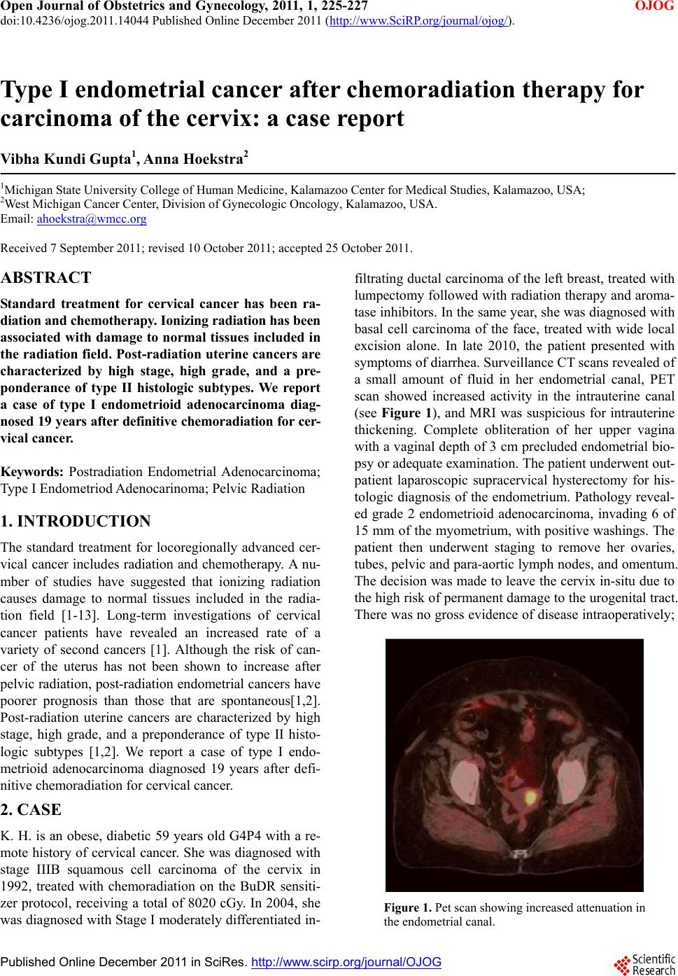 Open Journal of Obstetrics and Gynecology, 2011, 1, 225-227 OJOG doi:10.4236/ojog.2011.14044 Published Online December 2011 (http://www.SciRP.org/journal/ojog/). Published Online December 2011 in SciRes. http://www.scirp.org/journal/OJOG Type I endometrial cancer after chemoradiation therapy for carcinoma of the cervix: a case report Vibha Kundi Gupta1, Anna Hoekstra2 1Michigan State University College of Human Medicine, Kalamazoo Center for Medical Studies, Kalamazoo, USA; 2West Michigan Cancer Center, Division of Gynecologic Oncology, Kalamazoo, USA. Email: [email protected] Received 7 September 2011; revised 10 October 2011; accepted 25 October 2011. ABSTRACT Standard treatment for cervical cancer has been ra- diation and chemotherapy. Ionizing radiation has been associated with damage to normal tissues included in the radiation field. Post-radiation uterine cancers are characterized by high stage, high grade, and a pre- ponderance of type II histologic subtypes. We report a case of type I endometrioid adenocarcinoma diag- nosed 19 years after definitive chemoradiation for cer- vical cancer. Keywords: Postradiation Endometrial Adenocarcinoma; Type I Endometriod Adenocarinoma; Pelvic Radiation 1. INTRODUCTION The standard treatment for locoregionally advanced cer- vical cancer includes radiation and chemotherapy. A nu- mber of studies have suggested that ionizing radiation causes damage to normal tissues included in the radia- tion field [1-13]. Long-term investigations of cervical cancer patients have revealed an increased rate of a variety of second cancers [1]. Although the risk of can- cer of the uterus has not been shown to increase after pelvic radiation, po st-radiation endometrial cancers h ave poorer prognosis than those that are spontaneous[1,2]. Post-radiation uterine cancers are characterized by high stage, high grade, and a preponderance of type II histo- logic subtypes [1,2]. We report a case of type I endo- metrioid adenocarcinoma diagnosed 19 years after defi- nitive chemoradiation for cervical cancer. 2. CASE K. H. is an obese, diabetic 59 years old G4P4 with a re- mote history of cervical cancer. She was diagnosed with stage IIIB squamous cell carcinoma of the cervix in 1992, treated with chemoradiation on the BuDR sensiti- zer protocol, receiving a total of 8020 cGy. In 2004, she was diagnosed with Stage I moderately differentiated in- filtrating ductal carcinoma of the left breast, treated with lumpectomy followed with radiation therapy and aroma- tase inhibitors. In the same year, she was diagnosed with basal cell carcinoma of the face, treated with wide local excision alone. In late 2010, the patient presented with symptoms of diarrhea. Surveillance CT scans revealed of a small amount of fluid in her endometrial canal, PET scan showed increased activity in the intrauterine canal (see Figure 1), and MRI was suspicious for intrauterine thickening. Complete obliteration of her upper vagina with a vaginal depth of 3 cm precluded endometrial bio- psy or adequate examination. The patient underwent out- patient laparoscopic supracervical hysterectomy for his- tologic diagnosis of the endometrium. Pathology reveal- ed grade 2 endometrioid adenocarcinoma, invading 6 of 15 mm of the myometrium, with pos itive washings. The patient then underwent staging to remove her ovaries, tubes, pelvic and para-aortic lymph nodes, and omentum. The decision was made to leave the cervix in-situ due to the high risk of permanent damage to the urogenital tract. There was no gross evidence of disease intraoperatively; Figure 1. Pet scan showing increased attenuation in the endometrial canal.  V. K. Gupta et al. / Open Journal of Obstetrics and Gynecology 1 (2011) 225-227 226 pathology was negative, with the exception of micros- copic metastases to the omentum. She is currently under- going a planned 6 cycles of platinum and taxane based chemotherapy. 3. CONCLUSIONS The risk of uterine cancer after radiation for cervical cancer is not increased, compared to that of unradiated populations [1,3]. The estimated incidence of post-radia- tion uterine cancer is 0.5% - 0.8% [4]. International stu- dies [6,11-13] have reported a high number of combin- ed (type I and II) endometrial cancers. In the largest study of post radiation endometrial cancer, 308 cases of endometroid and endometroid cancer were reported over 40 years [1]. Of the 147 cases described in the US lite- rature, however, the vast majority were of non-endome- trioid histologic subtypes. Only 6 0 cases of endometrioid adenocarcinoma of the uterus are reported, many of whi- ch are grade 3 [2,4]. Although a large number of studies have demonstrated that radiotherapy can lead to secon- dary cancers [2-13], it appears that well or moderately differentiated endometrioid adenocarcinoma is exceedingly rare after pelvic radiation for cervical cancer. Diagnosis of radiation-associated endometrial cancer varies from sporadic cancers, which are most often diagnosed by ab- normal vaginal bleeding. Cervical stenosis and oblitera- tion of the upper vagina due to radiation damage may prevent vaginal bleeding. Evaluation after abnormal ima- ging can be accomplished with endometrial curettage, ultrasound guided aspiration, or hysterectomy. Differen- ces in diagnosis are associated with significant delays of diagnosis, contributing to the poorer prognosis associa- ted with post-radiation endometrial cancer. Advanced s- tage and aggressive histology of most uterine tumors af- ter radiation for cervical cancer may require more aggre- ssive surgical staging and adjuvant treatment than spo- radic endometrial cancers. Many patients reported in ca- se series have been surgically treated without extensive lymph node sampling or omentectomy, which may signi- ficantly affect adjuvant treatment recommendations. Rou- tine omentectomy, pelvic and para-aortic lymphadenec- tomy may be both diagnostic, to guide adjuvant treat- ment, and therapeutic to remove microscopic disease. In the case reported here, the microscopically po sitive ome- ntum changed her treatment recommendation to include cytotoxic chemotherapy. More aggressive treatment both surgically, and adjuvantly, may be considered for this particular patient population to improve prognosis. The etiology of endo metrial cancer after radiation therap y for cervical cancer has been the subject of multiple reviews [1-4]. Evaluation of this patien ts history and curren t eva- luation suggests a multifactorial etio logy for her disease, including traditional risk factors, genetic predisposition, and radiation effect. The patient is obese with a modera- tely differentiated endometrioid tumor. Microscopic spread outside the uterus may be explained by trans-tubal s- pread after a significant delay in diagnosis due to obli- teration of the upper vagina. The contribution of genetic predisposition is suggested by her personal history of 3 prior cancers. Lastly, the direct effect of ionizing ra- diation cannot be ignored. Pelvic radiation for cervical cancer has been implicated in the development of a va- riety of second cancers 1, often temporally distant from the radiation (5 years - 20 years). This is consistent with the 19 years gap between treatment of cervical cancer an d diagnosis of uterine cancer. This case suggests endome- trioid post-radiation tumors may have a complex origin. REFERENCES [1] Chaturvedi, A., Engels, E., Gilbert, E., Chen, B., Storm, H., Lynch, C., et al. (2007) Second cancers among 104, 760 survivors of cervical cancer: Evaluation of long- term risk. Journal of National Cancer Institute, 99, 1634- 1643. doi:10.1093/jnci/djm201 [2] Kumar, S., Shah, J., Bryant, C., Seward, S., Ali-Fehmi, R., Morris, R., et al. (2009) Radiation-associated endo- metrial cancer. Obstetrics and Gynecology, 11 3, 319-325. [3] Gallion, H., Van Nagell, J., Donaldson, E. and Powell, D. (1987) Endometrial cancer following radiation therapy for cervical cancer. Gynecologic Oncology, 27, 76-83. doi:10.1016/0090-8258(87)90232-0 [4] Pothuri, B., Ramondetta, L., Martino, M., Alektiar, K., Eifel, P., Deavers, M., et al. (2003) Development of en- dometrial cancer after radiation treatment for cervical carcinoma. Obstetrics and Gynecology, 101, 941-945. doi:10.1016/S0029-7844(03)00234-5 [5] Parkash, V. and Carcangiu, M.L. (1992) Uterine Papillary serous carcinoma after radiation therapy for carcinoma of the cervix. Cancer, 69, 496-501. doi:10.1002/1097-0142(19920115)69:2<496::AID-CNC R2820690237>3.0.CO;2-Q [6] Hagiwara, T., Mori, T. and Kaku, T. (2005) Development of endometrial cancer following radiation therapy for cervical carcinoma. European Journal of Gynaecologic Oncology, 26, 191-195. [7] Buchholz, T.A. (2003) Lung carcinoma development after radiotherapy for breast carcinoma. Cancer, 98, 1331-1333. doi:10.1002/cncr.11657 [8] Travis, L.B., Hill, D.A., Dores, G.M., et al. (2003) Breast cancer following radiotherapy and chemotherapy among young women with Hodgkin disease. Journal of the American Medical Association, 290, 465-475. doi:10.1001/jama.290.4.465 [9] Pride, G.L. and Buchler, D.A. (1997) Carcinoma of the vagina 10 or more years following pelvic irradiation therapy. American Journal of Obstetrics Gynecology, 127, 513-517. [10] Jereczek-Fossa, B.A. , Alterio, D., Jassem, J., et al. (2004) Radiotherapy-induced thyroid disorders. Cancer Treat- ment Review, 30, 369-384. doi:10.1016/j.ctrv.2003.12.003 [11] Rodriguez, J. and Hart, W. (1982) Endometrial cancers o- C opyright © 2011 SciRes. OJOG  V. K. Gupta et al. / Open Journal of Obstetrics and Gynecology 1 (2011) 225-227 Copyright © 2011 SciRes. OJOG 227 ccurring 10 or more years after pelvic irradiation for car- cinoma. International Journal of Gynecological Path- ology, 1, 135-144. doi:10.1097/00004347-198202000-00002 [12] Seidman, J.D., Kumar, D., Cosin, J.A., et al. (2006) Car- cinomas of the female genital tract occurring after pelvic irradiation: A report of 15 cases. International Journal of Gynecologic Pathology, 25, 293-297. doi:10.1097/01.pgp.0000189240.03780.b1 [13] De Hullu, J.A., Pras, E., Hollema, H., et al. (2005) Pres- entations of endometrial activity after curative radio- therapy for cervical cancer. Maturitas, 51, 172-176. doi:10.1016/j.maturitas.2004.07.005 |