

Paper Menu >>
Journal Menu >>
 Open Journal of Obstetrics and Gynecology, 2011, 1, 228-233 doi:10.4236/ojog.2011.14045 Published Online December 2011 (http://www.SciRP.org/journal/ojog/ OJOG ). Published Online December 2011 in SciRes. http://www.scirp.org/journal/OJOG Increased free androgen index is associated with hypertension in premenopausal women Liselott Andersson1, Mats Eliasson2,3, Inger Sundström Poromaa4 1Department of Obstetrics and Gynecology, Sunderby Hospital, Luleå, Sweden; 2Department of Internal Medicine, Sunderby Hospital, Luleå, Sweden; 3Department of Public Health and Clinical Medicine, Umeå University, Luleå, Sweden; 4Department of Women’s and Children’s Health, Uppsala University, Uppsala, Sweden. Email: [email protected] Received 15 August 2011; revised 20 September 2011; accepted 4 October 2011. ABSTRACT Objective: Increased testosterone and decreased sex hormone-binding globulin (SHBG) are associated with a number of adverse cardiovascular risk factors in postmenopausal women. The aim of this popula- tion-based study of women aged 25 to 50 was to as- sess the relationship between free androgen index (FAI) and cardiovascular risk factors in premeno- pausal women. Methods: A population-based survey of 396 premenopausal women with no hormonal trea- tment was undertaken as part of the Northern Swe- den MONICA study. The study involved question- naires, anthropometry and assays of testoste rone and SHBG. Results: Increased FAI was associated with a number of cardiovascular risk factors in premeno- pausal women but this relationship was strongly af- fected by body mass index (BMI). After adjustment for age and BMI, FAI was significantly associated with increased systolic and diastolic blood pressures. Conclusion: Hyperandrogenism is associated with increased blood pressure and these findings empha- size the need to assess cardiovascular risk factors in women with hyperandrogenism of all ages. Keywords: Free Androgen Index; Blood Pressure 1. INTRODUCTION Testosterone is carried in peripheral blood bound to sex hormone binding globulin, with approximately 1% - 2% of testosterone free and the remaining testosterone loo- sely bound to albumin [1]. The free androgen index (FAI), i.e. the ratio between testosterone and sex hormone bin- ding globulin (SHBG), may be used as an approximation of biologically available androgens in women, and lon- gitudinal studies in aging women indicate that SHBG levels decrease with increasing age, thus resulting in su- ccessively increasing amounts of biologically available androgen in healthy women [2]. Increased testosterone and decreased sex hormone- binding globulin (SHBG) have been strongly associated with a number of adverse cardiovascular disease (CVD) risk factors in postmenopausal women, such as central adiposity, decreased high-density lipoprotein (HDL) cholesterol levels and increased systolic and diastolic blood pressures [3-6]. A nested case-control study of po- stmenopausal women also suggested that higher free androgen index was associated with CVD events such as first occurrence of nonfatal myocardial infarction, coro- nary revascularization, nonfatal stroke, coronary disease, or stroke death, although this association was not inde- pendent of body mass index and other cardiovascular risk factors [7]. Besides a possible role of androgen hor- mones and SHBG in cardiovascular disease, low levels of SHBG have consistently been linked to higher rates of diabetes [8-9] and might contribute to a more adverse car- diovascular risk profile in women with diabetes [10-11]. Sex hormone levels may also be associated with CVD risk factors in pre- and perimenopausal women, although fewer studies have been conducted in this age-group [12-14]. In younger women most interest has instead been devoted to patients with polycystic ovary syndrome (PCOS), which is a hyperandrogenic population with nu- merous risk factors for later development of CVD [15]. However, in contrast to healthy women, we have previ- ously shown that testosterone levels gradually decrease with increasing age in PCOS women [16], although le- vels remain elevated in comparison with age-matched controls [17]. Outside the PCOS patient population, no population- based studies have been conducted in premenopausal women concerning the association between free andro- gen index and cardiovascular risk factors. Thus, the pur- pose of this population-based study of premenopausal  L. Andersson et al. / Open Journal of Obstetrics and Gynecology 1 (2011) 228-233 229 women aged 25 to 50 was to assess whether free andro- gen index is associated with risk factors for cardiovas- cular disease, even in younger women. 2. MATERIALS AND METHODS 2.1. Patients This study used data from the Northern Sweden compo- nent of the WHO MONICA study, originally designed to evaluate risk factors for cardiovascular disease and type 2 diabetes. Briefly, information was collected during a population-based survey during January to April 2004 [18]. Subjects were randomly selected from population registers, stratified for age (25 years - 74 years) and gen- der, in the two most northern counties of Sweden (target population 312,000). For the entire MONICA study 1250 men and 1250 women were invited, but for the purpose of this study, only women below age 50 years were considered. Details of sampling and selection ap- pear elsewhere and participation rate was 78% [18]. A total of 500 women 50 years or younger were eligible for this study. Exclusion criteria were postmenopausal status (more than 12 months since last menses), ongoing pregnancy, and use of hormone replacement therapy or combined oral contraceptives during the last year. The Research Ethics Committee of Umeå University and the National Computer Data Inspection Board ap- proved the Northern Sweden MONICA study. A specific approval for this sub-study was also obtained from The Research Ethics Committee of Umeå University. Parti- cipants gave written consent. 2.2. Study Procedures Subjects were weighed on an electronic scale. The sub- jects wore light clothes and no shoes and weight was measured to the nearest 0.2 kg. Height without shoes was measured to the nearest centimeter. Body mass in- dex (BMI) was calculated as weight (kg) divided by height (m2). To record waist circumference, measure- ment was performed midway between the lower rib mar- gin and iliac crest to the nearest 0.0 cm or 0.5 cm. The blood pressure was measured twice in every person after a five-minute rest in a sitting position with Hawksley’s random zero sphygmomanometer. The mean value of the two measurements was used. Blood samples were drawn after at least a 4-h fast (in approximately 65% of subjects blood samples were dra- wn after an overnight fast). The participants were asked to complete question- naires including items about smoking habits, sociodemo- graphic data and previously known cardiovascular dis- ease. The questionnaire also included a number of ques- tions for assessment of reproductive status including menstrual pattern, last menstrual period, use of oral con- traceptives and hormone replacement therapy during the past year. The highest attained educational level was classified as primary school (up to 9 years of school), secondary school (10 years - 12 years of school) and university stu- dies. Regular smokers smoked at least one cigarette a day; all other subjects were considered as nonsmokers. Participants were classified as physically active if re- porting physical activity more than two hours per week; all other subjects were considered not physically active. A positive family history of diabetes, stroke and cardio- vascular disease was only reported when occurring in first-degree relatives. A history of gestational diabetes was only assessed in parous subjects. Hypertension and elevated serum cholesterol were considered prevalent in women on medication for these reasons. 2.3. Assays Total cholesterol was determined by a dry chemistry method (Vitros 950; Kodak Echtachem, Rochester, NY, USA). The measurement of total cholesterol is accredi- ted by the national accreditation body, SWEDAC, with coefficients of variation of 3.6% at 3.91 mmol·L–1 and 3.1% at 6.66 mmol·L–1. An oral glucose tolerance test (OGTT; 75 g glucose) was performed in a random sub-sample (approximately 65%) of the non-diabetic participants after an overnight fast. A venous blood sample was taken immediately be- fore the glucose load and after two hours. Glucose levels were analyzed without delay on a HemoCue Glucose 201 device (HemoCue AB, Angelholm, Sweden) and converted to plasma glucose levels by a correction factor of 1.11. According to previous validation of this proce- dure, the adjusted data for glucose showed a high corre- lation to plasma glucose simultaneously measured by the hexokinase method in a sub-sample [18]. SHBG and testosterone were analyzed on a Modular E170 (Roche Diagnostics, Mannheim, Germany). The total coefficients of variation of the instrument for the analytes were 1.5% at 43 nmol/L for SHBG and 6.8% at 3.9 nmol/L for tes- tosterone. Free androgen index was calculated as testos- terone (nmol/L)/SHBG (nmol/L) × 100. Physical and biochemical parameters were analyzed using partial correlation test with adjustment for age and BMI and with logarithmic FAI. The women were grou- ped into quartiles depending on level of FAI. Dichoto- mized and continuous variables were compared between groups using linear or logistic regression analysis and adjusted for age and BMI. The software package SPSS (version 12.0) was used for statistical analyses. 3. RESULTS Fifteen postmenopausal women, 53 women on combined oral contraceptives, 16 women on hormone replacement C opyright © 2011 SciRes. OJOG 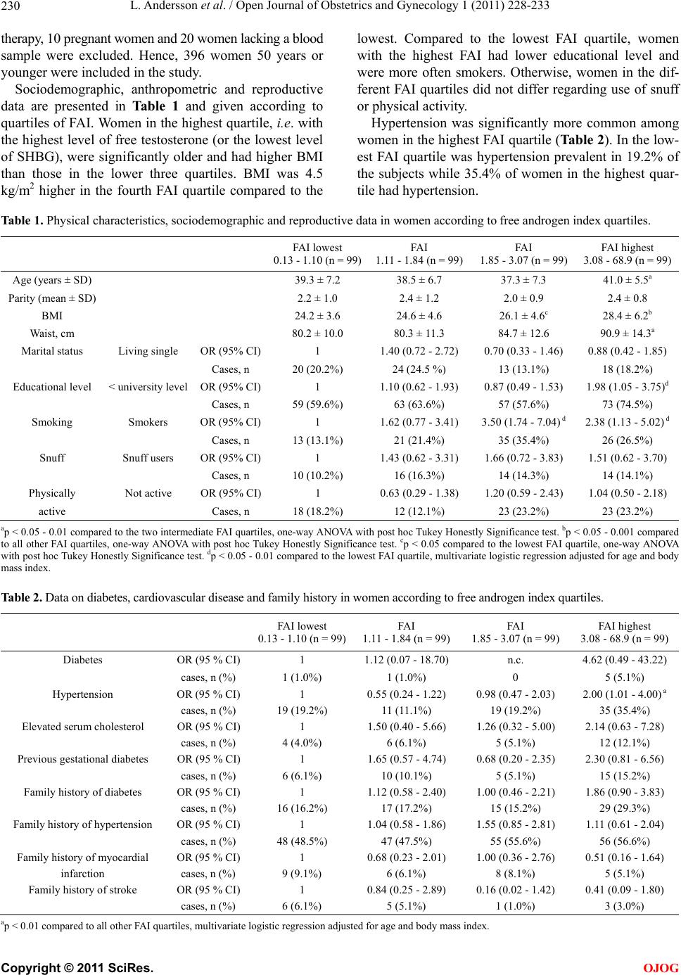 L. Andersson et al. / Open Journal of Obstetrics and Gynecology 1 (2011) 228-233 Copyright © 2011 SciRes. 230 OJOG therapy, 10 pregnant women and 20 women lacking a blood sample were excluded. Hence, 396 women 50 years or younger were included in the study. Sociodemographic, anthropometric and reproductive data are presented in Table 1 and given according to quartiles of FAI. Women in the highest quartile, i.e. with the highest level of free testosterone (or the lowest level of SHBG), were significantly older and had higher BMI than those in the lower three quartiles. BMI was 4.5 kg/m2 higher in the fourth FAI quartile compared to the lowest. Compared to the lowest FAI quartile, women with the highest FAI had lower educational level and were more often smokers. Otherwise, women in the dif- ferent FAI quartiles did not differ regarding use of snuff or physical activity. Hypertension was significantly more common among women in the highest FAI quartile (Table 2). In the low- est FAI quartile was hypertension prevalent in 19.2% of the subjects while 35.4% of women in the highest quar- tile had hypertension. Table 1. Physical characteristics, sociodemographic and reproductive data in women according to free androgen index quartiles. FAI lowest 0.13 - 1.10 (n = 99) FAI 1.11 - 1.84 (n = 99) FAI 1.85 - 3.07 (n = 99) FAI highest 3.08 - 68.9 (n = 99) Age (years ± SD) 39.3 ± 7.2 38.5 ± 6.7 37.3 ± 7.3 41.0 ± 5.5a Parity (mean ± SD) 2.2 ± 1.0 2.4 ± 1.2 2.0 ± 0.9 2.4 ± 0.8 BMI 24.2 ± 3.6 24.6 ± 4.6 26.1 ± 4.6c 28.4 ± 6.2b Waist, cm 80.2 ± 10.0 80.3 ± 11.3 84.7 ± 12.6 90.9 ± 14.3a Marital status Living single OR (95% CI) 1 1.40 (0.72 - 2.72) 0.70 (0.33 - 1.46) 0.88 (0.42 - 1.85) Cases, n 20 (20.2%) 24 (24.5 %) 13 (13.1%) 18 (18.2%) Educational level < university level OR (95% CI) 1 1.10 (0.62 - 1.93) 0.87 (0.49 - 1.53) 1.98 (1.05 - 3.75)d Cases, n 59 (59.6%) 63 (63.6%) 57 (57.6%) 73 (74.5%) Smoking Smokers OR (95% CI) 1 1.62 (0.77 - 3.41) 3.50 (1.74 - 7.04) d 2.38 (1.13 - 5.02) d Cases, n 13 (13.1%) 21 (21.4%) 35 (35.4%) 26 (26.5%) Snuff Snuff users OR (95% CI) 1 1.43 (0.62 - 3.31) 1.66 (0.72 - 3.83) 1.51 (0.62 - 3.70) Cases, n 10 (10.2%) 16 (16.3%) 14 (14.3%) 14 (14.1%) Physically Not active OR (95% CI) 1 0.63 (0.29 - 1.38) 1.20 (0.59 - 2.43) 1.04 (0.50 - 2.18) active Cases, n 18 (18.2%) 12 (12.1%) 23 (23.2%) 23 (23.2%) ap < 0.05 - 0.01 compared to the two intermediate FAI quartiles, one-way ANOVA with post hoc Tukey Honestly Significance test. bp < 0.05 - 0.001 compared to all other FAI quartiles, one-way ANOVA with post hoc Tukey Honestly Significance test. cp < 0.05 compared to the lowest FAI quartile, one-way ANOVA with post hoc Tukey Honestly Significance test. dp < 0.05 - 0.01 compared to the lowest FAI quartile, multivariate logistic regression adjusted for age and body mass index. Table 2. Data on diabetes, cardiovascular disease and family history in women according to free androgen index quartiles. FAI lowest 0.13 - 1.10 (n = 99) FAI 1.11 - 1.84 (n = 99) FAI 1.85 - 3.07 (n = 99) FAI highest 3.08 - 68.9 (n = 99) Diabetes OR (95 % CI) 1 1.12 (0.07 - 18.70) n.c. 4.62 (0.49 - 43.22) cases, n (%) 1 (1.0%) 1 (1.0%) 0 5 (5.1%) Hypertension OR (95 % CI) 1 0.55 (0.24 - 1.22) 0.98 (0.47 - 2.03) 2.00 (1.01 - 4.00) a cases, n (%) 19 (19.2%) 11 (11.1%) 19 (19.2%) 35 (35.4%) Elevated serum cholesterol OR (95 % CI) 1 1.50 (0.40 - 5.66) 1.26 (0.32 - 5.00) 2.14 (0.63 - 7.28) cases, n (%) 4 (4.0%) 6 (6.1%) 5 (5.1%) 12 (12.1%) Previous gestational diabetes OR (95 % CI) 1 1.65 (0.57 - 4.74) 0.68 (0.20 - 2.35) 2.30 (0.81 - 6.56) cases, n (%) 6 (6.1%) 10 (10.1%) 5 (5.1%) 15 (15.2%) Family history of diabetes OR (95 % CI) 1 1.12 (0.58 - 2.40) 1.00 (0.46 - 2.21) 1.86 (0.90 - 3.83) cases, n (%) 16 (16.2%) 17 (17.2%) 15 (15.2%) 29 (29.3%) Family history of hypertension OR (95 % CI) 1 1.04 (0.58 - 1.86) 1.55 (0.85 - 2.81) 1.11 (0.61 - 2.04) cases, n (%) 48 (48.5%) 47 (47.5%) 55 (55.6%) 56 (56.6%) Family history of myocardial OR (95 % CI) 1 0.68 (0.23 - 2.01) 1.00 (0.36 - 2.76) 0.51 (0.16 - 1.64) infarction cases, n (%) 9 (9.1%) 6 (6.1%) 8 (8.1%) 5 (5.1%) Family history of stroke OR (95 % CI) 1 0.84 (0.25 - 2.89) 0.16 (0.02 - 1.42) 0.41 (0.09 - 1.80) cases, n (%) 6 (6.1%) 5 (5.1%) 1 (1.0%) 3 (3.0%) ap < 0.01 compared to all other FAI quartiles, multivariate logistic regression adjusted for age and body mass index. 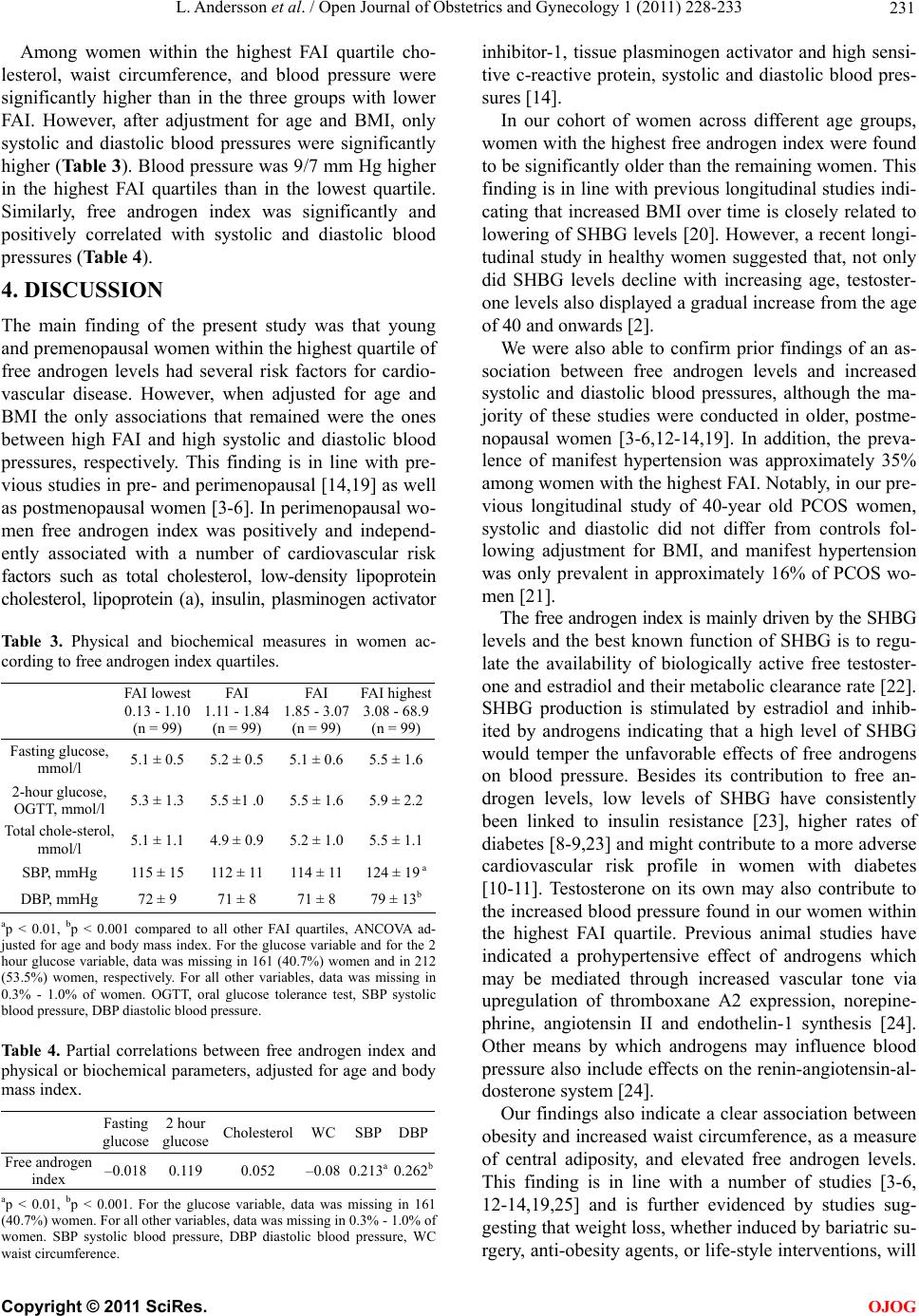 L. Andersson et al. / Open Journal of Obstetrics and Gynecology 1 (2011) 228-233 231 Among women within the highest FAI quartile cho- lesterol, waist circumference, and blood pressure were significantly higher than in the three groups with lower FAI. However, after adjustment for age and BMI, only systolic and diastolic blood pressures were significantly higher (Table 3). Blood pressure was 9/7 mm Hg higher in the highest FAI quartiles than in the lowest quartile. Similarly, free androgen index was significantly and positively correlated with systolic and diastolic blood pressures (Table 4). 4. DISCUSSION The main finding of the present study was that young and premenopausal women within the highest quartile of free androgen levels had several risk factors for cardio- vascular disease. However, when adjusted for age and BMI the only associations that remained were the ones between high FAI and high systolic and diastolic blood pressures, respectively. This finding is in line with pre- vious studies in pre- and perimenopausal [14,19] as well as postmenopausal women [3-6]. In perimenopausal wo- men free androgen index was positively and independ- ently associated with a number of cardiovascular risk factors such as total cholesterol, low-density lipoprotein cholesterol, lipoprotein (a), insulin, plasminogen activator Table 3. Physical and biochemical measures in women ac- cording to free androgen index quartiles. FAI lowest 0.13 - 1.10 (n = 99) FAI 1.11 - 1.84 (n = 99) FAI 1.85 - 3.07 (n = 99) FAI highest 3.08 - 68.9 (n = 99) Fasting glucose, mmol/l 5.1 ± 0.5 5.2 ± 0.5 5.1 ± 0.6 5.5 ± 1.6 2-hour glucose, OGTT, mmol/l 5.3 ± 1.3 5.5 ±1 .0 5.5 ± 1.6 5.9 ± 2.2 Total chole-sterol, mmol/l 5.1 ± 1.1 4.9 ± 0.9 5.2 ± 1.0 5.5 ± 1.1 SBP, mmHg 115 ± 15 112 ± 11 114 ± 11 124 ± 19 a DBP, mmHg 72 ± 9 71 ± 8 71 ± 8 79 ± 13b ap < 0.01, bp < 0.001 compared to all other FAI quartiles, ANCOVA ad- justed for age and body mass index. For the glucose variable and for the 2 hour glucose variable, data was missing in 161 (40.7%) women and in 212 (53.5%) women, respectively. For all other variables, data was missing in 0.3% - 1.0% of women. OGTT, oral glucose tolerance test, SBP systolic blood pressure, DBP diastolic blood pressure. Ta b l e 4 . Partial correlations between free androgen index and physical or biochemical parameters, adjusted for age and body mass index. Fasting glucose 2 hour glucose Cholesterol WC SBPDBP Free androgen index –0.018 0.119 0.052 –0.08 0.213a0.262b ap < 0.01, bp < 0.001. For the glucose variable, data was missing in 161 (40.7%) women. For all other variables, data was missing in 0.3% - 1.0% of women. SBP systolic blood pressure, DBP diastolic blood pressure, WC waist circumference. inhibitor-1, tissue plasminogen activator and high sensi- tive c-reactive protein, systolic and diastolic blood pres- sures [14]. In our cohort of women across different age groups, women with the highest free androgen index were found to be significantly older than the remaining women. This finding is in line with previous longitudinal studies indi- cating that increased BMI over time is closely related to lowering of SHBG levels [20]. However, a recent longi- tudinal study in healthy women suggested that, not only did SHBG levels decline with increasing age, testoster- one levels also displayed a gradual increase from the age of 40 and onwards [2]. We were also able to confirm prior findings of an as- sociation between free androgen levels and increased systolic and diastolic blood pressures, although the ma- jority of these studies were conducted in older, postme- nopausal women [3-6,12-14,19]. In addition, the preva- lence of manifest hypertension was approximately 35% among women with the highest FAI. Notably, in our pre- vious longitudinal study of 40-year old PCOS women, systolic and diastolic did not differ from controls fol- lowing adjustment for BMI, and manifest hypertension was only prevalent in approximately 16% of PCOS wo- men [21]. The free androgen index is mainly driven by the SHBG levels and the best known function of SHBG is to regu- late the availability of biologically active free testoster- one and estradiol and their metabolic clearance rate [22]. SHBG production is stimulated by estradiol and inhib- ited by androgens indicating that a high level of SHBG would temper the unfavorable effects of free androgens on blood pressure. Besides its contribution to free an- drogen levels, low levels of SHBG have consistently been linked to insulin resistance [23], higher rates of diabetes [8-9,23] and might contribute to a more adverse cardiovascular risk profile in women with diabetes [10-11]. Testosterone on its own may also contribute to the increased blood pressure found in our women within the highest FAI quartile. Previous animal studies have indicated a prohypertensive effect of androgens which may be mediated through increased vascular tone via upregulation of thromboxane A2 expression, norepine- phrine, angiotensin II and endothelin-1 synthesis [24]. Other means by which androgens may influence blood pressure also include effects on the renin-angiotensin-al- dosterone system [24]. Our findings also indicate a clear association between obesity and increased waist circumference, as a measure of central adiposity, and elevated free androgen levels. This finding is in line with a number of studies [3-6, 12-14,19,25] and is further evidenced by studies sug- gesting that weight loss, whether induced by bariatric su- rgery, anti-obesity agents, or life-style interventions, will C opyright © 2011 SciRes. OJOG  L. Andersson et al. / Open Journal of Obstetrics and Gynecology 1 (2011) 228-233 232 reduce androgen levels [25-26]. The major limitation of this study is the relatively small sample size, which might raise questions whether this study was adequately powered for some of the ana- lyses. However, the study has pointed out that increased FAI is associated with a number of cardiovascular risk factors but these relationships seem to be strongly af- fected by BMI. Free androgen index was significantly and independently associated with increased systolic and diastolic blood pressures, emphasizing the need to assess cardiovascular risk factors in women with hyperandro- genism across all ages. REFERENCES [1] Burger, H.G. (2002) Androgen production in women. Fertility and Sterility, 77, S3- S5. doi:10.1016/S0015-0282(02)02985-0 [2] Sowers, M.F., Zheng, H., McConnell, D., Nan, B., Kar- vonen-Gutierrez, C.A. and Randolph, J.F., Jr. (2009) Tes- tosterone, sex hormone-binding globulin and free andro- gen index among adult women: Chronological and ovar- ian aging. Human Reproduction, 24, 2276-2285. doi:10.1093/humrep/dep209 [3] Pugeat, M., Moulin, P., Cousin, P., Fimbel, S., Nicolas, M.H., Crave, J.C. and Lejeune, H. (1995) Interrelations between sex hormone-binding globulin (SHBG), plasma lipoproteins and cardiovascular risk. Journal of Steroid Biochemistry and Molecular Biology, 53, 567-572. doi:10.1016/0960-0760(95)00102-6 [4] Lambrinoudaki, I., Christodoulakos, G., Rizos, D., Eco- nomou, E., Argeitis, J., Vlachou, S., Creatsa, M., Kous- kouni, E. and Botsis, D. (2006) Endogenous sex hormo- nes and risk factors for atherosclerosis in healthy Greek postmenopausal women. European Journal of Endocri- nology, 154, 907-916. doi:10.1530/eje.1.02167 [5] Haffner, S.M., Katz, M.S., Stern, M.P. and Dunn, J.F. (1989) Association of decreased sex hormone binding globulin and cardiovascular risk factors. Arteriosclerosis, 9, 136-143. doi:10.1161/01.ATV.9.1.136 [6] Haffner, S.M., Newcomb, P.A., Marcus, P.M., Klein, B.E. and Klein, R. (1995) Relation of sex hormones and de- hydroepiandrosterone sulfate (DHEA-SO4) to cardio- vascular risk factors in postmenopausal women. Ameri- can Journal of Epidemiology, 142, 925-934. [7] Rexrode, K.M., Manson, J.E., Lee, I.M., Ridker, P.M., Sluss, P.M., Cook, N.R. and Buring, J.E. (2003) Sex hor- mone levels and risk of cardiovascular events in postme- nopausal women. Circulation, 108, 1688-1693. doi:10.1161/01.CIR.0000091114.36254.F3 [8] Haffner, S.M., Valdez, R.A., Morales, P.A., Hazuda, H.P. and Stern, M.P. (1993) Decreased sex hormone-binding globulin predicts noninsulin-dependent diabetes mellitus in women but not in men. Journal of Clinical Endocri- nology and Metabolism, 77, 56-60. doi:10.1210/jc.77.1.56 [9] Goodman-Gruen, D. and Barrett-Connor, E. (1997) Sex hormone-binding globulin and glucose tolerance in post- menopausal women. The Rancho Bernardo Study. Diabe- tes Care, 20, 645-649. doi:10.2337/diacare.20.4.645 [10] Maturana, M.A. and Spritzer, P.M. (2002) Association between hyperinsulinemia and endogenous androgen le- vels in peri- and postmenopausal women. Metabolism, 51, 238-243. doi:10.1053/meta.2002.29997 [11] Korytkowski, M.T., Krug, E.I., Daly, M.A., Deriso, L., Wilson, J.W. and Winters, S.J. (2005) Does androgen excess contribute to the cardiovascular risk profile in postmenopausal women with type 2 diabetes? Metaboli- sm, 54, 1626-1631. doi:10.1016/j.metabol.2005.06.011 [12] Sowers, M., Crawford, S.L., Cauley, J.A. and Stein, E. (2003) Association of lipoprotein(a), insulin resistance, and reproductive hormones in a multiethnic cohort of pre- and perimenopausal women (The SWAN Study). American Journal of Cardiology, 92, 533-537. doi:10.1016/S0002-9149(03)00720-3 [13] Sutton-Tyrrell, K., Wildman, R.P., Matthews, K.A., Chae, C., Lasley, B.L., Brockwell, S., Pasternak, R.C., Lloyd- Jones, D., Sowers, M.F. and Torrens, J.I. (2005) Sex-hor- mone-binding globulin and the free androgen index are related to cardiovascular risk factors in multiethnic pre- menopausal and perimenopausal women enrolled in the Study of Women Across the Nation (SWAN). Circulation, 111, 1242-1249. doi:10.1161/01.CIR.0000157697.54255.CE [14] Sowers, M.R., Jannausch, M., Randolph, J.F., McConnell, D., Little, R., Lasley, B., Pasternak, R., Sutton-Tyrrell, K. and Matthews, K.A. (2005) Androgens are associated with hemostatic and inflammatory factors among women at the mid-life. Journal of Clinical Endocrinology and Metabolism, 90, 6064-6071. doi:10.1210/jc.2005-0765 [15] Teede, H.J., Hutchison, S., Zoungas, S. and Meyer, C. (2006) Insulin resistance, the metabolic syndrome, dia- betes, and cardiovascular disease risk in women with PCOS. Endocrine, 30, 45-53. doi:10.1385/ENDO:30:1:45 [16] Hudecova, M., Holte, J., Moby, L., Olovsson, M., Stri- dsberg, M., Larsson, A., Berglund, L., Berne, C. and Sundstrom Poromaa, I. (2011) Androgen levels, insulin sensitivity, and early insulin response in women with polycystic ovary syndrome: A long-term follow-up study. Fertility and Sterility, 95, 1146-1148. doi:10.1016/j.fertnstert.2010.09.050 [17] Hudecova, M., Holte, J., Olovsson, M. and Sundstrom Poromaa, I. (2009) Long-term follow-up of patients with polycystic ovary syndrome: Reproductive outcome and ovarian reserve. Human Reproduction, 24, 1176-1183. doi:10.1093/humrep/den482 [18] Eliasson, M., Janlert, U., Jansson, J.H. and Stegmayr, B. (2006) Time trends in population cholesterol levels 1986- 2004: Influence of lipid-lowering drugs, obesity, smoking and educational level. The northern Sweden MONICA study. Journal of Internal Medicine, 260, 551-559. doi:10.1111/j.1365-2796.2006.01730.x [19] Mantzoros, C.S., Georgiadis, E.I., Young, R., Evagelo- poulou, C., Khoury, S., Katsilambros, N. and Sowers, J.R. (1995) Relative androgenicity, blood pressure levels, and cardiovascular risk factors in young healthy women. American Journal of Hypertension, 8, 606-614. doi:10.1016/0895-7061(95)00051-P [20] Sternfeld, B., Liu, K., Quesenberry, C.P., Jr., Wang, H., Jiang, S.F., Daviglus, M., Fornage, M., Lewis, C.E., Mahan, C opyright © 2011 SciRes. OJOG  L. Andersson et al. / Open Journal of Obstetrics and Gynecology 1 (2011) 228-233 Copyright © 2011 SciRes. 233 OJOG J., Schreiner, P.J., Schwartz, S.M., Sidney, S., Williams, O.D. and Siscovick, D.S. (2008) Changes over 14 years in androgenicity and body mass index in a biracial cohort of reproductive-age women. Journal of Clinical Endo- crinology and Metabolism, 93, 2158-2165. doi:10.1210/jc.2007-2203 [21] Hudecova, M., Holte, J., Olovsson, M., Lind, L. and Po- romaa, I.S. (2010) Endothelial function in patients with polycystic ovary syndrome: A long-term follow-up study. Fertility and Sterility, 94, 2654-2658. doi:10.1016/j.fertnstert.2010.02.048 [22] Rosner, W. (1991) Plasma steroid-binding proteins. En- docrinology and Metabolism Clinics of North America, 20, 697-720. [23] Ding, E.L., Song, Y., Malik, V.S. and Liu, S. (2006) Sex differences of endogenous sex hormones and risk of type 2 diabetes: A systematic review and meta-analysis. Jour- nal of the American Medical A ssociation, 295, 1288-1299. doi:10.1001/jama.295.11.1288 [24] Kienitz, T. and Quinkler, M. (2008) Testosterone and blood pressure regulation. Kidney and Blood Pressure Research, 31, 71-79. doi:10.1159/000119417 [25] Lindholm, A., Bixo, M., Bjorn, I., Wolner-Hanssen, P., E- liasson, M., Larsson, A., Johnson, O. and Poromaa, I.S. (2008) Effect of sibutramine on weight reduction in wo- men with polycystic ovary syndrome: A randomized, do- uble-blind, placebo-controlled trial. Fertility and Sterility, 89, 1221-1228. doi:10.1016/j.fertnstert.2007.05.002 [26] Kopp, H.P., Krzyzanowska, K., Schernthaner, G.H., Kri- wanek, S. and Schernthaner, G. (2006) Relationship of androgens to insulin resistance and chronic inflammation in morbidly obese premenopausal women: Studies before and after vertical banded gastroplasty. Obesity Surgery, 16, 1214-1220. doi:10.1381/096089206778392130 |