

Paper Menu >>
Journal Menu >>
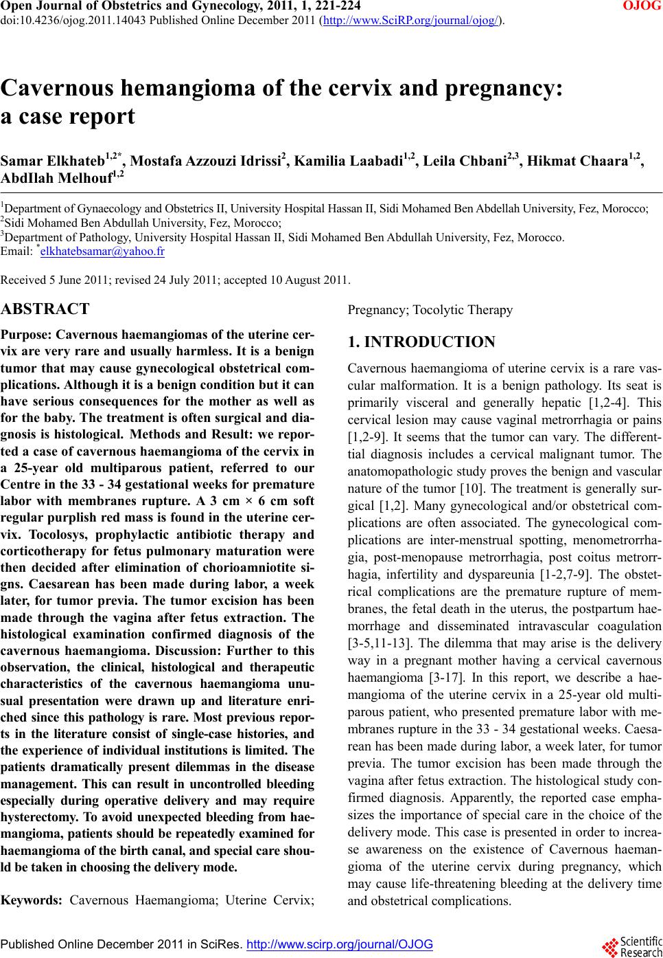 Open Journal of Obstetrics and Gynecology, 2011, 1, 221-224 doi:10.4236/ojog.2011.14043 Published Online December 2011 (http://www.SciRP.org/journal/ojog/ OJOG ). Published Online December 2011 in SciRes. http://www.scirp.org/journal/OJOG Cavernous hemangioma of the cervix and pregnancy: a case report Samar Elkhateb1,2*, Mostafa Azzouzi Idrissi2, Kamilia Laabadi1,2, Leila Chbani2,3, Hikmat Chaara1,2, AbdIlah Melhouf1,2 1Department of Gynaecology and Obstetrics II, University Hospital Hassan II, Sidi Mohamed Ben Abdellah University, Fez, Morocco; 2Sidi Mohamed Ben Abdullah University, Fez, Morocco; 3Department of Pathology, University Hospital Hassan II, Sidi Mohamed Ben Abdullah University, Fez, Morocco. Email: *[email protected] Received 5 June 2011; revised 24 July 2011; accepted 10 August 2011. ABSTRACT Purpose: Cavernous haemangiomas of the uterine cer- vix are very rare and usually harmless. It is a benign tumor that may cause gynecological obstetrical com- plications. Although it is a benign condition but it can have serious consequences for the mother as well as for the baby. The treatment is often surgical and dia- gnosis is histological. Methods and Result: we repor- ted a case of cavernous haemangioma of the cervix in a 25-year old multiparous patient, referred to our Centre in the 33 - 34 gestational weeks for premature labor with membranes rupture. A 3 cm × 6 cm soft regular purplish red mass is found in the uterine cer- vix. Tocolosys, prophylactic antibiotic therapy and corticotherapy for fetus pulmonary maturation were then decided after elimination of chorioamniotite si- gns. Caesarean has been made during labor, a week later, for tumor previa. The tumor excision has been made through the vagina after fetus extraction. The histological examination confirmed diagnosis of the cavernous haemangioma. Discussion: Further to this observation, the clinical, histological and therapeutic characteristics of the cavernous haemangioma unu- sual presentation were drawn up and literature enri- ched since this pathology is rare. Most previous repor- ts in the literature consist of single-case histories, and the experience of individual institutions is limited. The patients dramatically present dilemmas in the disease management. This can result in uncontrolled bleeding especially during operative delivery and may require hysterectomy. To avoid unexpected bleeding from hae- mangioma, patient s should be repeatedly examined for haemangioma of the birth canal, and special care shou- ld be taken in choosing the delivery mode. Keywords: Cavernous Haemangioma; Uterine Cervix; Pregnancy; Tocolytic Therapy 1. INTRODUCTION Cavernous haemangioma of uterine cervix is a rare vas- cular malformation. It is a benign pathology. Its seat is primarily visceral and generally hepatic [1,2-4]. This cervical lesion may cause vaginal metrorrhagia or pains [1,2-9]. It seems that the tumor can vary. The different- tial diagnosis includes a cervical malignant tumor. The anatomopathologic study proves the benign and vascular nature of the tumor [10]. The treatment is generally sur- gical [1,2]. Many gynecological and/or obstetrical com- plications are often associated. The gynecological com- plications are inter-menstrual spotting, menometrorrha- gia, post-menopause metrorrhagia, post coitus metrorr- hagia, infertility and dyspareunia [1-2,7-9]. The obstet- rical complications are the premature rupture of mem- branes, the fetal death in the uterus, the postpartum hae- morrhage and disseminated intravascular coagulation [3-5,11-13]. The dilemma that may arise is the delivery way in a pregnant mother having a cervical cavernous haemangioma [3-17]. In this report, we describe a hae- mangioma of the uterine cervix in a 25-year old multi- parous patient, who presented premature labor with me- mbranes rupture in the 33 - 34 gestational weeks. Caesa- rean has been made during labor, a week later, for tumor previa. The tumor excision has been made through the vagina after fetus extraction. The histological study con- firmed diagnosis. Apparently, the reported case empha- sizes the importance of special care in the choice of the delivery mode. This case is presented in order to increa- se awareness on the existence of Cavernous haeman- gioma of the uterine cervix during pregnancy, which may cause life-threatening bleeding at the delivery time and obstetrical complications. 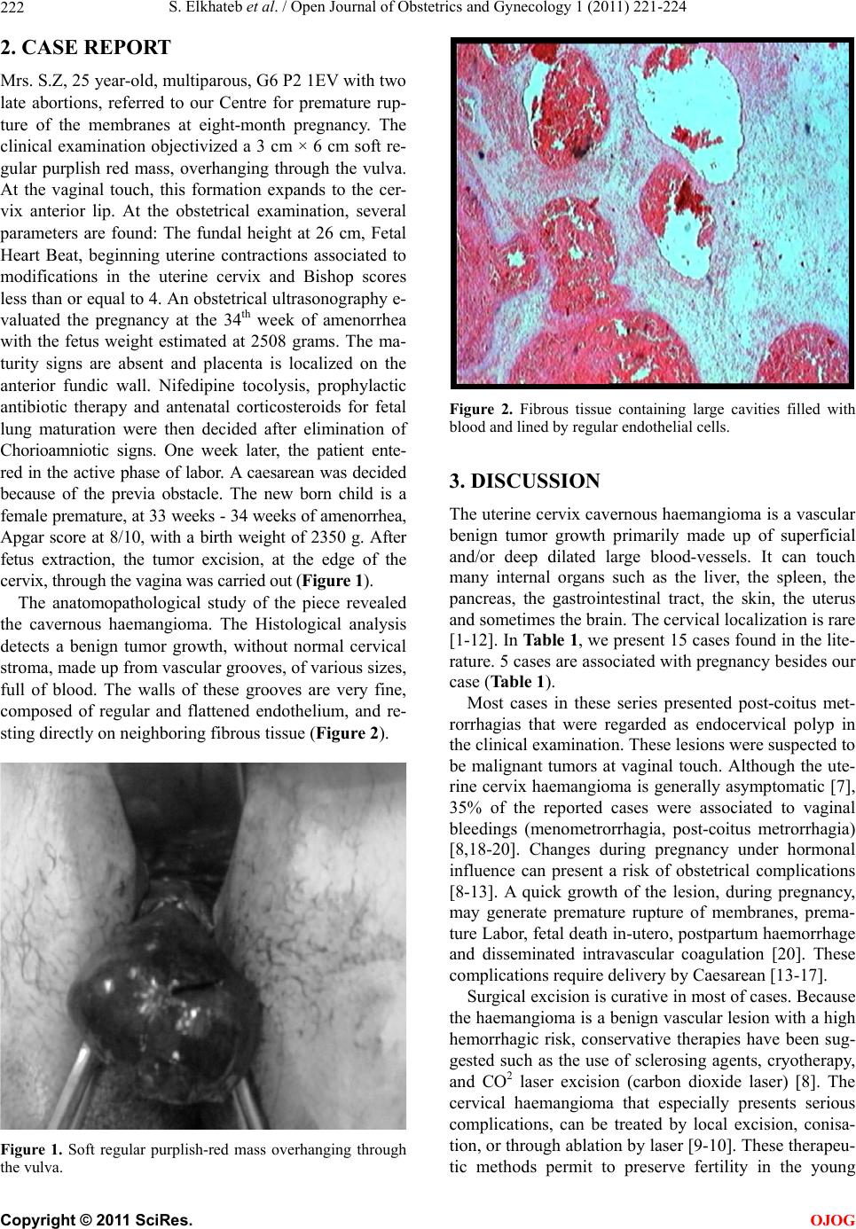 S. Elkhateb et al. / Open Journal of Obstetrics and Gynecology 1 (2011) 221-224 222 2. CASE REPORT Mrs. S.Z, 25 year-old, multiparous, G6 P2 1EV with two late abortions, referred to our Centre for premature rup- ture of the membranes at eight-month pregnancy. The clinical examination objectivized a 3 cm × 6 cm soft re- gular purplish red mass, overhanging through the vulva. At the vaginal touch, this formation expands to the cer- vix anterior lip. At the obstetrical examination, several parameters are found: The fundal height at 26 cm, Fetal Heart Beat, beginning uterine contractions associated to modifications in the uterine cervix and Bishop scores less than or equal to 4. An obstetrical ultrasonography e- valuated the pregnancy at the 34th week of amenorrhea with the fetus weight estimated at 2508 grams. The ma- turity signs are absent and placenta is localized on the anterior fundic wall. Nifedipine tocolysis, prophylactic antibiotic therapy and antenatal corticosteroids for fetal lung maturation were then decided after elimination of Chorioamniotic signs. One week later, the patient ente- red in the active phase of labor. A caesarean was decided because of the previa obstacle. The new born child is a female premature, at 33 weeks - 34 weeks of amenorrhea, Apgar score at 8/10, with a birth weight of 2350 g. After fetus extraction, the tumor excision, at the edge of the cervix, through the vagina was carried out (Figur e 1). The anatomopathological study of the piece revealed the cavernous haemangioma. The Histological analysis detects a benign tumor growth, without normal cervical stroma, made up from vascular grooves, of various sizes, full of blood. The walls of these grooves are very fine, composed of regular and flattened endothelium, and re- sting directly on neighboring fibrous tissue (Figure 2). Figure 1. Soft regular purplish-red mass overhanging through the vulva. Figure 2. Fibrous tissue containing large cavities filled with blood and lined by regular endothelial cells. 3. DISCUSSION The uterine cervix cavernous haemangioma is a vascular benign tumor growth primarily made up of superficial and/or deep dilated large blood-vessels. It can touch many internal organs such as the liver, the spleen, the pancreas, the gastrointestinal tract, the skin, the uterus and sometimes the brain. The cervical localization is rare [1-12]. In Table 1, we present 15 cases found in the lite- rature. 5 cases are associated with pregnancy besides our case (Table 1). Most cases in these series presented post-coitus met- rorrhagias that were regarded as endocervical polyp in the clinical examination. These lesions were suspected to be malignant tumors at vaginal touch. Although the ute- rine cervix haemangioma is generally asymptomatic [7], 35% of the reported cases were associated to vaginal bleedings (menometrorrhagia, post-coitus metrorrhagia) [8,18-20]. Changes during pregnancy under hormonal influence can present a risk of obstetrical complications [8-13]. A quick growth of the lesion, during pregnancy, may generate premature rupture of membranes, prema- ture Labor, fetal death in-utero, postpartum haemorrhage and disseminated intravascular coagulation [20]. These complications require delivery by Caesarean [13-17]. Surgical excision is curative in most of cases. Because the haemangioma is a benign vascular lesion with a high hemorrhagic risk, conservative therapies have been sug- gested such as the use of sclerosing agents, cryotherapy, and CO2 laser excision (carbon dioxide laser) [8]. The cervical haemangioma that especially presents serious complications, can be treated by local excision, conisa- tion, or through ablation by laser [9-10]. These therapeu- tic methods permit to preserve fertility in the young C opyright © 2011 SciRes. OJOG 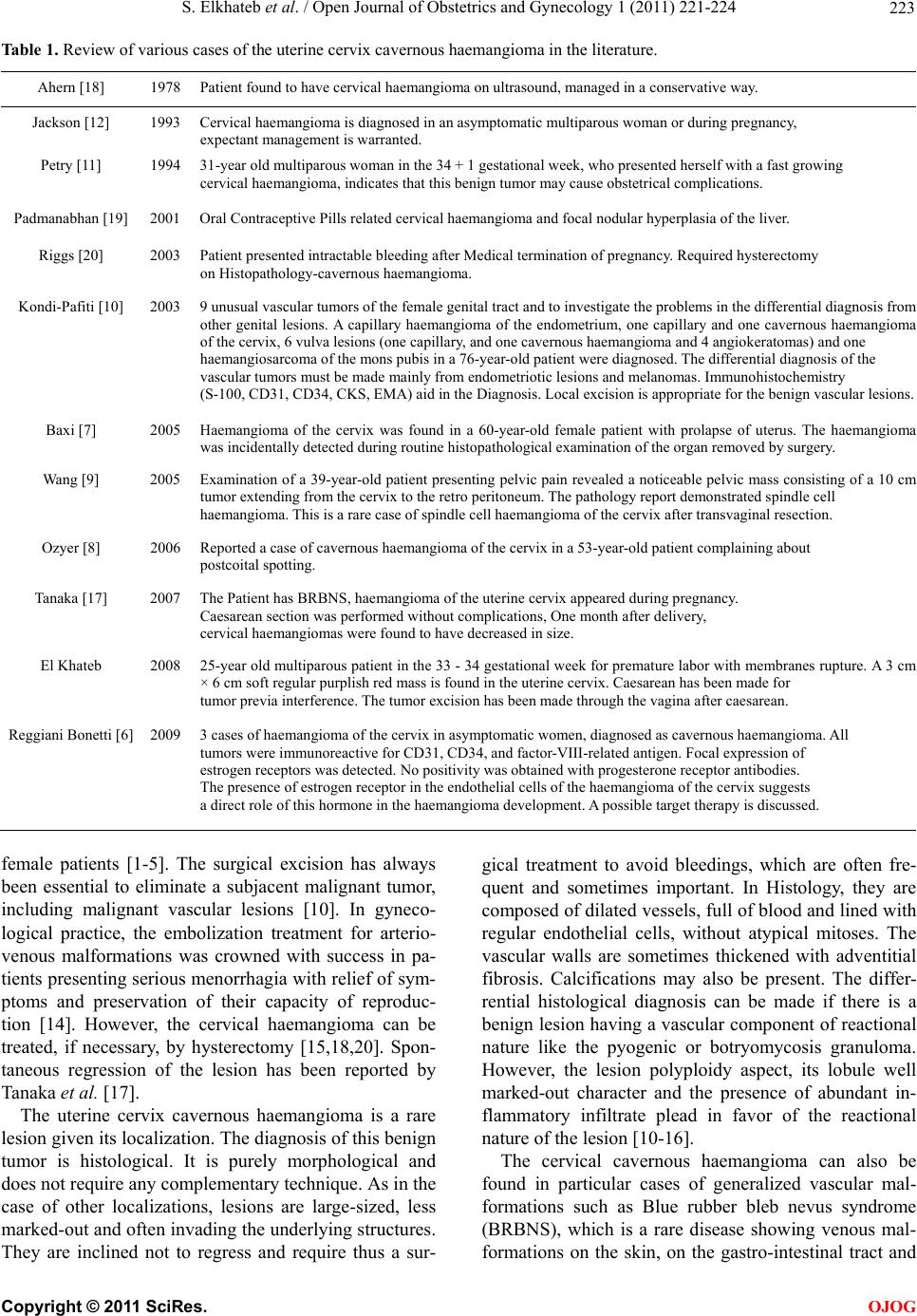 S. Elkhateb et al. / Open Journal of Obstetrics and Gynecology 1 (2011) 221-224 223 Table 1. Review of various cases of the uterine cervix cavernous haemangioma in the literature. Ahern [18] 1978 Patient found to have cervical haemangioma on ultrasound, managed in a conservative way. Jackson [12] 1993 Cervical haemangioma is diagnosed in an asymptomatic multiparous woman or during pregnancy, expectant management is warranted. Petry [11] 1994 31-year old multiparous woman in the 34 + 1 gestational week, who presented herself with a fast growing cervical haemangioma, indicates that this benign tumor may cause obstetrical complications. Padmanabhan [19] 2001 Oral Contraceptive Pills related cervical haemangioma and focal nodular hyperplasia of the liver. Riggs [20] 2003 Patient presented intractable bleeding after Medical termination of pregnancy. Required hysterectomy on Histopathology-cavernous haemangioma. Kondi-Pafiti [10] 2003 9 unusual vascular tumors of the female genital tract and to investigate the problems in the differential diagnosis from other genital lesions. A capillary haemangioma of the endometrium, one capillary and one cavernous haemangioma of the cervix, 6 vulva lesions (one capillary, and one cavernous haemangioma and 4 angiokeratomas) and one haemangiosarcoma of the mons pubis in a 76-year-old patient were diagnosed. The differential diagnosis of the vascular tumors must be made mainly from endometriotic lesions and melanomas. Immunohistochemistry (S-100, CD31, CD34, CKS, EMA) aid in the Diagnosis. Local excision is appropriate for the benign vascular lesions. Baxi [7] 2005 Haemangioma of the cervix was found in a 60-year-old female patient with prolapse of uterus. The haemangioma was incidentally detected during routine histopathological examination of the organ removed by surgery. Wang [9] 2005 Examination of a 39-year-old patient presenting pelvic pain revealed a noticeable pelvic mass consisting of a 10 c m tumor extending from the cervix to the retro peritoneum. The pathology report demonstrated spindle cell haemangioma. This is a rare case of spindle cell haemangioma of the cervix after transvaginal resection. Ozyer [8] 2006 Reported a case of cavernous haemangioma of the cervix in a 53-year-old patient complaining about postcoital spotting. Tanaka [17] 2007 The Patient has BRBNS, haemangioma of the uterine cervix appeared during pregnancy. Caesarean section was performed without complications, One month after delivery, cervical haemangiomas were found to have decreased in size. El Khateb 2008 25-year old multiparous patient in the 33 - 34 gestational week for premature labor with membranes rupture. A 3 cm × 6 cm soft regular purplish red mass is found in the uterine cervix. Caesarean has been made for tumor previa interference. The tumor excision has been made through the vagina after caesarean. Reggiani Bonetti [6] 2009 3 cases of haemangioma of the cervix in asymptomatic women, diagnosed as cavernous haemangioma. All tumors were immunoreactive for CD31, CD34, and factor-VIII-related antigen. Focal expression of estrogen receptors was detected. No positivity was obtained with progesterone receptor antibodies. The presence of estrogen receptor in the endothelial cells of the haemangioma of the cervix suggests a direct role of this hormone in the haemangioma development. A possible target therapy is discussed. female patients [1-5]. The surgical excision has always been essential to eliminate a subjacent malignant tumor, including malignant vascular lesions [10]. In gyneco- logical practice, the embolization treatment for arterio- venous malformations was crowned with success in pa- tients presenting serious menorrhagia with relief of sym- ptoms and preservation of their capacity of reproduc- tion [14]. However, the cervical haemangioma can be treated, if necessary, by hysterectomy [15,18,20]. Spon- taneous regression of the lesion has been reported by Tanaka et al. [17]. The uterine cervix cavernous haemangioma is a rare lesion given its localization. The diagnosis of this benign tumor is histological. It is purely morphological and does not require any complementary technique. As in the case of other localizations, lesions are large-sized, less marked-out and often invading the underlying structures. They are inclined not to regress and require thus a sur- gical treatment to avoid bleedings, which are often fre- quent and sometimes important. In Histology, they are composed of dilated vessels, full of blood and lined with regular endothelial cells, without atypical mitoses. The vascular walls are sometimes thickened with adventitial fibrosis. Calcifications may also be present. The differ- rential histological diagnosis can be made if there is a benign lesion having a vascular component of reactional nature like the pyogenic or botryomycosis granuloma. However, the lesion polyploidy aspect, its lobule well marked-out character and the presence of abundant in- flammatory infiltrate plead in favor of the reactional nature of the lesion [10-16]. The cervical cavernous haemangioma can also be found in particular cases of generalized vascular mal- formations such as Blue rubber bleb nevus syndrome (BRBNS), which is a rare disease showing venous mal- formations on the skin, on the gastro-intestinal tract and C opyright © 2011 SciRes. OJOG 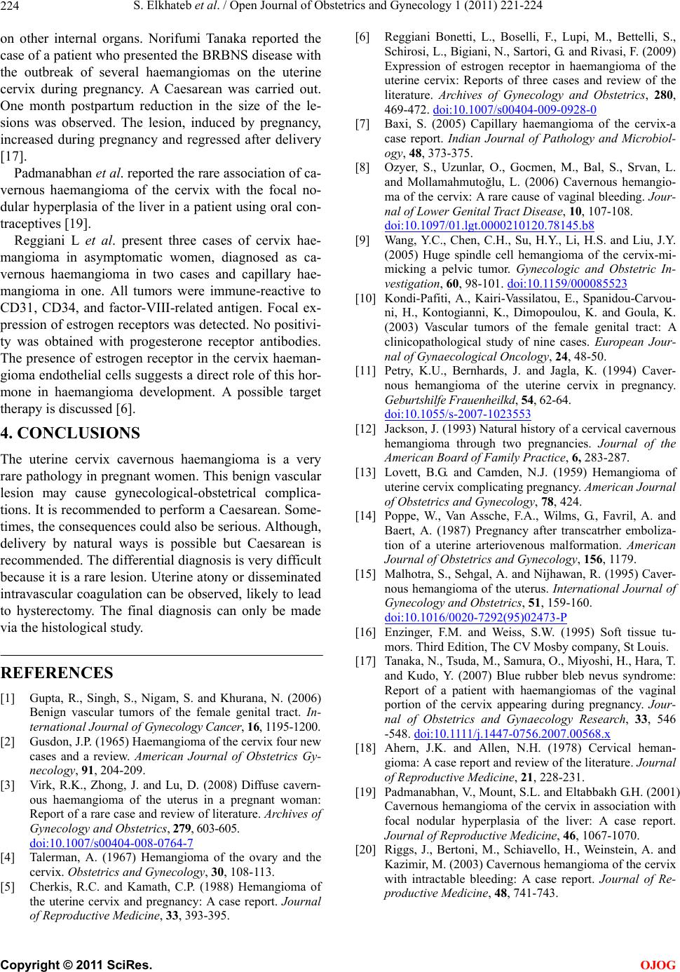 S. Elkhateb et al. / Open Journal of Obstetrics and Gynecology 1 (2011) 221-224 224 on other internal organs. Norifumi Tanaka reported the case of a patient who presented the BRBNS disease with the outbreak of several haemangiomas on the uterine cervix during pregnancy. A Caesarean was carried out. One month postpartum reduction in the size of the le- sions was observed. The lesion, induced by pregnancy, increased during pregnancy and regressed after delivery [17]. Padmanabhan et al. reported the rare association of ca- vernous haemangioma of the cervix with the focal no- dular hyperplasia of the liver in a patient using oral con- traceptives [19]. Reggiani L et al. present three cases of cervix hae- mangioma in asymptomatic women, diagnosed as ca- vernous haemangioma in two cases and capillary hae- mangioma in one. All tumors were immune-reactive to CD31, CD34, and factor-VIII-related antigen. Focal ex- pression of estrogen receptors was detected. No positivi- ty was obtained with progesterone receptor antibodies. The presence of estrogen receptor in the cervix haeman- gioma endothelial cells suggests a direct role of this hor- mone in haemangioma development. A possible target therapy is discussed [6]. 4. CONCLUSIONS The uterine cervix cavernous haemangioma is a very rare pathology in pregnant women. This benign vascular lesion may cause gynecological-obstetrical complica- tions. It is recommended to perform a Caesarean. Some- times, the consequences could also be serious. Although, delivery by natural ways is possible but Caesarean is recommended. The differential diagnosis is very difficult because it is a rare lesion. Uterine atony or disseminated intravascular coagulation can be observed, likely to lead to hysterectomy. The final diagnosis can only be made via the histological study. REFERENCES [1] Gupta, R., Singh, S., Nigam, S. and Khurana, N. (2006) Benign vascular tumors of the female genital tract. In- ternational Journal of Gynecology Cancer, 16, 1195-1200. [2] Gusdon, J.P. (1965) Haemangioma of the cervix four new cases and a review. American Journal of Obstetrics Gy- necology, 91, 204-209. [3] Virk, R.K., Zhong, J. and Lu, D. (2008) Diffuse cavern- ous haemangioma of the uterus in a pregnant woman: Report of a rare case and review of literature. Archives of Gynecology and Obstetrics, 279, 603-605. doi:10.1007/s00404-008-0764-7 [4] Talerman, A. (1967) Hemangioma of the ovary and the cervix. Obstetrics and Gynecology, 30, 108-113. [5] Cherkis, R.C. and Kamath, C.P. (1988) Hemangioma of the uterine cervix and pregnancy: A case report. Journal of Reproductive Medicine, 33, 393-395. [6] Reggiani Bonetti, L., Boselli, F., Lupi, M., Bettelli, S., Schirosi, L., Bigiani, N., Sartori, G. and Rivasi, F. (2009) Expression of estrogen receptor in haemangioma of the uterine cervix: Reports of three cases and review of the literature. Archives of Gynecology and Obstetrics, 280, 469-472. doi:10.1007/s00404-009-0928-0 [7] Baxi, S. (2005) Capillary haemangioma of the cervix-a case report. Indian Journal of Pathology and Microbiol- ogy, 48, 373-375. [8] Ozyer, S., Uzunlar, O., Gocmen, M., Bal, S., Srvan, L. and Mollamahmutoğlu, L. (2006) Cavernous hemangio- ma of the cervix: A rare cause of vaginal bleeding. Jour- nal of Lower Genital Tract Disease, 10, 107-108. doi:10.1097/01.lgt.0000210120.78145.b8 [9] Wang, Y.C., Chen, C.H., Su, H.Y., Li, H.S. and Liu, J.Y. (2005) Huge spindle cell hemangioma of the cervix-mi- micking a pelvic tumor. Gynecologic and Obstetric In- vestigation, 60, 98-101. doi:10.1159/000085523 [10] Kondi-Pafiti, A., Kairi-Vassilatou, E., Spanidou-Carvou- ni, H., Kontogianni, K., Dimopoulou, K. and Goula, K. (2003) Vascular tumors of the female genital tract: A clinicopathological study of nine cases. European Jour- nal of Gynaecological Oncology, 24, 48-50. [11] Petry, K.U., Bernhards, J. and Jagla, K. (1994) Caver- nous hemangioma of the uterine cervix in pregnancy. Geburtshilfe Frauenheilkd, 54, 62-64. doi:10.1055/s-2007-1023553 [12] Jackson, J. (1993) Natural history of a cervical cavernous hemangioma through two pregnancies. Journal of the American Board of Family Practice, 6, 283-287. [13] Lovett, B.G. and Camden, N.J. (1959) Hemangioma of uterine cervix complicating pregnancy. American Journal of Obstetrics and Gynecology, 78, 424. [14] Poppe, W., Van Assche, F.A., Wilms, G., Favril, A. and Baert, A. (1987) Pregnancy after transcatrher emboliza- tion of a uterine arteriovenous malformation. American Journal of Obstetrics and Gynecology, 156, 1179. [15] Malhotra, S., Sehgal, A. and Nijhawan, R. (1995) Caver- nous hemangioma of the uterus. International Journal of Gynecology and Obstetrics, 51, 159-160. doi:10.1016/0020-7292(95)02473-P [16] Enzinger, F.M. and Weiss, S.W. (1995) Soft tissue tu- mors. Third Edition, The CV Mosby company, St Louis. [17] Tanaka, N., Tsuda, M., Samura, O., Miyoshi, H., Hara, T. and Kudo, Y. (2007) Blue rubber bleb nevus syndrome: Report of a patient with haemangiomas of the vaginal portion of the cervix appearing during pregnancy. Jour- nal of Obstetrics and Gynaecology Research, 33, 546 -548. doi:10.1111/j.1447-0756.2007.00568.x [18] Ahern, J.K. and Allen, N.H. (1978) Cervical heman- gioma: A case report and review of the literature. Journal of Reproductive Medicine, 21, 228-231. [19] Padmanabhan, V., Mount, S.L. and Eltabbakh G.H. (2001) Cavernous hemangioma of the cervix in association with focal nodular hyperplasia of the liver: A case report. Journal of Reproductive Medicine, 46, 1067-1070. [20] Riggs, J., Bertoni, M., Schiavello, H., Weinstein, A. and Kazimir, M. (2003) Cavernous hemangioma of the cervix with intractable bleeding: A case report. Journal of Re- productive Medicine, 48, 741-743. C opyright © 2011 SciRes. OJOG |