

Paper Menu >>
Journal Menu >>
 World Journal of Neuroscience, 2011, 1, 49-54 doi:10.4236/wjns.2011.13008 Published Online November 2011 (http://www.SciRP.org/journal/wjns/ WJNS ). Published Online November 2011 in SciRes. http://www.scirp.org/journal/WJNs The antinociceptive role of central arginine vasopressin is involved in the endogenous opiate peptide, serotonin and acetylcholine systems Xiang-Yong Li1,2, Jun Yang1*, Xi-Qing Yan1, Yan-Juan Pan1, Ying Zhao1, Pei-Yong Qiu1, Xi-Jian Zhou2, Da-Xin Wang3 1College of Pharmacy, Xinxiang Medical University, Xinxiang, Henan, China; 2101 Hospital of PLA, Wuxi, Jiangsu, China; 3Jiangsu Su Bei People’s Hospital, Yangzhou University, Yangzhou, Jiangsu, China. Email: *[email protected] Received 19 August 2011; revised 5 October 2011; accepted 24 October 2011. ABSTRACT Our previous work has demonstrated that arginine vasopressin (AVP) plays a role in pain modulation. The present study investigated which kinds of neu- ropeptides and neurotransmitters in central nervous system might be involved in AVP antinociceptive role in the rat. The results showed that (1) intraventricu- lar injection (icv) of V1 receptor antagonist [d(CH2)5- Tyr(Me)AVP] and V2 receptor antagonist [d(CH2)5- [D-Ile2, Ile4, Ala9-NH2]AVP] blocked the antinocicep- tive effect induced by AVP (icv), (2) the opiate recap- tor antagonist (naloxone) reversed the antinociceptive effect induced by AVP (icv), and (3) both the sero- tonin receptor antagonist (cypoheptadine) and M re- ceptor antagonist (atropine) could attenuate the anti- nociceptive effect induced by AVP (icv); but (4) oxyto- cin, dopamine, N-methyl-D-aspartate (NMDA), γ-amino- butyric acid (GABA), N, α or β receptor antagonist did not influence the antinociceptive effect induced by AVP (icv). The data suggested that AVP antinoci- ceptive role was involved in the endogenous opiate peptide, serotonin and acetylcholine systems in cen- tral nervous system. Keywords: Arginine Vasopressin; Antinociception; En- dorgenous Opiate Peptide; Serotonin; Acetycholine 1. INTRODUCTION Arginine vasopressin (AVP), a nonapeptide posterior pi- tuitary hormone, is synthesized in the paraventricular and supraoptic nuclei of hypothalamus [1]. This hormone, combined with an apparent carrier protein (neurophysin), is transported along the hypothalamo-hypophyseal pathway to the neurohypophysis, where it is stored for subsequent release [2]. The remarkable functions of AVP include body fluid homeostasis, hormone probation, cardiovascular con- trol, learning and memory [3]. Many studies have showed that AVP influences antinociception in both human and nonhuman species [1,4-7]. Intraventricular injection (icv) of AVP increases the pain threshold, while anti-AVP se- rum (icv) decreases the pain threshold, but intrathecal injection (ith) or intravenous injection (iv) of either AVP or anti-AVP serum does not influence the pain threshold [8,9]. Pain stimulation could change AVP concentration in some brain nuclei, but did not change AVP concentra- tion in the spinal cord and serum [8,9]. The antinocicep- tive effect of AVP is limited to the brain nuclei, not the spinal cord and peripheral organs. Many studies have proven that most of neuropeptides (such as endogenous opiate peptides) and neurotransmit- ters (such as serotonin, acetylcholine, norepinephine and epinephrine) are involved in pain modulation [10]. For example, oxytocin (icv) could increase the pain threshold and enhance acupuncture analgesia, while anti-oxytocin serum (icv) decreases the pain threshold and weakens acupuncture analgesia [11-13]. However, it is not clear the interaction between AVP and other neuropeptides or neurotransmitters in pain modulation. The present study investigated which neuropeptides and neurotransmitters in central nerve system might be involved in AVP anti- nociceptive effect in the rat. 2. MATERIALS AND METHODS 2.1. Animals Adult male Sprague-Dawley rats weighing 180-220 g, which were obtained from Animal Center of Yangzhou University, Yangzhou, Jiangsu, China, were housed with food and water available ad libitum in a colony room under controlled temperature, humidity and a 12 hours  X.-Y. Li et al. / World Journal of Neuroscience 1 (2011) 49-54 50 light/dark cycle (light at 6:00 AM and dark at 6:00 PM). All the procedures were approved by Animal Care Com- mittee of Yangzhou University and conducted according to the guidelines of the International Association for the Study of Pain [14]. 2.2. Materials AVP, d(CH2)5Tyr(Me)AVP, d(CH2)5[D-Ile2, Ile4, Ala9- NH2]AVP and [1-D(CH2)5,Tyr(ME)2,Thr4,Tyr-NH2(9)] ornithine vasotocin were obtained from Peninsula Lab, San Carlos, CA, USA. Naloxone, cypoheptadine, atropine, 6-OH gallamine, fluperidol, phentolamine, propranolol, MK801, bicuculline, 5-amino valeric acid (5AVA), 3-ami- noproyl phossphonic acid (3APPA). and the other chemi- cals were bought from Sigma Co., St. Louis, MO, USA. 2.3. Surgery With Pellegrino L.J. rat brain atlas as reference, we used the stereotaxic apparatus (Jiangwan I-C, Shanghai, China) to implant a stainless steel guide cannula of 0.5 mm outer diameter into the right lateral ventricle (AP 0.3 mm, LR 0.5 mm, H 3.0 mm) for icv under the pentobarbital so- dium (35 mg/kg, intraperitoneal injection) anaesthesia. The guide cannula was fixed to the skull by dental acrylic. All operations were carried out in the aseptic condition and the animals were allowed to recover for at least 14 days after the surgery. 2.4. Intraventricular Injection (Icv) On the day of experiment, a stainless steel needle with 0.3 mm diameter for icv was directly inserted into the guide cannula, with 1mm beyond the tip of the latter. The 10 ml of antiserum or solution was injected into the lateral ventricle gently over 10 min. 2.5. Nociceptive Tests All animals were tested under the condition of free ac- tivity in the small cages (30 cm in diameter, 25 cm in height) from 8:00 to 10:00 am. Depending on the 30-year experience of studying pain in our laboratory, we used the potassium iontophoresis inducing tail-flick served as pain stimulus. The small wet cotton with the potassium iontophoresis was set on the skin of the tail. The cotton was exposed to direct electrical current, and the anode led the potassium iontophoresis to permeate the skin of the tail. If the current was strong enough, the permeated potassium iontophoresis resulted in the animal feeling the pain stimulation. The intensity of current at the mo- ment of the response was recorded as the pain threshold, which was expressed as mA (WQ-9E Pain Threshold Measurer, Shanghai, China). The duration between con- secutive stimuli was 10 min, and the pain stimulus was terminated at once when the rat showed response to this stimulus. 2.6. Histological Verification At the end of the experiments, the rat was sacrificed un- der the high dose of pentobarbital sodium (80 mg/kg, in- traperitoneal injection), and the histological location of icv was ascertained. The data were excluded from analy- sis if the positions were not accurate. 2.7. Statistical Analysis All values were expressed as mean ± standard error of the mean (SEM) and were analyzed between groups by analysis of variance (ANOVA) and χ2 test. P < 0.05 was considered statistically significant. 3. RESULTS 3.1. Effect of the Neuropeptide Receptor Antagonist on Pain Threshold Increase Induced by AVP (icv) Table 1 showed that 100 ng AVP (icv) could increase the pain threshold from 0.52 ± 0.03 mA to 0.77 ± 0.04 mA (P < 0.001). Although icv of 2 μg d(CH2)5Tyr(Me)AVP (V1 recep- tor antagonist), 2 μg d(CH2)5[D-Ile2, Ile4, Ala9-NH2]AVP (V2 receptor antagonist), 2 μg [1-D(CH2)5,Tyr(ME)2,Thr4, Tyr-NH2(9)] ornithine vasotocin (oxytocin receptor an- tagonist) or 2 μg naloxone (opiate receptor antagonist) decreased the pain threshold (all p < 0.01 ~ 0.001), ven- tricular pretreatment with V1 receptor antagonist, V2 re- ceptor antagonis, opiate receptor antagonist could reverse the antinociceptive effect induced by 100 ng AVP ad- ministration (icv), and ventricular pretreatment with oxy- tocin receptor antagonist did not influence the antino- ciceptive effect induced by 100 ng AVP administration (icv) (Table 1). 3.2. Effect of the Neurotransmitter Receptor Antagonist on Pain Threshold Increase Induced by AVP (icv) Table 2 showed that icv of 2 μg 5-HT receptor antago- nist (cypoheptadine), 2 μg M receptor antagonist (atro- pine), 2 μg N receptor antagonist (6-OH gallamine), 2 μg α receptor antagonist (phentolamine) or 2 μg β receptor antagonist (propranolol) decreased the pain threshold (all p < 0.01 ~ 0.001), but icv of 2 μg dopamine receptor an- tagonist (fluperidol), 2 μg N-methyl-D-aspartate (NMDA) receptor antagonist (MK801), 2 μg γ-aminobutyric acid (GABA)a receptor antagonist (bicuculline), 2 μg GABAb receptor antagonist (5-amino valeric acid) or 2 μg GABAc receptor antagonist (3-aminoproyl phossphonic acid) did not influence the pain threshold. Pretreatment with either 5-HT receptor antagonist or M receptor antagonist (icv) could attenuate the antinociceptive C opyright © 2011 SciRes. WJNS 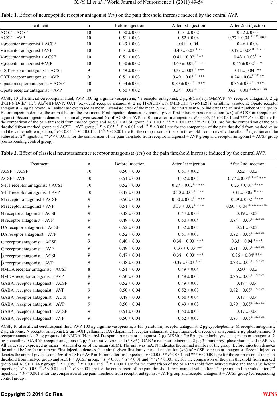 X.-Y. Li et al. / World Journal of Neuroscience 1 (2011) 49-54 51 Table 1. Effect of neuropeptide receptor antagonist (icv) on the pain threshold increase induced by the central AVP. Treatment n Before injection After 1st injection After 2nd injection ACSF + ACSF 10 0.50 ± 0.03 0.51 ± 0.02 0.52 ± 0.03 ACSF + AVP 10 0.51 ± 0.03 0.52 ± 0.04 0.77 ± 0.04111 222 *** V1receptor antagonist + ACSF 10 0.49 ± 0.03 0.41 ± 0.041 0.46 ± 0.04 V1receptor antagonist +AVP 10 0.51 ± 0.04 0.40 ± 0.0311 °°° 0.49 ± 0.04111 2 °°° V2receptor antagonist + ACSF 10 0.51 ± 0.03 0.41 ± 0.02111 ** 0.43 ± 0.0311 * V2receptor antagonist +AVP 10 0.50 ± 0.02 0.40 ± 0.02111 °°° 0.45 ± 0.021 °°° OXT receptor antagonist + ACSF 9 0.49 ± 0.03 0.39 ± 0.0311 *** 0.41 ± 0.041 ** OXT receptor antagonist + AVP 9 0.51 ± 0.03 0.40 ± 0.03111 °°° 0.74 ± 0.0411 222 aaa Opiate receptor antagonist + ACSF 10 0.54 ± 0.04 0.37 ± 0.01111 *** 0.35 ± 0.03111 *** Opiate receptor antagonist + AVP 10 0.50 ± 0.02 0.34 ± 0.03111 °°° 0.62 ± 0.0311 222 °°° aaa ACSF, 10 μl artificial cerebrospinal fluid; AVP, 100 ng arginine vasopressin; V1 receptor antagonist, 2 μg d(CH2)5Tyr(Me)AVP; V2 receptor antagonist, 2 μg d(CH2)5[D-Ile2, Ile4, Ala9-NH2]AVP; OXT (oxytocin) receptor antagonist, 2 μg [1-D(CH2)5,Tyr(ME)2,Thr4,Tyr-NH2(9)] ornithine vasotocin; Opiate receptor antagonist, 2 μg naloxone. All values are expressed as mean ± standard error of the mean (SEM). The unit was mA. N indicates the animal number of the group. Before injection denotes the animal before the treatment; First injection denotes the animal given first intraventricular injection (icv) of ACSF or receptor an- tagonist; Second injection denotes the animal given second icv of ACSF or AVP in 10 min after first injection. P < 0.05, ** P < 0.01 and *** P < 0.001 are for the comparison of the pain threshold from marked group and ACSF + ACSF group; ° P < 0.05, °° P < 0.01 and °°° P < 0.001 are for the comparison of the pain threshold from marked group and ACSF + AVP group; 1 P < 0.05, 11 P < 0.01 and 111 P < 0.001 are for the comparison of the pain threshold from marked value and the value before injection; 2 P < 0.05, 22 P < 0.01 and 222 P < 0.001 are for the comparison of the pain threshold from marked value after 1st injection and the value after 2nd injection; ªªª P < 0.001 is for the comparison of the pain threshold from receptor antagonist + AVP group and receptor antagonist + ACSF group (corresponding control group). Table 2. Effect of classical neurotransmitter receptor antagonists (icv) on the pain threshold increase induced by the central AVP. Treatment n Before injection After 1st injection After 2nd injection ACSF + ACSF 10 0.50 ± 0.03 0.51 ± 0.02 0.52 ± 0.03 ACSF + AVP 10 0.51 ± 0.03 0.52 ± 0.04 0.77 ± 0.04111 222 *** 5-HT receptor antagonist + ACSF 10 0.52 ± 0.03 0.27 ± 0.02111 *** 0.23 ± 0.01111*** 5-HT receptor antagonist + AVP 10 0.47 ± 0.03 0.30 ± 0.03111 °°° 0.31 ± 0.05111 °°° M receptor antagonist + ACSF 9 0.50 ± 0.03 0.30 ± 0.02111 *** 0.29 ± 0.02111*** M receptor antagonist + AVP 9 0.51 ± 0.03 0.33 ± 0.02111 °°° 0.60 ± 0.04111 222 °°° aaa N receptor antagonist + ACSF 9 0.48 ± 0.03 0.47 ± 0.03 0.49 ± 0.03 N receptor antagonist + AVP 9 0.49 ± 0.03 0.50 ± 0.04 0.84 ± 0.06111 222 aaa DA receptor antagonist + ACSF 9 0.52 ± 0.03 0.52 ± 0.04 0.51 ± 0.03 DA receptor antagonist + AVP 9 0.52 ± 0.03 0.51 ± 0.03 0.82 ± 0.05111 222 aaa α receptor antagonist + ACSF 9 0.48 ± 0.03 0.38 ± 0.031 *** 0.33 ± 0.0411 *** α receptor antagonist + AVP 9 0.49 ± 0.03 0.37 ± 0.031 °°° 0.81 ± 0.06111 222 aaa β receptor antagonist + ACSF 9 0.47 ± 0.04 0.38 ± 0.031 *** 0.36 ± 0.041 *** β receptor antagonist + AVP 9 0.48 ± 0.03 0.39 ± 0.0311 °°° 0.78 ± 0.05111 222 aaa NMDA receptor antagonist + ACSF 8 0.51 ± 0.03 0.49 ± 0.04 0.50 ± 0.03 NMDA receptor antagonist + AVP 8 0.50 ± 0.03 0.48 ± 0.03 0.76 ± 0.05111 222 aaa GABAa receptor antagonist + ACSF 9 0.52 ± 0.03 0.49 ± 0.03 0.48 ± 0.04 GABAa receptor antagonist + AVP 9 0.50 ± 0.04 0.52 ± 0.03 0.82 ± 0.05111 222 aaa GABAb receptor antagonist + ACSF 9 0.48 ± 0.03 0.50 ± 0.04 0.47 ± 0.04 GABAb receptor antagonist + AVP 9 0.50 ± 0.04 0.49 ± 0.03 0.79 ± 0.05111 222 aaa GABAc receptor antagonist + ACSF 9 0.51 ± 0.03 0.50 ± 0.03 0.47 ± 0.04 GABAc receptor antagonist + AVP 9 0.50 ± 0.04 0.52 ± 0.03 0.83 ± 0.05111 222 aaa ACSF, 10 μl artificial cerebrospinal fluid; AVP, 100 ng arginine vasopressin; 5-HT (serotonin) receptor antagonist, 2 μg cypoheptadine; M receptor antagonist, 2 μg atropine; N receptor antagonist, 2 μg 6-OH gallamine; DA (dopamine) receptor antagonist, 2 μg fluperidol; α receptor antagonist: 2 μg phentolamine; β receptor antagonist: 2 μg propranolol; NMDA (N-methyl-D-aspartate) receptor antagonist: 2 μg MK801; GABAa (γ-aminobutyric acid) receptor antagonist: 2 μg bicuculline; GABAb receptor antagonist: 2 μg 5-amino valeric acid (5AVA); GABAc receptor antagonist, 2 μg 3-aminoproyl phossphonic acid (3APPA). All values are expressed as mean ± standard error of the mean (SEM). The unit was mA. N indicates the animal number of the group. Before injection denotes the animal before the treatment; First injection denotes the animal given first intraventricular injection (icv) of ACSF or receptor antagonist; Second injection denotes the animal given second icv of ACSF or AVP in 10 min after first injection. P < 0.05, ** P < 0.01 and *** P < 0.001 are for the comparison of the pain threshold from marked group and ACSF + ACSF group; ° P < 0.05, °° P < 0.01 and °°° P < 0.001 are for the comparison of the pain threshold from marked group and ACSF + AVP group; 1 P < 0.05, 11 P < 0.01 and 111 P < 0.001 are for the comparison of the pain threshold from marked value and the value before injection; 2 P < 0.05, 22 P < 0.01 and 222 P < 0.001 are for the comparison of the pain threshold from marked value after 1st injection and the value after 2nd injection; ªªª P < 0.001 is for the comparison of the pain threshold from receptor antagonist + AVP group and receptor antagonist + ACSF group (corresponding control group). C opyright © 2011 SciRes. WJNS  X.-Y. Li et al. / World Journal of Neuroscience 1 (2011) 49-54 52 effect induced by 100 ng AVP administration (icv) (all P < 0.001), but the other studied neurotransmitter recap- tor antagonists did not influence the antinociceptive ef- fect induced by the administration of 100 ng AVP (icv) (Table 2). 4. DISCUSSION AVP is synthesized within cells located in the brain and in certain peripheral organs of the body. In the brain, AVP is synthesized in cell groups within the hypothala- mus; several of these cell groups release hormones into the systemic circulation or into the portal circulation of the anterior pituitary gland and others release neurotran- smitters at synaptic targets within the brain. AVP is also synthesized in certain extrahypothalamic brain sites, such as limbic system structures in the forebrain. In peripheral tissues, there is evidence that AVP is synthesized in the anterior pituitary, adrenal, and thymus glands and in male and female reproductive structures (ovaries, uterus, and testes) [3]. However, most of AVP is synthesized in hy- pothalamic paraventricular nucleus (PVN) and hypotha- lamic supraoptic nucleus (SON) [2,15]. It has been proven that PVN and SON play an important role in analgesia [16-20], and AVP, which may be from PVN and SON, is involved in pain modulation [21,22]. Our present study showed that (1) not only V1 recep- tor antagonist [d(CH2)5Tyr(Me)AVP] and V2 receptor antagonist [d(CH2)5[D -Ile2, Ile4, Ala9-NH2]AVP] blocked the antinociceptive effect induced by AVP (icv), but also the opiate receptor antagonist (naloxone), 5-HT receptor antagonist (cypoheptadine) and M receptor antagonist (atropine) could reserve the antinociceptive effect in- duced by AVP (icv); (2) oxytocin, dopamine, NMDA, GABA, N, α and β receptor antagonist did not influence the antinociceptive effect induced by AVP (icv). The data suggested that AVP antinociceptive effect was related with the endogenous opiate peptide, serotonin and ac- erycholine systems. Histological study has shown that there are many AVP containing fibers in the periaqueductal gray (PAG), which come from PVN neurons [23,24]. AVP enhances the syn- thesis and secretion of endogenous opiate peptides in the PAG [25,26]. The nucleus raphe magnus (NRM) is a serotonergic nucleus located in the rostral ventromedial medulla of the brainstem. Axons of the NRM project to the spinal cord [27], terminating primarily in the dorsal horn [28]. Brainstem nuclei that project to the dorsal horn of the spinal cord can function to inhibit afferent nociceptive transmission [29-31]. Activation of these descending an- tinociceptive pathways may be triggered by physiologi- cal stimuli [32] as well as by pharmacological agents [33]. Antinociception involving the NRM has been studied after either electrical stimulation or direct administration of pharmacological agents [34-36]. The NRM is a key neural structure for pain modulation, in which serotonin (5-HT) is a major site for pain regulation [10]. AVP and 5-HT interaction in the brain controls many animal be- haviors [37,38]. There are many bioactive substances in the caudate nucleus (CdN) including dopamine (DA) and acetylcho- line (Ach), which show interaction with AVP [35,39-41]. DA and Ach in CdN are important bioactive substances in pain modulation and the CdN is showing an important neural structure in pain modulation [38]. Our pervious study has shown that AVP in the PAG, NRM and CdN could regulate the pain process [18,42,43], and pain stimulation changes the AVP concentration in the PAG, NRM and CdN [15,40]. So we could imagine that AVP regulating the pain process might be involved in the endogenous opiate system in the PAG, serotonin system in the NRM and acetylcholine system in the CdN. However, it needs to be confirmed. 5. ACKNOWLEDGEMENTS This work was supported by Xinxiang Medical University, 101 Hospi- tal of PLA, Jiangsu Su Bei People’s Hospital and grants from National Basic Research Program of China (2007CB936104). REFERENCES [1] Aziz, H., Pearce, J. and Miller, E. (1968) Vasopressin in prevention of lumbar puncture headache. British Medical Journal, 4, 677-678. doi:10.1136/bmj.4.5632.677 [2] Martin, J.B., Reichlan, S. and Bick, K.L. (1981) Neu- rosecretion and brain peptides. Raven Press, New York. [3] McEwen, B.B. (2004) The role of vasopressin and oxy- tocin in memory processing. Elsevier, Amsterdam. [4] Berkowitz, B.A. and Sherman, S. (1982) Characteriza- tion of vasopressin analgesia. Journal of Pharmacology and Experimental Therapeutics, 220, 329-334. [5] Berson, B.S., Berntson, G.G., Zipf, W., Torello, M.W. and Kirk, W.T. (1983) Vasopressin-induced antinociception: An investigation into its physiological and hormonal ba- sis. Endocrinology, 113, 337-343. doi:10.1210/endo-113-1-337 [6] Kendler, K.S., Weitzman, R.E. and Fisher, D.A. (1978) The effect of pain on plasma arginine vasopressin con- centrations in man. Clinical Endocrinology (Oxf), 8, 89- 94. doi:10.1111/j.1365-2265.1978.tb02156.x [7] Madrazo, I., Franco-Bourland, R.E., Leon-Meza, V.M. and Mena, I. (1987) Intraventricular somatostatin-14, ar- ginine vasopressin, and oxytocin: analgesic effect in a patient with intractable cancer pain. Applied Neurophysi- ology, 50, 427-431. [8] Yang, J., Song, C.Y., Liu, W.Y. and Lin, B.C. (2006) Only through the brain nuclei, arginine vasopressin regulates antinociception in the rat. Peptides, 27, 3341-3346. doi:10.1016/j.peptides.2006.08.019 [9] Yang, J., Yang, Y., Wang, C.H., Wang, G., Xu, H.T., Liu, W.Y. and Lin, B.C. (2009) Effect of arginine vasopressin C opyright © 2011 SciRes. WJNS  X.-Y. Li et al. / World Journal of Neuroscience 1 (2011) 49-54 53 on acupuncture analgesia in the rat. Peptides, 30, 241- 247. doi:10.1016/j.peptides.2008.10.013 [10] Weng, N.Q. (1988) Pain and analgesia. Shanghai Science Press, Shanghai. [11] Yang, J. (1994) Intrathecal administration of oxytocin induces analgesia in low back pain involving the en- dogenous opiate peptide system. Spine, 19, 867-871. doi:10.1097/00007632-199404150-00001 [12] Yang, J., Yang, Y., Chen, J.M., Liu, W.Y., Wang, C.H. and Lin, B.C. (2007) Central oxytocin enhances antino- ciception in the rat. Peptides, 28, 1113-1119. doi:10.1016/j.peptides.2007.03.003 [13] Yang, J., Yang, Y., Chen, J.M., Liu, W.Y., Wang, C.H. and Lin, B.C. (2007) Effect of oxytocin on acupuncture analgesia in the rat. Neuropeptides, 41, 285-292. doi:10.1016/j.npep.2007.05.004 [14] Zimmermann, M. (1983) Ethical guidelines for investi- gations of experimental pain in conscious animal. Pain, 16, 109-110. doi:10.1016/0304-3959(83)90201-4 [15] Dorner, G. and Kawakamin, M. (1978) Hormones and brain development. Elsevier, Amsterdam. [16] Yang, J. and Lin, B.C. (1992) Hypothalamic paraven- tricular nucleus plays a role in acupuncture analgesia through the central nervous system in the rat. Acupunc- ture Electro-Therapeutics Research, 17, 209-220. [17] Shiraishi, T., Onoe, M., Kojima, T., Sameshima, Y. and Kageyama, T. (1995) Effects of hypothalamic paraven- tricular nucleus: Electrical stimulation produce marked nalgesia in rats. Neurobiology (Bp), 3, 393-403. [18] Yang, J., Chen, J.M., Liu, W.Y., Song, C.Y. and Lin, B.C. (2008) Investigating the role of hypothalamic paraven- tricular nucleus in nociception of the rat. International Journal of Neuroscience, 118, 473-485. doi:10.1080/00207450601123563 [19] Yang, J., Yang, Y., Chen, J.M., Liu, W.Y., Wang, C.H. and Lin, B.C. (2008) Effect of hypothalamic supraoptic nucleus on acupuncture analgesia in the rat. Brain Re- search Bull, 75, 681-686. doi:10.1016/j.brainresbull.2007.11.004 [20] Yang, J., Yang, Y., Chen, J.M., Liu, W.Y., Wang, C.H. and Lin, B.C. (2008) Investigating the role of the hypo- thalamic supraoptic nucleus in nociception in the rat. Life Science, 82, 166-173. doi:10.1016/j.lfs.2007.10.023 [21] Bodnar, R.J., Nilaver, G., Wallace, M.M., Badillo-Mar- tinez, D. and Zimmerman, E.A. (1984) Pain threshold changes in rats following central injection of beta-en- dorphin, met-enkephalin, vasopressin or oxytocin antisera. International Journal of Neuroscience, 24, 149-160. doi:10.3109/00207458409089803 [22] Yang, J., Yang, Y., Chen, J.M., Xu, H.T., Liu, W.Y., Wang, C.H. and Lin, B.C. (2007) Arginine vasopressin is an im- portant regulator in antinociceptive modulation of hypo- thalamic paraventricular nucleus in the rat. Neuropep- tides, 41, 165-176. doi:10.1016/j.npep.2006.12.005 [23] Antunes, J.L. and Zimmerman, E.A. (1978) The hypotha- lamic magnocellular system of the rhesus monkey: An immunocytochemical study. Journal of Comparative Neu- rology, 81, 539-565. doi:10.1002/cne.901810306 [24] Swanson, L.W. and Sawchenko, P.E. (1980) Separate neurons in the paraventricular nucleus project to the me- dian eminence and to the medulla or spinal cord. Brain Research, 198, 190-195. doi:10.1016/0006-8993(80)90354-6 [25] Yang, J., Yang, Y., Xu, H.T., Chen, J.M., Liu, W.Y. and Lin, B.C. (2006) Arginine vasopressin enhances periaq- ueductal grey synthesis and secretion of enkephalin and endorphin in the rat. Brain Research Bull, 71, 193-199. doi:10.1016/j.brainresbull.2006.09.003 [26] Yang, J., Yang, Y., Chen, J.M., Xu, H.T., Liu, W.Y. and Lin, B.C. (2007) Arginine vasopressin in periaqueductal gray, which relates to antinociception, comes from hypo- thalamic paraventricular nucleus in the rat. Neur oscience Letters, 412, 154-158. doi:10.1016/j.neulet.2006.10.049 [27] Bowker, R.M., Westlund, K.N., Sullivan, M.C. and Coulter, J.D. (1982) Organization of descending seroton- ergic projections to the spinal cord. Prog Brain Res earch, 57, 239-265. doi:10.1016/S0079-6123(08)64132-1 [28] Jones, S.L. and Light, A.R. (1990) Electrical stimulation in the medullary nucleus raphe magnus inhibits noxious heat-evoked protein-like immunoreactivity in the rat lum- bar spinal cord. Brain Research, 530, 335-538. doi:10.1016/0006-8993(90)91306-2 [29] Basbaum, A.I. and Fields, H.L. (1979) The origin of de- scending pathways in the dorsolateral funiculus of the spinal cord of the cat and rat: Further studies on the ana- tomy of pain modulation. Journal of Comparative Neurol- ogy, 187, 513-532. doi:10.1002/cne.901870304 [30] Fields, H.L. and Besson, J.M. (1988) Progress in brain research, Pain modulation. Elsevier, Amsterdam, 77. [31] Sandkuhler, J. (1996) The organization and function of endogenous antinociceptive systems. Prog Neurobiology, 50, 49-81. doi:10.1016/0301-0082(96)00031-7 [32] Myers, R.D., Rezvani, A.H. and Gurley-Orkin, L.A. (1985) New doublelumen polyethylene cannula for push-pull perfusion of brain tissue in vivo. Journal of Neuroscience Methods, 12, 205-218. doi:10.1016/0165-0270(85)90003-2 [33] Gogas, K.R., Presley, R.W., Levine, J.D. and Basbaum, A.I. (1991) The antinociceptive action of supraspinal opioids results from an increase in descending inhibitory control: Correlation of nociceptive behavior and c-fos expression. Neuroscience, 42, 617-628. doi:10.1016/0306-4522(91)90031-I [34] Proudfit, H.K. and Anderson, E.G. (1975) Morphine an- algesia: Blockade by raphe magnus lesions. Brain Re- search, 98, 612-618. doi:10.1016/0006-8993(75)90380-7 [35] Newman, M.E. (1985) Vasopressin inhibits cyclic AMP accumulation and adenylate activity cerebral preparation. FEBS Letters, 181, 203-206. doi:10.1016/0014-5793(85)80260-X [36] Brodie, M.S. and Proudfit, H.K. (1986) Antinociception induced by local injections of carbachol into the nucleus raphe magnus in rats: Alteration by intrathecal injection of monoaminergic antagonists. Brain Research, 371, 70-79. doi:10.1016/0006-8993(86)90811-5 [37] Albers, H.E., Karom, M. and Smith, D. (2002) Serotonin and vasopressin interact in the hypothalamus to control communicative behavior. NeuroReport, 13, 931-933. doi:10.1097/00001756-200205240-00006 [38] Ferris, C.F. and Delville, Y. (1994) Vasopressin and se- rotonin interactions in the control of agonistic behavior. Psychoneuroendocrinology, 19, 593-601. C opyright © 2011 SciRes. WJNS  X.-Y. Li et al. / World Journal of Neuroscience 1 (2011) 49-54 Copyright © 2011 SciRes. 54 WJNS doi:10.1016/0306-4530(94)90043-4 [39] Yang, J., Chen, J.M., Liu, W.Y., Song, C.Y. and Lin, B.C. (2006) Arginine vasopressin in the caudate nucleus plays an antinociceptive role in the rat. Life Science, 79, 2086-2090. doi:10.1016/j.lfs.2006.07.005 [40] Courtney, N. and Raskind, M. (1983) Vasopressin affects adenylate cyclase activity in rat brain: A possible neuro- modulator. Life Science, 7, 591-596. doi:10.1016/0024-3205(83)90203-5 [41] Pefracca, F.M., Baskin, D.G., Diaz, J. and Dorsa, O.M. (1986) Ontogenetic changes in vasopressin binding site distribution in rat brain: An autoradiographic study. Brain Research, 393, 63-68. [42] Yang, J., Chen, J.M., Liu, W.Y., Song, C.Y., Lin, B.C. (2006) Effect of arginine vasopressin in the nucleus raphe mag- nus on antinociception in the rat. Peptides , 27, 2224-2229. [43] Yang, J., Chen, J.M., Liu, W.Y., Song, C.Y., Lin, B.C. (2006) Through V2, not V1 receptor relating to endogenous opi- ate peptides, arginine vasopressin in periaqueductal gray regulates antinociception in the rat. Regulatory Peptides, 137, 156-161. |