

Paper Menu >>
Journal Menu >>
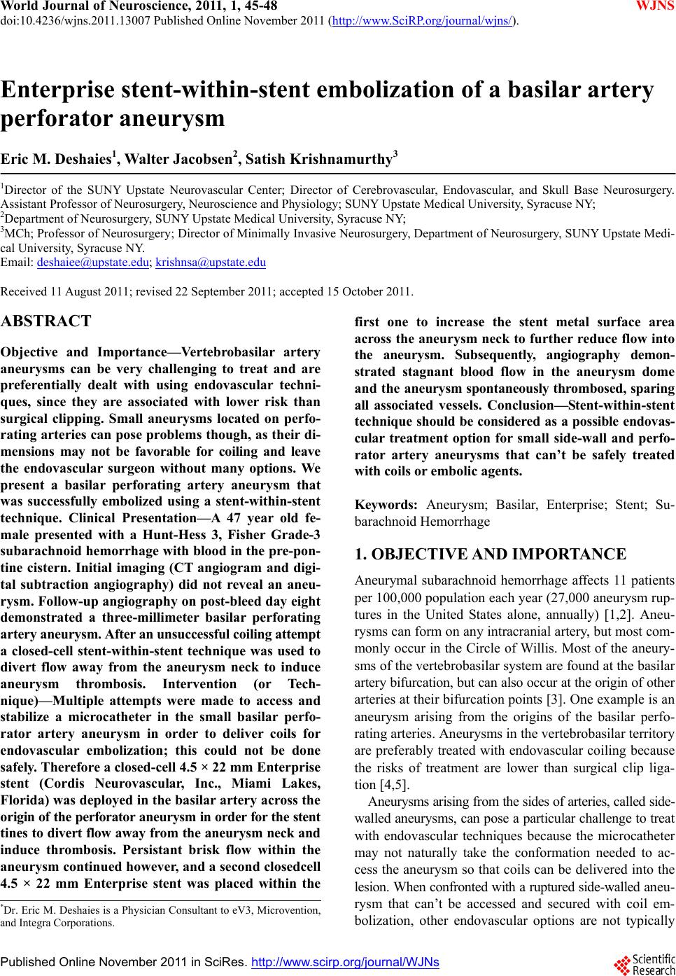 World Journal of Neuroscience, 2011, 1, 45-48 doi:10.4236/wjns.2011.13007 Published Online November 2011 (http://www.SciRP.org/journal/wjns/ WJNS ). Published Online November 2011 in SciRes. http://www.scirp.org/journal/WJNs Enterprise stent-within-stent embolization of a basilar artery perforator aneurysm Eric M. Deshaies1, Walte r Jacobsen2, Satish Krishnamurthy3 1Director of the SUNY Upstate Neurovascular Center; Director of Cerebrovascular, Endovascular, and Skull Base Neurosurgery. Assistant Professor of Neurosurgery, Neuroscience and Physiology; SUNY Upstate Medical University, Syracuse NY; 2Department of Neurosurgery, SUNY Upstate Medical University, Syracuse NY; 3MCh; Professor of Neurosurgery; Director of Minimally Invasive Neurosurgery, Department of Neurosurgery, SUNY Upstate Medi- cal University, Syracuse NY. Email: [email protected]; [email protected] Received 11 August 2011; revised 22 September 2011; accepted 15 October 2011. ABSTRACT Objective and Importance—Vertebrobasilar artery aneurysms can be very challenging to treat and are preferentially dealt with using endovascular techni- ques, since they are associated with lower risk than surgical clipping. Small aneurysms located on perfo- rating arteries can pose problems though, as their di- mensions may not be favorable for coiling and leave the endovascular surgeon without many options. We present a basilar perforating artery aneurysm that was successfully embolized using a stent-within-stent technique. Clinical Presentation—A 47 year old fe- male presented with a Hunt-Hess 3, Fisher Grade-3 subarachnoid hemorrhage with blood in the pre-pon- tine cistern. Initial imaging (CT angiogram and digi- tal subtraction angiography) did not reveal an aneu- rysm. Follow-up angiography on post-bleed day eight demonstrated a three-millimeter basilar perforating artery aneurysm. After an unsuccessful coiling attempt a closed-cell stent-within-stent technique was used to divert flow away from the aneurysm neck to induce aneurysm thrombosis. Intervention (or Tech- nique)—Multiple attempts were made to access and stabilize a microcatheter in the small basilar perfo- rator artery aneurysm in order to deliver coils for endovascular embolization; this could not be done safely. Therefore a closed-cell 4.5 × 22 mm Enterprise stent (Cordis Neurovascular, Inc., Miami Lakes, Florida) was deployed in the basilar artery across the origin of the perforator aneurysm in order for the stent tines to divert flow away from the aneurysm neck and induce thrombosis. Persistant brisk flow within the aneurysm continued however, and a second closedcell 4.5 × 22 mm Enterprise stent was placed within the first one to increase the stent metal surface area across the aneurysm neck to further reduce flow into the aneurysm. Subsequently, angiography demon- strated stagnant blood flow in the aneurysm dome an d the aneurysm spontaneously thrombosed, sparing all associated vessels. Conclusion—Stent-within-stent technique should be considered as a possible endovas- cular treatment option for small side-wall and perfo- rator artery aneurysms that can’t be safely treated with coils or embolic agents. Keywords: Aneurysm; Basilar, Enterprise; Stent; Su- barachnoid Hemorrhage 1. OBJECTIVE AND IMPORTANCE Aneurymal subarachnoid hemorrhage affects 11 patients per 100,000 population each year (27,000 aneurysm rup- tures in the United States alone, annually) [1,2]. Aneu- rysms can form on any intracranial artery, but most com- monly occur in the Circle of Willis. Most of the aneury- sms of the vertebrobasilar system are found at the basilar artery bifurcation, but can also occur at the origin of other arteries at their bifurcation points [3]. One example is an aneurysm arising from the origins of the basilar perfo- rating arteries. Aneurysms in the vertebrobasilar territory are preferably treated with endovascular coiling because the risks of treatment are lower than surgical clip liga- tion [4,5] . Aneurysms arising from the sides of arteries, called side- walled aneurysms, can pose a particular challenge to trea t with endovascular techniques because the microcatheter may not naturally take the conformation needed to ac- cess the aneurysm so that coils can be delivered into the lesion. When confronted with a ruptured side-walled an eu - rysm that can’t be accessed and secured with coil em- bolization, other endovascular options are not typically *Dr. Eric M. Deshaies is a Physicia n Consultant to eV3, Microvention, and Integra Corporations. 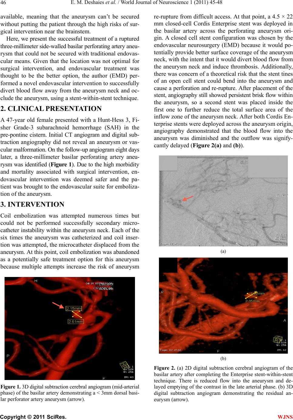 E. M. Deshaies et al. / World Journal of Neuroscience 1 (2011) 45-48 46 available, meaning that the aneurysm can’t be secured without putting the patien t through the high risks of sur- gical intervention near the brainstem. Here, we present the successful tre atment of a ruptured three-millimeter side-walled basilar perforating artery aneu- rysm that could not be secured with traditional endovas- cular means. Given that the location was not optimal for surgical intervention, and endovascular treatment was thought to be the better option, the author (EMD) per- formed a novel endovascular intervention to successfully divert blood flow away from the aneurysm neck and oc- clude the aneurysm, using a stent-within-stent techniqu e. 2. CLINICAL PRESENTATION A 47-year old female presented with a Hunt-Hess 3, Fi- sher Grade-3 subarachnoid hemorrhage (SAH) in the pre-pontine cistern. Initial CT angiogram and digital sub- traction angiography did not reveal an aneurysm or vas- cular malformation. On the follow-up angiogram eight days later, a three-millimeter basilar perforating artery aneu- rysm was identified (Figure 1). Due to the high morbid ity and mortality associated with surgical intervention, en- dovascular intervention was deemed safer and the pa- tient was brought to the endovascular suite for emboliza- tion of the aneurysm. 3. INTERVENTION Coil embolization was attempted numerous times but could not be performed successfully secondary micro- catheter instab ility within the aneurys m neck. Each of the six times the aneurysm was catheterized and coil inser- tion was attempted, the microcatheter disp laced from the aneurysm. At this point, coil embolization was abandoned as a potentially safe treatment option for this aneurysm because multiple attempts increase the risk of aneurysm Figure 1. 3D digital subtraction cerebral angiogram (mid-arterial phase) of the basilar artery demonstrating a < 3mm dorsal basi- lar perforator artery aneurysm (arrow). re-rupture from difficult access. At that point, a 4.5 × 22 mm closed-cell Cordis Enterprise stent was deployed in the basilar artery across the perforating aneurysm ori- gin. A closed cell stent configuration was chosen by the endovascular neurosurgery (EMD) because it would po- tentially provide better surface coverage of the aneurysm neck, with the intent that it would divert blood flow from the aneurysm neck and induce thrombosis. Additionally, there was concern of a theoretical risk that the stent tines of an open cell stent could bend into the aneurysm and cause a perforation and re-rupture. After placement of the st ent, angiography still showed persistent brisk flow withi n the aneurysm, so a second stent was placed inside the first one to further reduce the total surface area of the inflow zone of the aneurysm neck. After both Cordis En- terprise stents were deployed acros s the aneurysm origin, angiography demonstrated that the blood flow into the aneurysm was diminished and the outflow was signify- cantly delayed (Figure 2(a) and (b)). (a) (b) Figure 2. (a) 2D digital subtraction cerebral angiogram of the basilar artery after completing the Enterprise stent-within-stent technique. There is reduced flow into the aneurysm and de- layed emptying of the contrast in the late arterial phase. (b) 3D digital subtraction angiogram demonstrating the residual an- eurysm (arrow). C opyright © 2011 SciRes. WJNS  E. M. Deshaies et al. / World Journal of Neuroscience 1 45-48 47 The patient was placed on aspirin therapy alone to simultaneously lower the risk of in-stent thrombosis yet allow the aneurysm to thrombose, and also allow for potentially needed external ventricular drain placement in the future if needed. Repeated cerebra l angiography the next day demonstrated that the aneurysm dome was al- most completely thrombosed. At this point she was placed on an intravenous drip of abciximab (ReoPro, Centocor, Philadelphia, PA), a sho rt-acting glycoprotein IIb /IIIa re- ceptor antagonist, to minimize the risk of in-stent throm- bosis. A second angiogram on day three showed that the aneurysm was filling again, likely secondary to the ab- ciximab. Hence abciximab was stopped again and she was continued on aspirin alone until it was decided that a ventriculosotomy was not needed, after which time clopi- dogrel (Plavix 75 mg oral, daily) was added to her anti- platelet regimen. The patient recovered well and CT an- giogram performed prior to discharge showed no resid- ual aneurysm dome filling. The patient was discharged home on post-bleed day 20 with a standard antiplatelet regimen of aspirin (325 mg oral, daily) and clopidogrel (75 mg oral, daily) therapy for 12 weeks. Six month fol- low-up digital subtraction angiography showed that the aneurysm had thrombosed and the stents remained pat- ent without any evidence of stent migration or stenosis (Figure 3). 4. CONCLUSIONS Ruptured basilar perforating artery aneurysms can pose challenges both in diagnosis and treatment. We could find three articles that reported four patients with similar an- eurysms [6-8]. All five patients (including the present Figure 3. Six month follow-up 3D digital subtraction cerebral angiogram (mid-arterial phase) of the basilar artery demon- strating complete occlusion of the basilar perforator artery aneurysm (arrow), no in-stent stenosis, and no stent migration (gray). one reported here) had no aneurysm found on the initial angiogram and the aneurysms were demonstrated on a subsequent study. A perimesencephalic location of the SAH, particularly in conjunction with an angiographically ne- gative study, may lull the treating neurosurgeon into thinking that these patients have non-aneurysmal SAH. Furthermore, all of the reported cases treated prior to our report, were treated surgically as endovascular coiling was not possible. Flow diverting sten ts, such as the Pipe- line Embolization Device (eV3, Irvine, California) and the Silk Stent (Balt, Montmorency, France), have been shown to be unsafe for use in the basilar artery since they are associated with a high risk of basilar perforating artery occlusion and brainstem stroke. Traditional intracranial arterial stents can b e divided into “open” and “closed” cell designs. The “open” cell design has holes in the stent wall that do not have metal around the entire circumference of the cells, whereas the “closed” cell stent has metal e ncircling the entire cell cir- cumference (Figure 4(a) and (b)). The Cordis Enterprise stent is a closed-cell nitinol stent, with each cell diameter measuring less than 1.3 French. We had postulated that placing the closed-cell stent across the aneurysm neck would divert flow away from the aneurysm neck and impede outflow of blood from the aneurysm, both pro- moting thrombosis [9]. An additional theoretical advantage of using the closed-cell design is th at is would lessen the risk of the stent tines perforating th e aneurysm. A second stent was placed within the first to further reduce the total stent cell size and hence, the cross-sectional area of the aneurysm inflow zone. In vitro and in vivo studies on aneurysm hemodynam- ics after stent placement have consistently demonstrated reduced flow and shear-stress inside the aneurysm after stent placement [9-15]. Stents also reduce the pressure in the distal zone of the aneurysm neck and dome [10]. All of these hemodynamic changes are favorable for pro- moting intra-aneurysmal flow stasis and thrombosis. The (a) (b) Figure 4. (a) Enterprise (Cordis, Corporation, Johnson and John- son) stent with closed-cell technology. (b) Neuroform microde- livery stent (Boston Scientific) stent with open-cell technology. C opyright © 2011 SciRes. WJNS  E. M. Deshaies et al. / World Journal of Neuroscience 1 (2011) 45-48 Copyright © 2011 SciRes. 48 [6] Ghogawala Z, S.J. and Ogilvy, C.S. (1996) Distal basilar perforator artery aneurysm: Case report. Neurosurgery, 39, 393-396. doi:10.1097/00006123-199608000-00034 stent surface provides scaffolding for neo-intimal prolif- eration and healing of the aneurysm neck, which can fur- ther reduce flo w into the aneurysm once the stent “he als” into place. [7] Mathieson CS, B.P., Jenkins, S. and Hanzely, Z. (2010) An unusual case of spontaneous subarachnoid hemor- rhage—a ruptured aneurysm of a basilar perforator artery. British Journal of Neurosurgery, 24, 291-293. doi:10.3109/02688690903572095 In conclusion, stent-within-stent placement across the neck of a side-walled aneurysm should be considered an alternative treatment for aneurysms that cannot be treated with coil embolization, flow-div erting stents, or embolic glue agents. This technique may also apply to endovas- cular treatment of small blister aneurysms in other arte- rial locations that are difficult to coil within the Circle of Willis. Acute in-stent thro mbosis fro m the metal-to-blo od interface and delayed in-stent stenosis from neointimal hyperplasia, are real risks that must be monitored closely. [8] Sanchez-Mejia RO, L.M. (2007) Distal aneurysms of basilar perforating and circumferential arteries. Report of three cases. Journal of Neurosurgery, 107, 654-659. doi:10.3171/JNS-07/09/0654 [9] Wanke, I. and Forsting, M. (2008) Stents for intracranial wide-necked aneurysms: More than mechanical protec- tion. Neuroradiology, 50, 991-998. doi:10.1007/s00234-008-0460-0 [10] Aenis, M., et al. (1997) Modeling of flow in a straight stented and nonstented side wall aneurysm model. Jour- nal of Biomechanical Engineering, 119, 206-212. doi:10.1115/1.2796081 REFERENCES [11] Geremia, G., Haklin, M. and Brennecke, L. (1994) Em- bolization of experimentally created aneurysms with in- travascular stent devices. American Journal of Neurora- diology, 15, 1223-1231. [1] Schievink, W.I. (1997) Intracranial aneurysms. New England Journal of Medicine, 336, 28-40. doi:10.1056/NEJM199701023360106 [2] Wijdicks, E.F., Kallmes, D.F., Manno, E.M., Fulgham, J.R. and Piepgras, D.G. (2005) Subarachnoid hemorrhage: Neurointensive care and aneurysm repair. Mayo Clinic Proceedings, 80, 550-559. doi:10.4065/80.4.550 [12] Henkes, H., et al. (2004) Coil treatment of a fusiform up- per basilar trunk aneurysm with a combination of “kiss- ing” neuroform stents, TriSpan-, 3D- and fibered coils, and permanent implantation of the micro-guidewires. Neuroradiology, 46, 464-468. [3] Rhoton, A.J. (2002) Aneurysms. Neurosurgery, 51, 121- 158. doi:10.1097/00006123-200210001-00004 [13] Liou, T.M., Liou, S.N. and Chu, K.L. (2004) Intra-an- eurysmal flow with helix and mesh stent placement across side-wall aneurysm pore of a straight par e nt v esse l. Journal of Biomechanical Engineering, 126, 36-43. doi:10.1115/1.1644566 [4] Leusseveld, E.B.E., Nijssen, P.C., Van Rooij, W.J., Sluze- wski, M., Tulleken, C.A., Wijnalda, D., Schellens, R.L., Van der Graaf, Y. and Rinkel, G.J. (2002) Endovascular coiling versus neurosurgical clipping in patients with a ruptured basilar tip aneurysm. Journal of Neurology Neu- rosurgery and Psychiatry, 73, 591-593. doi:10.1136/jnnp.73.5.591 [14] Sadasivan, C., et al. (2002) Angiographic quantification of contrast medium washout from cerebral aneurysms after stent placement. American Journal of Neuroradiol- ogy, 23, 1214-1221. [5] Van der Schaaf I, A.A., Wermer, M., Molyneux, A., Clarke, M., Van Gijn, J. and Rinkel, G. (2005) Endovas- cular coiling versus neurosurgical clipping for patients with aneurysmal subarachnoid hemorrhage. Cochrane Database of Systematic Reviews, Octobe r 19 . [15] Wanke, I. (2008) Stents for intracranial aneurysms: indi- cations, limits and non indications. Interventional Neu- roradiologic, 14, 60-62. WJNS |