

Paper Menu >>
Journal Menu >>
 Open Journal of Ophthalmol ogy, 2011, 1, 1-3 doi:10.4236/ojoph.2011.11001 Published Online November 2011 (http://www.SciRP.org/journal/ojoph) Copyright © 2011 SciRes. OJOph 1 Fraser Syndrome: Case Report with Review of Literature Saruban Pasu1*, Luna Dhir2, Sarah Mackenzie3, Graham Thompson3 1Moorfields Eye Hospital NHS Foundation Trust, London, England; 2Chelsea and Westminster Hospital, London, England; 3Moor- fields at St. George’s Hospital, Duke Elder Eye Centre, Tooting, London, England. Email: [email protected] Received September 26th, 2011; revised October 23rd, 2011; accepted November 3rd, 2011. ABSTRACT Fraser syndrome is a rare autosomal recessive multisystem disorder with a reported incidence of 0.043 per 10,0 00 live born infants and 1.1 in 10,000 stillbirths [1]. The condition is cha racterised by cryptophtha lmos, cutaneous syndactyly, laryngeal and genitourinary malformations, craniofacial dysmorphism, orofacial clefting, musculoskeletal anomalies and mental retardation. The diagnosis can be made on prenatal scans, post natal clinical examination or on autopsy findings. We present a case of Fraser syndrome and review of the ocular manifestations of this co ndition. Keywords: Cryptophthalmos, Fraser Sy n dr o me 1. Case History A one day old male infant was referred to the ophthal- mology department for assessment of a fused right upper and lower eyelid. He was the first child born to noncon- sanguineous parents of south East Asian origin. He had a birth weight of 2.8 Kg and was born via vaginal ventouse delivery at 39 + 2 weeks. Antenatal scans at 20 weeks had revealed unilateral right renal agenesis (see Figure 1(a)). An apparently normal left kidney was seen at this stage (see Figure 1(b)). No other abnormalities were noted on antenatal scanning. Clinical examination revealed a normal anterior and posterior segment of the left eye. Right complete cryp- tophthalmos was noted with a palpable eyeball beneath (see Figure 1(c)). Systemic examination findings were an umbilical hernia, widely spaced fontanelle, bulbous nose and clinodactyly of the right fourth and fifth toes. The genitalia were normal, and no hypertelorism or oro- facial clefting were noted. Further ultrasound scans and biochemical investigations showed the left kidney to be cystic, dyplastic and poorly functioning. An MRI orbits and brain confirmed right cryptoph- thalmos (see Figure 1(d)). A diagnosis of Fraser syndrome was made based on the clinical findings. 2. Discussion The earliest reports of what is now known as cryptoph- thalmos (hidden eye) date back to the first century A.D. Pliny the Elder described the Lepidus family in which three children were born with a membrane over the eye, typical of this rare anomaly. The term cryptophthalmos was introduced by Ze- hender [2] et al. in 1872 who described a child whose eyes were covered by continuous sheets of skin from forehead to cheek, associated with additional malforma- tions including hypertelorism, syndactyly, abnormal genitalia, umbilical hernia , anal stenosis and hoarse voice. George Fraser [3] in 1962 was the first to group these features together under the term “cryptophthalmos syn- drome”. In fact, cryptophthalmos is not always a feature of this syndrome, and thus, the eponym Fraser syndrome is preferable for the condition. Fraser syndrome is a multiple malformation syndrome with a probable autosomal recessive inheritance. It may be caused by mutations in two genes FRAS1 [4] (chro- mosome 4) and FREM2 [4]. The FRAS1 extracellular matrix protein regulates epidermal-basement membrane adhesion and organogenesis during development. The FREM2 gene (chromosome 13) encodes the FRAS1- related extracellular matrix protein 2. Thomas et al. in 1986 were the first to publish diag- nostic criteria (see Table 1) for Fraser syndrome. The criteria were based upon a study of 124 cases. Two major criteria and one minor criterion or one major and at least four minor criteria were required for the diagnosis of 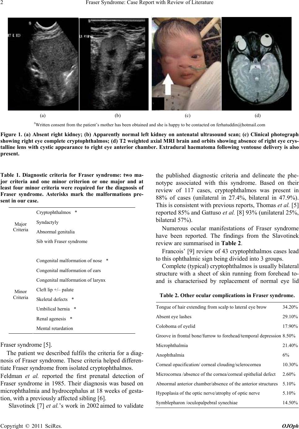 Fraser Syndrome: Case Report with Review of Literature 2 (a) (b) (c) (d) #Written consent from the patient’ s mother has been obtaine d and she is happy to be contacted on ferhatuddin@hot mail.com Figure 1. (a) Absent right kidney; (b) Apparently normal left kidney on antenatal ultrasound scan; (c) Clinical photograph showing right eye complete cryptophthalmos; (d) T2 weighted axial MRI brain and orbits showing absence of right eye crys- talline lens with cystic appearance to right eye anterior chamber. Extradural haematoma following ventouse delivery is also present. Table 1. Diagnostic criteria for Fraser syndrome: two ma- jor criteria and one minor criterion or one major and at least four minor criteria were required for the diagnosis of Fraser syndrome. Asterisks mark the malformations pre- sent in our case. Cryptophthalmos * Syndactyly Abnormal genitali a Major Criteria Sib with Fraser syndrome Congenital malformation of nose * Congenital malformation of ears Congenital malformation of larynx Cleft lip +/– palate Skeletal defects * Umbilical hernia * Renal agenesis * Minor Criteria Mental retardation Fraser syndrome [5]. The patient we described fulfils the criteria for a diag- nosis of Fraser syndrome. These criteria helped differen- tiate Fraser syndrome from isolated cryptophthalmos. Feldman et al. reported the first prenatal detection of Fraser syndrome in 1985. Their diagnosis was based on microphthalmia and hydrocephalus at 18 weeks of gesta- tion, with a previous ly affected sibling [6]. Slavotinek [7] et al.’s work in 2002 aimed to validate the published diagnostic criteria and delineate the phe- notype associated with this syndrome. Based on their review of 117 cases, cryptophthalmos was present in 88% of cases (unilateral in 27.4%, bilateral in 47.9%). This is consistent with previous reports, Thomas et al. [5] reported 85% and Gattuso et al. [8] 93% (unilateral 25%, bilateral 57%). Numerous ocular manifestations of Fraser syndrome have been reported. The findings from the Slavotinek review are summarised in Table 2. Francois’ [9] review of 43 cryptophthalmos cases lead to this ophthalmic sign being divided into 3 groups. Complete (typical) cryptophthalmos is usually bilateral structure with a sheet of skin running from forehead to- and is characterised by replacement of normal eye lid Table 2. Other ocular complications in Fraser syndr ome . Tongue of hair extending from scalp to lateral eye brow 34.20% Absent eye lashes 29.10% Coloboma of eyelid 17.90% Groove in frontal bone/fur row to forehead/temporal depression8.50% Microphthalmia 21.40% Anophthalmia 6% Corneal opacification/ corneal clouding/sclerocornea 10.30% Microcornea /absence of the cornea/corneal epithelial defect 2.60% Abnormal anteri or chamber/absence of the anterior str uctu res 5.10% Hypoplasia of the optic nerve/atrophy of optic nerve 5.10% Symblepharon /oculop a lp e b ral synechiae 14.50% Copyright © 2011 SciRes. OJOph  Fraser Syndrome: Case Report with Review of Literature 3 cheek. There can be absence or poor development of the eyebrows, eyelashes, gland structures and conjunctival sac. The skin is adherent to the underlying cornea. Mi- crophthalmia, symblepharon and abnormalities of the anterior chamber are common. Incomplete (atypical) cryptophthalmos refers to rudi- mentary lid structures that are present with small con- junctival sacs placed laterally, Small palpebral fissures, micophthalmia and symblepharon [10]. Abortive cryptophthalmos or congenital symblepharon described an upper lid without a well defined margin that is adherent to the cornea. Slavotinek also suggests sym- plepharon is part of the ocular manifestations of Fraser syndrome [7]. We suggest utmost vigilance when clinicians are en countered with one fetal anomaly o n antenatal ultrasou nd. Rare multisystem disorders like Fraser syndrome should be considered, so adequate parental counselling can be delivered. 3. Literature Search A MEDLINE search covering the years 1950 to the pre- sent was conducted using the following terms: Fraser syndrome, cryptophthalmos syndrome, and cryptoph- thalmos. Articles cited in the reference lists of other arti- cles were also searched. The authors declare no conflicts of interest. REFERENCES [1] M. Narang, M. Kumar and D. Shah, “Fraser-Crypto- phthalmos Syndrome with Colonic Atresia,” Indian Jour- nal of Pediatrics, Vol. 75, No. 2, 2008, pp. 89-91. doi:10.1007/s12098-008-0030-9 [2] W. Zehender, “Eine Missge burt mit Hautueberwachsenen Augen oder Kryptophthalmus,” Klin Monatsbl Augen- heilkd, Vol. 10, 1872, pp. 225-234. doi:10.1007/s12098-008-0030-9 [3] G. R. Fraser, “Our Genetical ‘Load’: A Review of Some Aspects of Genetic Variation,” Annals of Human Genetics, Vol. 25, 1962, pp. 387-415. [4] I. Smyth and P. Scambler, “The Genetics of Fraser Syn- drome and the Blebs Mouse Mutants,” Human Molecular Genetics, Vol. 14, No. 2, 2005, pp. R269-274. doi:10.1093/hmg/ddi262 [5] I. T. Thomas, J. L. Frias, V. Felix, L. Sanchex de Leon, R. A. Hernandex and M. C. Jones, “Isolated and Syndromic Cryptophthalmos,” American Journal of Medical Genet- ics, Vol. 25, 1986, pp. 85-98. doi:10.1002/ajmg.1320250111 [6] E. Feldman, E. Shalev, E. Weiner, H. Cohen and H. Zuckerman, “Microphthalmia B Prenatal Ultrasonic Di- agnosis: A Case Report,” Prenatal Diagnosis, Vol. 5, 1985, pp. 205-207. doi:10.1002/pd.1970050307 [7] A. M. Slavotinek and C. J. Tifft, “Fraser Syndrome and Cryptophthalmos: A Review of the Diagnostic Criteria and Evidence of Phenotypic Modules,” Journal of Medi- cal Genetics, Vol. 39, 2002, pp. 623-633. doi:10.1136/jmg.39.9.623 [8] J. Gattuso, M. A. Patton and M. Baraitser, “The Clinical Spectrum of Fraser Syndrome: Report of Three New Cases and Review, ” Journal of Medical Genetics, Vol. 24, 1987, pp. 549-555. doi:10.1136/jmg.24.9.549 [9] J. Francois, “Syndrome Malformatif avec Cryptophthal- mie,” Annals Genertics Medical Gemellol, Vol. 18, 1969, pp. 18-50. [10] D. J. Brazier, S. J. Hardman-Lea and J. R. O. Collin, “Cryp- tophthalmos: Surgical Treatment of the Congenital Symblepharon Variant,” British Journal of Ophthalmol- ogy, Vol. 70, 1986, pp. 391-395. doi:10.1136/bjo.70.5.391 Copyright © 2011 SciRes. OJOph |