

Paper Menu >>
Journal Menu >>
 Open Journal of Gastroenterology, 2011, 1, 7-12 doi:10.4236/ojgas.2011.12002 Published Online November 2011 (http://www.SciRP.org/journal/ojgas/ OJGas ). Published Online November 2011 in SciRes. http://www.scirp.org/journal/OJGas The pathogenesis of primary pouchitis following ileal pouch-anal anastomosis: a review of current hypotheses Sally Bath1, Christian P. Selinger1,2, Rupert W. L. Leong1,3 1Concord Repatriation General Hospital, Gastroenterology and Liver Services, Hospital Road, Concord, Australia; 2Salford Royal Hospital, Department of Gastroenterology, Stott Lane, Salford, UK; 3The Faculty of Medicine, The University of New South Wales, Sydney, Australia. Email: [email protected] Received 3 October 2011; revised 8 November 2011; accepted 19 November 2011. ABSTRACT Primary pouchitis is a common complication of ileal pouch-anal anastomosis following proctocolectomy in patients treated for ulcerative colitis (UC), but is un- usual for those treated for familial adenomatous polyposis (FAP). While a number of theories as to the pathogenesis of this inflammatory condition have been proposed, no single one has been wholly satis- factory. Much research has been devoted to investi- gating a link between the pathogenic factors involved in UC, but not FAP, and those underlying pouchitis. The contribution of sulfate-producing bacteria has also been explored. The role of other intraluminal factors, such as short chain fatty acids and unconju- gated bile salts, has also been investigated. A unifying theory of a multi-step process might explain the pathogenesis of pouchitis, but further research is re- quired to proof causation. It is likely that pouchitis develops as a result of a combination of genetic, im- munological, microbial and metabolic factors. Future insight into the causes of pouchitis may eventually allow for the development of more effective treat- ments. Keywords: Pouchitis; Ileo-Pouch Anal Anastomosis; Pathogenesis 1. INTRODUCTION Prior to the development of the ileal pouch-anal anasto- mosis technique (IPAA) patients requiring proctocolec- tomy were mandated to have an end-ileostomy. IPAA restore the continuity of the lower GI tract by creating a pouch of ileal loops directly anastomosed to the anal canal, and preserving the anal sphincter function. Re- storative proctocolecto my followed by IPAA is currently the treatment of choice in the surgical management of refractory ulcerative colitis (UC) and familial adenoma- tous polyposis (FAP) [1,2]. Epidemiological studies show that at least 10% of patients with UC will undergo proctocolectomy during the course of their illness [3]. IPAA has been fou nd to have a positive impact on global quality of life score as it provides symptomatic relief while preserving fecal continence [4,5]. However, the procedure is associated with significant short and long- term morbidity, including immediate post-operative com- ations, pouch failure, small bowel obstruction, sexual dysfunction, irritable pouch syndro me and pouchitis [2]. Of these, pouchitis is the most common long-term complication, occurring at a rate of 48% at 10 years and 70% at 20 years in patients with UC [6]. For patients who have had one episode of pouchitis, there is a 64% risk of recurrence [2]. In FAP, pouchitis is uncommon and approximated 5% in those treated with restorative proctoco lectomy with pouch [7]. Overall similar rates of adverse post-operative outcomes have been reported in patients treated for UC and those treated for FAP [8]. There is however a marked difference in the risk of fis- tulisation and pouchitis [8]. Pouchitis is significantly more common in patients treated for UC than FAP, and this observation has formed the basis for a number of hypotheses of pathogenesis. Pouchitis is an inflammatory condition of the ileal reservoir that is formed during IPAA [9]. It occurs as a single acute episode in a third of cases but most have recurrent acute episodes or a chronic course of disease [10]. Acutely, it can be distressing with symptoms of increased stool frequency, urgency, nocturnal inconti- nence and abdomino-pelvic pain, often accompanied by fever, weight-loss and bloody stools [5]. In chronic cases, pouchitis may be associated with reduced quality of life and the need for further surgery [9]. Currently, there are no consensus guidelines for diagnosis and in most cases a combination of clinical signs and symptoms and pou ch 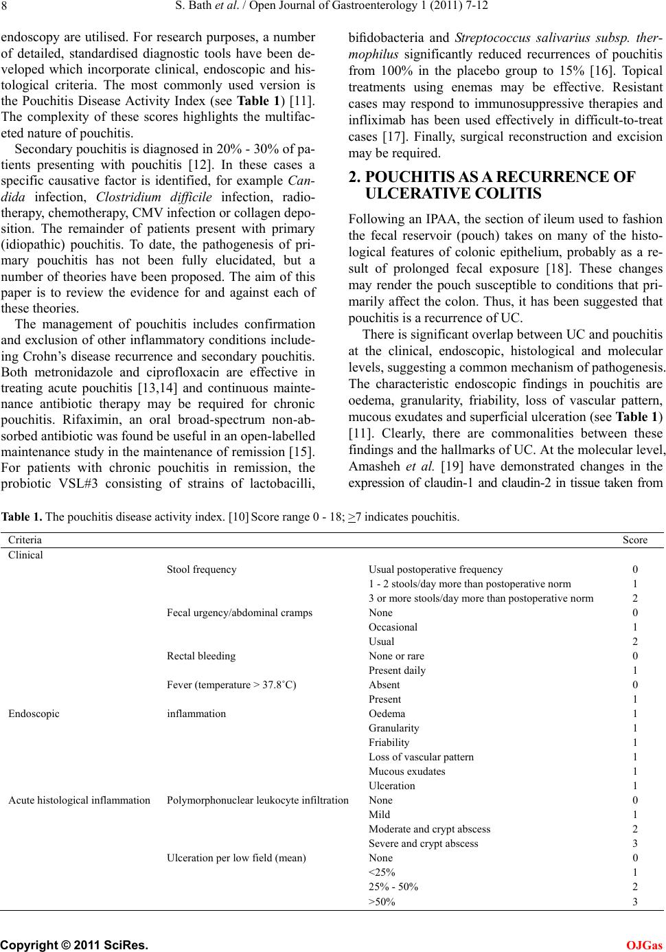 S. Bath et al. / Open Journal of Gastroenterology 1 (2011) 7-12 8 endoscopy are utilised. For research purposes, a number of detailed, standardised diagnostic tools have been de- veloped which incorporate clinical, endoscopic and his- tological criteria. The most commonly used version is the Pouchitis Disease Activity Index (see Ta bl e 1 ) [11]. The complexity of these scores highlights the multifac- eted nature of pouchitis. Secondary pouchitis is diagnosed in 20% - 30% of pa- tients presenting with pouchitis [12]. In these cases a specific causative factor is identified, for example Can- dida infection, Clostridium difficile infection, radio- therapy, chemotherapy, CMV infection or collagen depo- sition. The remainder of patients present with primary (idiopathic) pouchitis. To date, the pathogenesis of pri- mary pouchitis has not been fully elucidated, but a number of theories have been proposed. The aim of this paper is to review the evidence for and against each of these theories. The management of pouchitis includes confirmation and exclusion of other inflammatory conditions include- ing Crohn’s disease recurrence and secondary pouchitis. Both metronidazole and ciprofloxacin are effective in treating acute pouchitis [13,14] and continuous mainte- nance antibiotic therapy may be required for chronic pouchitis. Rifaximin, an oral broad-spectrum non-ab- sorbed antib iotic was found be useful in an open-labelled maintenance study in the maintenance of remission [15]. For patients with chronic pouchitis in remission, the probiotic VSL#3 consisting of strains of lactobacilli, bifidobacteria and Streptococcus salivarius subsp. ther- mophilus significantly reduced recurrences of pouchitis from 100% in the placebo group to 15% [16]. Topical treatments using enemas may be effective. Resistant cases may respond to immunosuppressive therapies and infliximab has been used effectively in difficult-to-treat cases [17]. Finally, surgical reconstruction and excision may be required. 2. POUCHITIS AS A RECURRENCE OF ULCERATIVE COLITIS Following an IPAA, the section of ileum used to fashion the fecal reservoir (pouch) takes on many of the histo- logical features of colonic epithelium, probably as a re- sult of prolonged fecal exposure [18]. These changes may render the pouch susceptible to conditions that pri- marily affect the colon. Thus, it has been suggested that pouchitis is a recurrence of UC. There is significant overlap between UC and pouchitis at the clinical, endoscopic, histological and molecular levels, suggesting a common mechanism of pathogenesis. The characteristic endoscopic findings in pouchitis are oedema, granularity, friability, loss of vascular pattern, mucous exudates and superficial ulceration (see Table 1) [11]. Clearly, there are commonalities between these findings and the hallmarks of UC. At the molecular level, Amasheh et al. [19] have demonstrated changes in the expression of claudin-1 and claudin-2 in tissue taken from Table 1. The pouchitis disease activity index. [10] Score range 0 - 18; >7 indicates pouchitis. Criteria Score Clinical Stool frequency Usual postoperative frequency 0 1 - 2 stools/day more than postoperative norm 1 3 or more stools /day more than po stoperative norm 2 Fecal urgenc y/abdominal cramps None 0 Occasional 1 Usual 2 Rectal bleeding None or rare 0 Present daily 1 Fever (temp erature > 37.8˚C) Absent 0 Present 1 Endoscopic inflammation Oedema 1 Granularity 1 Friability 1 Loss of vascular pattern 1 Mucous exudates 1 Ulceration 1 Acute histological inflamm ation Polymorphonuclear leukocyte infiltration None 0 Mild 1 Moderate and crypt abscess 2 Severe and crypt abscess 3 Ulceration per low field (mean) None 0 <25% 1 25% - 50% 2 >50% 3 C opyright © 2011 SciRes. OJGas 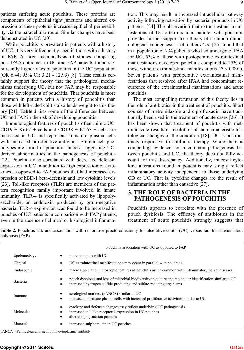 S. Bath et al. / Open Journal of Gastroenterology 1 (2011) 7-12 9 patients suffering acute pouchitis. These proteins are components of epithelial tight junctions and altered ex- pression of these proteins increases epithelial permeabil- ity via the paracellular route. Similar changes have been demonstrated in UC [20]. While pouchitis is prevalent in patients with a history of UC, it is very infrequently seen in those with a history of FAP. A large meta-analysis of studies comparing post-IPAA outcomes in UC and FAP patients found sig- nificantly higher rates of pouchitis in the UC population (OR 6.44; 95% CI: 3.21 - 12.93) [8]. These results cer- tainly support the theory that the pathological mecha- nisms underlying UC, but not FAP, may be responsible for the development of pouchitis. That pouchitis is more common in patients with a history of pancolitis than those with left-sided colitis also len ds weight to this the- ory [21]. Ta bl e 2 lists some of the differences between UC and FAP in the risk of developing pouchitis. Immunological features of pouchitis often mimic UC. CD19 + Ki-67 + cells and CD138 + Ki-67 + cells are increased in UC and represent immature plasma cells with increased proliferative activities. Similar cell phe- notypes are found in pouchitis mucosa suggesting UC- derived abnormalities in the pathogenesis of pouchitis [22]. Pouchitis also correlated with decreased defensin expression in UC in addition to high expr ession of cyto- kines as opposed to FAP pouches that had increased ex- pression of hBD-1 beta-defensin and low cytokine levels [23]. Toll-like receptors (TLR) are members of the pat- tern recognition family important involved in innate immunity. TLR-4 is specifically activated by lipopoly- saccharide, an endotoxin produced by gram-negative bacteria. TLR-4 expression was found to be increased in pouches of UC patients in comparison with FAP patients, even in the absence of clin ical or histological inflamma- tion. This may result in increased intracellular pathway activity following activation by bacterial products in UC patients. [24] The observation that extraintestinal mani- festations of UC often occur in parallel with pouchitis provides further support to a theory of common immu- nological pathogenesis. Lohmuller et al. [25] found that in a population of 734 pa tients who had undergone IPAA for UC, 53% of those with postoperative extraintestinal manifestations developed pouchitis compared to 25% of those without extraintestinal manifestations (P < 0.001). Seven patients with preoperative extraintestinal mani- festations that resolved after IPAA had concomitant re- currence of the extraintestinal manifestations and acute pouchitis. The most compelling refutation of this theory lies in the role of antibio tics in the treatment of pouchitis. Short courses of metronidazole and ciprofloxacin have tradi- tionally been used in the treatment of acute cases [26]. It has been shown that treatment of pouchitis with met- ronidazole results in resolution of the characteristic his- tological changes of the condition [18]. UC is not rou- tinely responsive to antibiotic therapy. While there is compelling evidence for a common pathogenesis be- tween pouchitis and UC, the theory does not fully ac- count for this discrepancy. Additionally, mucosal cyto- kine alterations found in pouchitis may simply reflect inflammatory activity independent to those underlying CD or UC. That is, cytokine changes are the result of inflammation rather than causative [27]. 3. THE ROLE OF BACTERIA IN THE PATHOGENESIS OF POUCHITIS Pouchitis appears to correlate with the presence of pouch dysbiosis. The efficacy of antibiotics in the treatment of acute pouchitis strongly suggests that Table 2. Pouchitis risk and association with restorative procto-colectomy for ulcerative colitis (UC) versus familial adenomatous polyposis (FAP). Pouchitis association with UC a s opposed to FAP Epidemiology more common wit h U C Clinical UC extraintestinal manifestations may occur in parallel with pouchitis Endoscopic macroscopic and microscopic features of pouchitis are in common with inflammatory bowel diseases Bacteria pouch dysbiosis and loss of microbial biodivers ity in culture and molecular identification similar to UC increased hydrogen sulfide-producing and sulfate-reducing organisms Immune serological markers (pANCA) similar to UC increased immature plasma cells with increased proliferative activities similar to UC Molecular cytokine and defensin changes may reflect underlying UC pathogenesis increased toll-like receptor 4 expression in UC pouches altered tight junction proteins Mucosal increased sulphomucin in UC pouches pANCA = Perinuclear anti-neutrophil cytoplasmic antibody. C opyright © 2011 SciRes. OJGas  S. Bath et al. / Open Journal of Gastroenterology 1 (2011) 7-12 10 bacteria play a role in the pathogenesis of the condi- tion. Loss of biodiversity occurs in pouches of UC patients but not in pouches following FAP. UC pouches in contrast to FAP pouches, there was increase in Proteobacteria (P = 0.019), decrease in Bacteroide- tes (P = 0.001) and Faecalibacterium prausnitzii (P = 0.029). Furthermore, bacterial diversity was signifi- cantly greater in UC non-pouchitis compared with UC pouchitis (P = 0.009). [28] Mucosal Clostridiaceae spp colony forming units were significantly increased in patients with recurrent or chronic pouchitis compared to those with no- or single-episode pouchitis (OR:14. 95% CI: 0.887 - 224.021; P = 0.045) [29]. VSL#3, a probiotic, is effective in preventing relapse of chronic pouchitis and may prevent episodes of acute pouchitis [26]. Specific causative bacteria or differential bacte- rial count were not identified in the faeces of patients with or without pouchitis in another study [30]. How- ever, negative findings may reflect older techniques in defining the microbiome. Patients with pouchitis have been found to have a greater anaerobe to aerobe ratio than those without [30,31]. Thus, it may be qualitative rather than quantitative differences in microflora that lead to the development of pouchitis. More recently, a role for sulfate-reducing bacteria has been proposed [32,33]. These bacteria are native to the human colon and produce hydrogen sulfide as a by-product of me- tabolism. Hydrogen sulfide is believed to compete with normal colonic metabolic substrates, leading to disruption of colonocyte metabolism and injury to the intestinal mucosa [32]. The quantity of hydrogen sul- fide gas produced by the pouch contents of patients with active pouchitis is significantly greater than that produced by patients with no history of pouchitis and those receiving antibiotic therapy. In patients with FAP, the quantity of hydrogen sulfide produced is signifi- cantly less than in any group of UC patients [32]. Duffy et al. [33] report that sulfate-reducing bacteria are found in the pouches of patients with UC, but not those with FAP. Thus, this theory accounts for a num- ber of the observed features of pouchitis: the efficacy of antibiotics, the lack of a single identifiable causa- tive organism and the differences in incidence between UC and FAP patients. The chief argument against a bacterial theory of pathogenesis is that there is no correlation between the characteristic histological changes of pouchitis and fecal aerobic or anaerobic coun ts [18]. Future studies examin- ing the relationship between hydrogen sulfide production and mucosal morphology will, no doubt, be valuable. 4. THE ROLE OF OTHER INTRALUMINAL FACTORS Pouch mucosal mucin characteristics appear to differ between UC and FAP pouches. The expression of sul- phomucin is increased in the mucous gel layer of UC compared with FAP pouches. Differential mucin ex- pression favours colonization by different organisms and sulphomucin expression was associated with sul- phate-reducing bacteria and increased chronic inflam- mation [34]. Short-chain fatty acids (SCFAs) are produced by anaerobic bacteria through the fermentation of dietary fibre. They are the principle source of energy for colonocytes and are important for mucosal homeosta- sis. Clausen et al. [35] found that concentrations of SCFAs in the faeces of patients with pouchitis are lower than in those without pouchitis, albeit in a small sample of patients. This finding has led to the hy- pothesis that reduced availability of SCFAs in the ileal pouch plays a role in the development of pouchitis. However, this is unlike ly to be the pr imary mechan ism responsible for pouchitis for a number of reasons. Sandborn et al. [30] quan tified SCFA concen tration s in the faeces of UC patients, both with and without pouchitis, and FAP patients, finding no significant dif- ferences between the groups. This finding calls into question the role of SCFAs in the pathogenesis of pouchitis. Furthermore, it highlights the fact that this theory cannot account for the observed differences in incidence of pouchitis between patients with UC and FAP. Glutamine and butyrate suppositories have been trialled as therapy for chronic pouchitis on the basis that increasing concentrations of SCFAs in the pouch may reduce epithelial permeability, leading to symp- tomatic improvement. The clinical response rate in small, uncontrolled studies has been very low [26], further undermining this hypothesis. Unconjugated bile acids are released from primary bile salts by the actions of anaerobic bacteria. These unconjugated bile acids are toxic to lipid membranes. High levels of unconjugated bile acids have been found in the feces of patients with ileal pouches, as compared to those who have undergone conventional ileostomy [36]. It has been suggested that this increase may predispose to pouchitis but others have not con- firmed these findings. As for SCFAs, the concentra- tions of unconjugated bile acids are no different in UC and FAP patients [30]. If bile acids were the primary pathogenic factor, a similar incidence of pouchitis would be expected in the two groups. Thus, it is unlikely that bile acids are a major pathogenic factor in the development of pouchitis. 5. MULTI-FACTORIAL, MULTI-STEP HYPOTHESIS None of the theories discussed above can explain the development of pouchitis fully on its own merit. Cof- C opyright © 2011 SciRes. OJGas  S. Bath et al. / Open Journal of Gastroenterology 1 (2011) 7-12 11 fey et al. [37] have proposed that pouchitis is the re- sults of a multi-step process rather than anyone single factor. Their unifying theory is based on the following steps: First colonic metaplasia develops in the ileal pouch, which is followed by the production of sul- phomycin by the goblets cells. Sulphomycin then pro- vides the basis for sulfate-reducing bacteria colonisa- tion. Hydrogen sulfide production by sulfate-reducing bacteria may cause apoptosis and reactive crypt cell hyperplasia. Hydrogen sulphide will then also cause infl ammat io n and asso c iated s ympto ms [37]. While the multi-step theory unifies current isolated findings, it does not fully explain the pathogenesis. It remains unclear why greater rates of colonic metapla- sia occur in UC compare to FAP patients. So far only an association between sulfate-reducing bacteria colo- nisation and colonic metaplasia has been found and any causality is speculative. 6. CONCLUSIONS Understanding of the pathogenesis of primary pouchitis remains incomplete. While a number of theories have been proposed, no single one fully explains the histo- logical findings and the efficacy of certain treatments. The evidence for a shared pathological basis between UC and pouchitis is compelling, but it can not explain th e therapeutic benefit of antibiotics. There is mounting evidence that sulfate-reducing bacteria play a major role in the condition, while it seems unlikely that SCFAs or bile acid concentrations are the chief pathological cul- prits. Certainly it seems plausible that all of these factors may contribute to the development of pouchitis, in the context of unknown immunological factors unique to patients with UC. The multi-step process theory pro- posed by Coffey et al. aims to unify the current find ings. Future studies may elicit a unifying link between the single hypotheses and proof or disproof the proposed multi-step model. Regardless, further investigation into the pathogenesis of primary pouchitis is warranted in the search for more efficacious treatments and preventative strategies. REFERENCES [1] Doherty, G.A. and Chiefetz, A.S. (2009) Management of acute severe ulcerative colitis. Expert Review of Gastro- enterology and Hepatology, 3, 395-405. doi:10.1586/egh.09.24 [2] Meagher, A.P., Farouk, R., Dozois, R.R., et al. (1998) Ji- leal pouch-anal anastomosis for chronic ulcerative colitis: Complications and long-term outcome in 1310 patients. British Journal of Surgery, 85, 800-803. doi:10.1046/j.1365-2168.1998.00689.x [3] Soberg, I.C, Lygren, I., Jahnsen, J., et al. (2009) Clinical course during the first ten years of ulcerative colitis: Re- sults from a population-based inception cohort (IBSE Study). Scandinavian Journal of Gastroenterology, 44, 431-440. doi:10.1080/00365520802600961 [4] Somashekar, U., Gupta, S., Soin, A., et al. (2010) Func- tional outcome and quality of life following restorative protocolectomy for ulcerative colitis in Indians. Interna- tional Journal of Colorectal Disease, 25, 967-973. doi:10.1007/s00384-010-0974-x [5] Coffey, J.C., Winter, D.C., Neary, P., et al. (2002) Quality of life after ileal pouch-anal anastomosis: An evaluation of diet and other factors using the Cleveland Global Quality of Life instrument. Diseases of the Colon Rectum, 45, 30-38. [6] Hahnloser, D., Pemberton, J.H., Wolff, B.G. , et al. (2007) Results at up to 20 years after ileal pouch-anal anasto- mosis for chronic ulcerative colitis. British Journal of Surgery, 94, 333-340. doi:10.1002/bjs.5464 [7] Banasiewicz, T., Marciniak, R., Kaczmarek, E., et al. (2011) The prognosis of clinical course and the analysis of the frequency of the inflammation and dysplasia in the intestinal J-pouch at the patients after restorative procto- colectomy due to FAP. International Journal of Colorec- tal Disease, 26, 1197-1203. doi:10.1007/s00384-011-1241-5 [8] Lovegrove, R.E., Tilney, H.S., Heriot, A.G., et al. (2006) A comparison of adverse events and functional outcomes after restorative protocolectomy for familial adenoma- tous polyposis and ulcerative colitis. Diseases of the Co- lon & Rectum, 49, 1293-1306. doi:10.1007/s10350-006-0608-0 [9] Navaneethan, U. and Shen, B. (2010) Diagnosis and management of pouchitis and ileoanal pouch dysfunction. Current Gastroenterology Reports, 12, 485-494. doi:10.1007/s11894-010-0143-y [10] Beliard, A. and Prudhomme, M. (2010) Ileal reservoir with ileo-anal anastomosis: Long-term complications. Journal of Visceral Surgery, 147, E137-E144. doi:10.1016/j.jviscsurg.2010.07.001 [11] Sandborn, W.J., Tremaine, W.J., Batts, K.P., et al. (1994) Pouchitis after ileal pouch-anal anastomosis: A pouchitis disease activity index. Mayo Clinic Procedures, 69, 409-415. [12] Navaneethan, U. and Shen, B. (2010) Secondary pouchi- tis: Those with identifiable etiopathogenetic or triggering factors. American Journal of Gastroenterology, 105, 51-64. doi:10.1038/ajg.2009.530 [13] Madden, M.V., McIntyre, A.S. and Nicholls, R.J. (1994) Double-blind crossover trial of metronidazole versus placebo in chronic unremitting pouchitis. Digestive Dis- eases and Science, 39, 1193-1196. doi:10.1007/BF02093783 [14] Shen, B., Achkar, J.P., Lashner, B.A., et al. (2001) A randomized clinical trial of ciprofloxacin and metroni- dazole to treat acute pouchitis. Inflammatory Bowel Dis- eases, 7, 301-305. doi:10.1097/00054725-200111000-00004 [15] Shen, B., Remzi, F.H., Lopez, A.R. and Queener, E. (2008) Rifaximin for maintenance therapy in antibi- otic-dependent pouchitis. B M C G a s t roe n t ero l og y , 23, 26. doi:10.1186/1471-230X-8-26 [16] Gionchetti, P., Rizzello, F., Venturi, A., et al. (2000) Oral bacteriotherapy as maintenance treatment in patients with C opyright © 2011 SciRes. OJGas  S. Bath et al. / Open Journal of Gastroenterology 1 (2011) 7-12 Copyright © 2011 SciRes. 12 OJGas chronic pouchitis: A double-blind, placebo-controlled tri- al. G astroenterology, 119, 305-309. doi:10.1053/gast.2000.9370 [17] Ferrante, M., D'Haens, G., Dewit, O., et al. (2010) Bel- gian IBD Research Group. Efficacy of infliximab in re- fractory pouchitis and Crohn’s disease-related complica- tions of the pouch: A Belgian case series. Inflammatory Bowel Diseases, 16, 243-249. [18] Kmiot, W.A., Youngs, D., Tudor, R., et al. (1993) Muco- sal morphology, cell proliferation and faecal bacteriology in acute pouchitis. Br itish Journa l of Sur ger y, 80, 1445-1449 . doi:10.1002/bjs.1800801132 [19] Amasheh, S., Dullat, S., Fromm, M., et al. (2009) In- flamed pouch mucosa possesses altered tight junctions indicating recurrence of inflammatory bowel disease. In- ternational Journal of Colorectal Disease, 24, 1149- 1156. [20] Heller, F., Florian, P., Bojarski, C., et al. (2005) Inter- leukin-13 is the key effector Th2 cytokine in ulcerative colitis that affects epithelial tight junctions, apoptosis and cell restitution. Gastroenterology, 129, 550-564. doi:10.1016/j.gastro.2005.05.002 [21] Simchuk, E.J. and Thirlby, R.C. (2000) Risk factors and true incidence of pouchitis in patients after ileal pouch-anal anastomoses. World Journal of Surgery, 24, 851-856. doi:10.1007/s002680010136 [22] Hirata, N., Oshitani, N., Kamata, N., et al . (2008) Prolif- eration of immature plasma cells in pouchitis mucosa in patients with ulcerative colitis. Inflammatory Bowel Dis- eases, 14, 1084-1090. doi:10.1002/ibd.20447 [23] Kiehne, K., Brunke, G. and Wegner, F. (2006) Defensin expression in chronic pouchitis in patients with ulcerative colitis or familial adenomatous polyposis coli. World Journal of Gastroenterology, 12, 1056-1062. [24] de Paiva, N.M., Ayrizono, M.L., Milanski, M., et al. (2011) Differential expression of TLR2, TLR4 and JNK in mucosa of ileal pouches for ulcerative colitis. Is there a role for bacterial antigen pathway in asymptomatic pa- tients? International Journal of Clinical and Experimen- tal Medicine, 4, 179-186. [25] Lohmuller, J.L., Pemberton, J.H., Dozois, R.R., et al. (1990) Pouchitis and extraintestinal manifestations of in- flammatory bowel disease after ileal pouch-anal anasto- mosis. Annals of Surgery, 211, 622-627. [26] Holubar, S.D., Cima, R.R., Sandborn, W.J., et al. (2010) Treatment and prevention of pouchitis after ileal pouch-anal anastomosis for chronic ulcerative colitis. Cochrane Database of Systematic Reviews, 6, CD00- 1176. [27] Schmidt, C., Giese, T., Ludwig, B., Menges, M., Schil- ling, M., Meuer, S.C., Zeuzem, S. and Stallmach, A. (2006) Increased cytokine transcripts in pouchitis reflect the degree of inflammation but not the underlying entity. International Journal of Colorectal Disease, 21, 419-426. doi:10.1007/s00384-005-0024-2 [28] McLaughlin, S.D., Walker, A.W., Churcher, C. et al. (2010) The bacteriology of pouchitis: A molecular phylogenetic analysis using 16S rRNA gene cloning and sequencing. Annals of Surgery, 252, 90-98. doi:10.1097/SLA.0b013e3181e3dc8b [29] Scarpa, M., Grillo, A., Pozza, A., et al. (2011) TLR2 and TLR4 up-regulation and colonization of the ileal mucosa by Clostridiaceae spp. in chronic/relapsing pouchitis. Journal of Surgery Research, 169, E145-154. doi:10.1016/j.jss.2011.04.003 [30] Sandborn, W.J., Tremaine, W.J., Batts, K.P., et al. (1995) Fecal bile acids, short-chain fatty acids, and bacteria after ileal pouch-anal anastomosis do not differ in patients with pouchitis. Digestive Diseases and Sciences, 40, 1474-1483. doi:10.1007/BF02285195 [31] Nasmyth, D.G., Godwin, P.G., Dixon, M.F., et al. (1989) Ileal ecology after pouch-anal anastomosis or ileostomy. A study of mucosal morphology, fecal bacteriology, fecal volatile fatty acids, and their interrelationship. Gastroen- terology, 96, 817-824. [32] Ohge, H., Furne, J.K., Springfield, J., et al. (2005) Asso- ciation between fecal hydrogen sulphide production and pouchitis. Diseases of the Colon & Rectum, 48, 469-475. doi:10.1007/s10350-004-0820-8 [33] Duffy, M., O’Mahoney, L., Coffey, J.C., et al. (2002) Sulfate-reducing bacteria colonise pouches formed for ulcerative colitis but not familial adenomatous polyposis. Diseases of the Colon & Rectum, 45, 384-388. doi:10.1007/s10350-004-6187-z [34] Bambury, N., Coffey, J.C., Burke, J., Redmond, H.P. and Kirwan, W.O. (2008) Sulphomucin expression in ileal pouches: Emerging differences between ulcerative colitis and familial adenomatous polyposis pouches. Diseases of the Colon & Rectum, 51, 561-567. doi:10.1007/s10350-008-9200-0 [35] Clausen, M.R., Tvede, M. and Mortensen, P.B. (1992) Short-chain fatty acids in pouch contents from patients with and without pouchitis after ileal pouch-anal anas- tomosis. Gastroenterology, 103, 1144-1153. [36] Nasmyth, D.G., Johnston, D., Williams, N.S., et al. (1989) Changes in the absorption of bile acids after total colec- tomy in patients with ileostomy or pouch-anal anastomo- sis. Diseases of the Colon & Rectum, 32, 230-234. doi:10.1007/BF02554535 [37] Coffey, J.C., Rowan, F., Burke, J., et al. (2009) Patho- genesis of and Unifying Hypothesis for Idiopathic Pouchitis. American Journal of Gastroenterology, 104, 1013-1023. doi:10.1038/ajg.2008.127 |