

Paper Menu >>
Journal Menu >>
 Open Journal of Gastroenterology, 2011, 1, 23-27 doi:10.4236/ojgas.2011.12004 Published Online November 2011 (http://www.SciRP.org/journal/ojgas/ OJGas ). Published Online November 2011 in SciRes. http://www.scirp.org/journal/OJGAS HBs antigene prevalence in blood donors and the risk of transfusion of hepatitis B at the central hospital of yaounde, cameroon Dominique Noah Noah1, Richard Njouom 2, Aimé Bonny 3, Pirsou1, J. Meli4, M. Biwole Sida5 1Central Hospital of Yaounde, Gastroenterology Service, Yaounde, Cameroon; 2Centre Pasteur of Cameroon, Virology Laboratory, Ya ounde, Cameroon; 3Saint Camille Hospital, Cardiology Service, Bry sur Marne, Pitié Salpêtrière Hospital, Paris, France; 4Faculty of Medicine and Biomedical Sciences, University of Yaoundé 1, Yaoundé, Cameroon; 5Central Hospital of Yaoundé, Faculty of Medicine and Biomedical Sciences, University of Yaoundé 1, Ya oundé, Cameroon. Email: [email protected] Received 17 September 2011; revised 25 October 2011; accepted 3 November 2011. ABSTRACT Parenteral transmission of hepatitis B virus (HBV) during blood transfusion is not insignificant. Al- though blood transfusion safety has greatly im- proved over the last 15 years, the transfusion risk of HBV remains high in developing countries. In the context where blood transfusion safety is limited in some hospitals in Cameroon, the development of good and quality practice for blood donation, based on the use of the most sensitive techniques for the detection of infectious risk of blood donation should be a priority of health authorities. The aim of this paper is to document the epidemiology of HBV in- fection in the population of blood donors and to assess the risk of infection during blood transfu- sions at the Central Hospital of Yaoundé (CHY), Cameroon. Methods: During a seven months period, 1000 volunteer donors were recruited prospectively at the blood bank of the CHY. Those included in the study were people aged from 18 years to 55 years without any particular medical history. Data collec- tion was done through an investigation form. Sam- ples were first analysed at the CHY and then at Centre Pasteur of Cameroon. Results: Of the 1000 samples tested 108 (10.8%) were positive for HBs Ag. The male and the female sex represented 83.1% and 16.9% respectively. According to the age groups, 56.2% were 18 to 27 years, 30.5% were 28 years to 37 years, 10.4% were 38 years to 47 years and 29% were 48 years to 55 years. The 892 nega- tive sera were analyzed for total anti-HBc antibod- ies of which 75.78% were positive and 24.56% negative. The 676 samples positive for anti-HBc antibody were retested for HBs Ag using enzyme immunoassay with a confidence level of 95%, be- tween 52 and 82 positive tests were still positive, a proportion that vary between 7.64% and 12.14% (9.89% ± 2.25%). Conclusion: The prevalence rate of 10.8% found in our series is consistent with data in Cameroon. The infectious risk of transmission of HBV among blood donors remains a major problem (9.89% ± 2.25%) related to the test used. Keywords: Infectious Risk; HBV; CHY 1. INTRODUCTION Viral hepatitis B is a major public health problem in the world where the number of people infected is estimated at 2 billion and 350 million for chronic carriers. The B virus is responsible for approximately 500,000 deaths annually and 75% of cases of hepatocellular carcinoma [1,2]. The hepatitis B virus (HBV) is the second most common carcinogen agent known to humans after smok- ing [3]. HBV is also the first cause of hepatocellular carcinoma in South-East and Sub-Saharan Africa [4]. In Cameroon, HBV is by far considered as the leading cause of liver cirrhosis before chronic alcoholism [5]. The most common means of transmission of HBV are from mother to child, parenteral and sexual transmission. Besides these means of transmission, parenteral trans- mission of HBV during blood transfusion is not insig- nificant. In Greece, a study carried out in 1996 showed that the rate of post transfus ion hepatitis was higher than the prevalence of hepatitis C [6]. In Sub-Saharan Africa, the prevalence of HBs Ag among blood donors ranges between 9.2% in the DRC [7] and 10.7% in Cameroon [8]. The transfusion risk is even higher if the donation is made during the window period when the HBV infection  D. Noah Noah et al. /Open Journal of Gastroenterology 1 (2011) 23-27 24 cannot be detected by conventional screening techniques. This risk is considerably reduced by adding to the sero- logical screening, a viral genome screening. In this con- text, routine laboratory diagnosis appears to be a means of preventing HBV infection risk to be implemented in the frame work of blood safety that stands as a strong aspect to minimize the transmission post transfusion hepatitis [6]. This blood safety has greatly improved over the last 15 years in industrialized countries, due to the development of more efficient and innovative screening techniques. The prevalence rates of transfused viral infections have significantly decreased in these countries to reach negligible levels. Generally, in devel- oping countries and in sub-Saharan Africa in particular, the registered prevalence reflects some deficiencies re- lated to blood safety in blood banks and blood centres of hospitals. While most infections are detected inciden- tally during a blood donation, for example, other infec- tions are diagnosed in the hospital at the beginning of a complication. In Cameroon, the prevalence of HBV infection among blood donors is esti mated at 10.7% [8 ]. In this contex t of limited blood safety, the development of good and qual- ity practice of blood donation based on the use of the most sensitive techniques for detecting the risk of infec- tion should be a health priority being aware of the fact that approximately 14,000 blood donations are made annually at the Central Hospital of Yaoundé (Sources blood bank). In this paper, we propose to document the epidemiology of HBV infection in Cameroon by deter- mining the prevalence of HBs Ag in the population of blood donors and assessing the risk of infection of HBV during blood transfusions at the Central Hospital of Yaoundé (CHY), Cameroon. 2. MATERIALS AND METHODS A prospective study carried out between July 2007 and January 2008 (7 months) was used to examine blood samples from voluntary donors recruited at the blood bank of the CHY. Were included in the study participan ts with the following criteria: Age between 18 and 55 years; Took a meal before donating blood; Blood test before blood donation; Date of last blood donation > 3 months; Date of the last or next menses > 10 days; Satisfactory or normal physical condition; Body weight > 50 kg. Was excluded from the study, p articipants who did not meet the inclusion criteria listed above and those with the following peculiarities: Lactating Women; Patient who had undergone a surgery; Patient who had under- gone a blood transfusion during the last twelve months, Patient with chronic disease (hypotension or hyperten- sion, diabetes, heart disease, asthma, epilepsy, etc.); Pa- tient in poor physical condition (emaciation, weakness, jaundice, dyspnoea, pallor, obesity, etc.). Data was collected from the patients recruited in the study using a questionnaire after having read and signed informed consent. The random sampling method was used. The minimum sample size was calculated accord- ing to the Lorenz formula with a prevalence of HBV in the general population estimated at 14.85%. The detec- tion of HBs Ag in all blood samples was performed at the CHY laboratory using the immuno-chromatographic technique (Hexagon® HBs Ag). Sera samples testing negative immuno-chromatographic HEXAGON® tech- nique were further tested in the Virology Laboratory of Centre Pasteur of Cameroon using the Monolisa® tech- nique for the detection of anti-HBc antibodies. Finally, the samples positive for anti-HBc antibodies were re- tested for the detection of HBs Ag by enzyme immuno- assay (ELISA Monolisa® HBs Ag Ultra). Data entry was done using the Excel® 2007. Descriptive analysis was performed through the calculation of proportions for qualitative variables (frequency, percentage), means and standard deviations for continuous variables. The differ- ent frequency comparisons were made using Chi-square test (x2) of Pearson and Fisher test when necessary. 3. RESULTS 3.1. Characteristics of Donors From July 2007 to January 2008, a total of 1000 blood donors were recruited prosp ectively at the CHY, with an average daily recruitment of 6.49 volunteers. Old donors were 825 with 82.5% of donations and new donors 175 with 17.5% of the donations. According to sex we had 831 (83.1%) men and 169 (16.9%) women. The most represented age group was 18 years - 27 years with 562 (56.2%) donors, followed by the 28-37 years age gro up with 305 (30.5%) dono rs and that of 38 years - 47 years with 104 (10.4%) donors; the least rep- resented being the 48 year s - 55 years age group with 29 (2.9%) donors (Table 1). 3.2. Prevalence of HBs Antigen by the Rapid Immuno-Chromatographic Technique Of the 1000 donor samples tested in the CHY, 108 were positive to the rapid test (Hexagon® HBs Ag), with a se- roprevalence of 10.80% ± 1.92%. Male donors (95% or 87.96%) were more infected than female (13% or 12.04%) (P < 0.005). The prevalence of HBs Ag according to age groups was as follows: 18 years - 27 years: 63 (58.33% ± 1.92%); 28 years - 37 years: 36 (33.33% ± 1.92) and 38 years - 47 years 9 (08.33% ± 1.92 % ). The 48 years - 55 years age group was not represented in this seroprevalence. C opyright © 2011 SciRes. OJGas 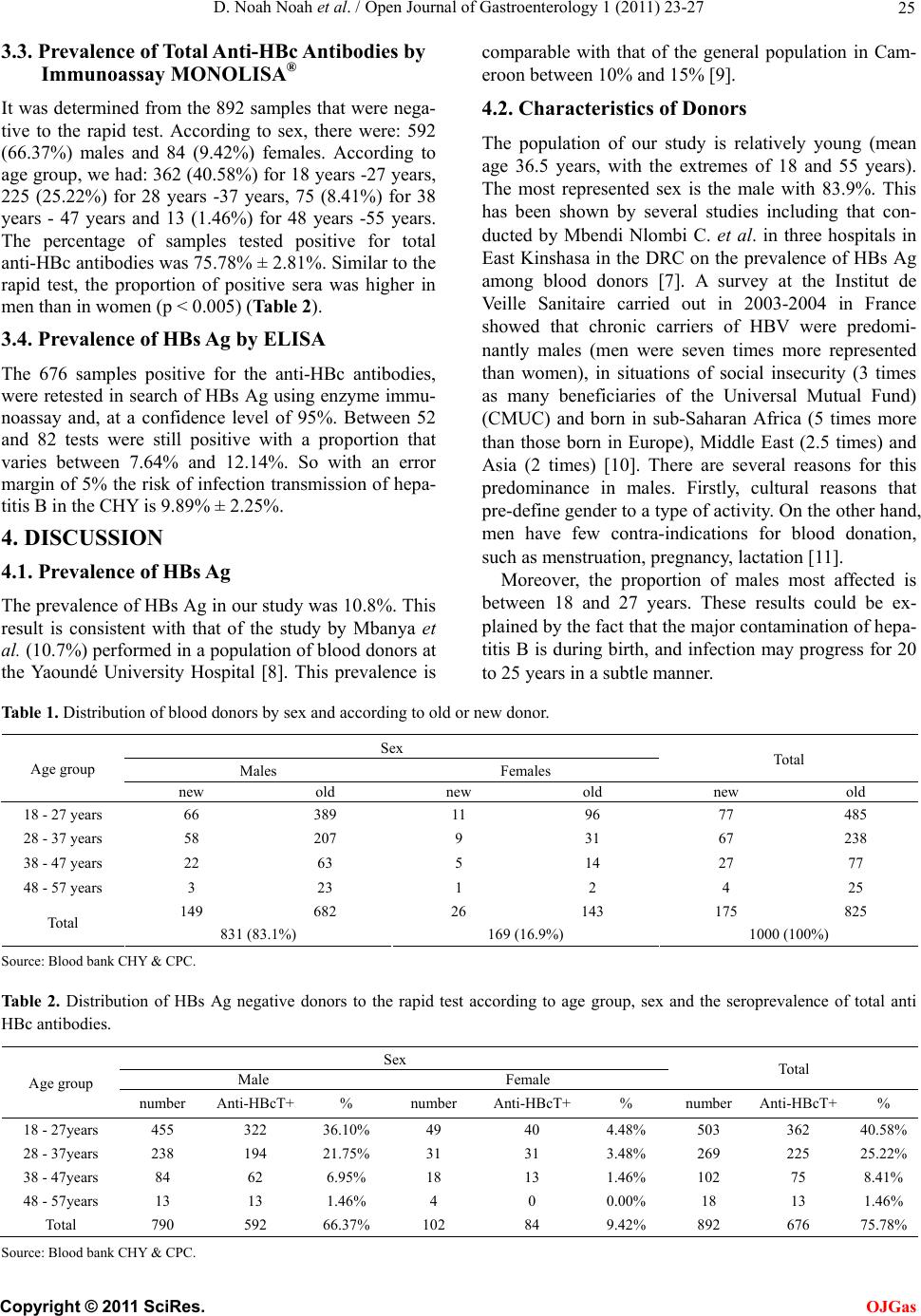 D. Noah Noah et al. / Open Journal of Gastroenterology 1 (2011) 23-27 Copyright © 2011 SciRes. 25 OJGas 3.3. Prevalence of To tal Anti-HBc Antibodies by Immunoassay MONOLISA® It was determined from the 892 samples that were nega- tive to the rapid test. According to sex, there were: 592 (66.37%) males and 84 (9.42%) females. According to age group, we had: 362 (40.58%) for 18 years -27 years, 225 (25.22%) for 28 years -37 years, 75 (8.41%) for 38 years - 47 years and 13 (1.46%) for 48 years -55 years. The percentage of samples tested positive for total anti-HBc antibodies was 75.78% ± 2.81%. Similar to th e rapid test, the proportion of positive sera was higher in men than in women (p < 0.005) (Table 2). 3.4. Prevalence of HBs Ag by ELISA The 676 samples positive for the anti-HBc antibodies, were retested in search of HBs Ag using enzyme immu- noassay and, at a confidence level of 95%. Between 52 and 82 tests were still positive with a proportion that varies between 7.64% and 12.14%. So with an error margin of 5% the risk of infection transmission of hepa- titis B in the CHY is 9.89% ± 2.25%. 4. DISCUSSION 4.1. Prevalence of HBs Ag The prevalence of HBs Ag in our stud y was 10.8 %. This result is consistent with that of the study by Mbanya et al. (10.7%) performed in a population of blood donors at the Yaoundé University Hospital [8]. This prevalence is comparable with that of the general population in Cam- eroon between 10% and 15% [9]. 4.2. Characteristics of Donors The population of our study is relatively young (mean age 36.5 years, with the extremes of 18 and 55 years). The most represented sex is the male with 83.9%. This has been shown by several studies including that con- ducted by Mbendi Nlombi C. et al. in three hospitals in East Kinshasa in the DRC on the prevalence of HBs Ag among blood donors [7]. A survey at the Institut de Veille Sanitaire carried out in 2003-2004 in France showed that chronic carriers of HBV were predomi- nantly males (men were seven times more represented than women), in situations of social insecurity (3 times as many beneficiaries of the Universal Mutual Fund) (CMUC) and born in sub-Saharan Africa (5 times more than those born in Europe), Middle East (2.5 times) and Asia (2 times) [10]. There are several reasons for this predominance in males. Firstly, cultural reasons that pre-define gender to a type of activity. On the other hand, men have few contra-indications for blood donation, such as menstruation, pregnancy, lactation [11]. Moreover, the proportion of males most affected is between 18 and 27 years. These results could be ex- plained by the fact that the major contamination of hepa- titis B is during birth, and infection may progress for 20 to 25 years in a subtle manner. Table 1. Distribution of blood donors by sex and according to old or new donor. Sex Males Females Total Age group new old new old new old 18 - 27 years 66 389 11 96 77 485 28 - 37 years 58 207 9 31 67 238 38 - 47 years 22 63 5 14 27 77 48 - 57 years 3 23 1 2 4 25 149 682 26 143 175 825 Total 831 (83.1%) 169 (16.9%) 1000 (100%) Source: B l ood bank CHY & CPC. Table 2. Distribution of HBs Ag negative donors to the rapid test according to age group, sex and the seroprevalence of total anti HBc antibodies. Sex Male Female Total Age group number Anti-HBcT+ % number Anti-HBcT+ % number Anti-HBcT+% 18 - 27years 455 322 36.10% 49 40 4.48% 503 362 40.58% 28 - 37years 238 194 21.75% 31 31 3.48% 269 225 25.22% 38 - 47years 84 62 6.95% 18 13 1.46% 102 75 8.41% 48 - 57years 13 13 1.46% 4 0 0.00% 1 8 13 1.46% Total 790 592 66.37% 102 84 9.42% 892 676 75.78% Source: B l ood bank CHY & CPC.  D. Noah Noah et al. /Open Journal of Gastroenterology 1 (2011) 23-27 26 According to the criterion of old or new donor , we had 82.5% of old donors and 17.5% of new donors. These results can be explained by the fact that the CHY has successfully implemented a loyalty program for volun- tary donors program with the help of the Red Cross. Otherwise, the major reason for blood donation is to support a friend or family member [11]. 4.3. Transmission and Evolution of HBV Diop T. et al. [12] in a study in Senegal showed that ver- tical transmission from mother to child represents 50 to 75% of all transmissions of the B virus. Other factors equally play a role during birth: the use of contaminated material during clamping of the umbilical cord, circum- cision and scarification rituals. The major problem of infection with hepatitis B virus is the risk of developing chronic complications. If 5% - 10% of immunocompe- tent adults are likely to become chronic carriers after infection, this percentage is much higher among infants born to mothers carrying HBs Ag (close to 90%). In case of prenatal contamination immune tolerance phase is extended and the transition to the immune response phase is rare and late. Half of chronic carriers develop cirrhosis and/or with a risk of progression to HCC, which varies from 2% to 3% per year [6]. Some studies have shown that in chronic carriers of HBs Ag, HCC is likely to occur in approximately 40 % of cases before the stage of cirrhosis [4,12]. 4.4. Role of Blood Transfusion Blood transfusion is a major risk of HBV infection in most health facilities in Cameroon, first because of the increased demand for blood to treat certain emergency conditions such as acute anemia due to malaria, sickle cell anemia, traffic accidents (with the current inflation motorcycle taxis), different gynecology bleeding... and also the lack of blood banks in most rural health facili- ties. If the blood safety techniques have infinitely re- duced the risk of contamination through transfusion in developed countries, this progress must be weighed against the continuous emergence of new pathogens po- tentially transmissible during transfusion and the dispar- ity with safety blood transfusions in developing coun- tries where resources are insufficient for a baseline screening for infectious diseases in donors. 4.5. Risk of Infection Transmission In this study we aimed at evaluating in a central level university hospital, the risk of infection transmission after blood transfusion related to the type of reagent usually used. We did not consider patient samples that tested positive since they were immediately eliminated. The infectious risk of transmission of hepatitis B thro ugh transfusion of a pine of blood declared negative to HBs Ag was 9.89% ± 2.25% at a confidence level of 95%. This rate is close to the prevalence of HBs Ag in the general population of Cameroon [9]. Considering the fact that the blood bank of the CHY delivers an average more than 14,000 pines of blood yearly, this implies that there are between 1070 and 1670 induced infections of HBs Ag annually. The Immunochromatographic method (rapid test) used in the CHY is less sensitive than the immunoassay used at Centre Pasteur of Cameroon (CPC) which served as a reference test or immunoradiometry (IRMA) recommended in countries such as Tunisia [13]. The 892 sera that tested negative for HBs Ag were re- tested by the immunoenzymatic method for the detection of anti-HBc antibodies. Two hundred and sixteen of these tests were negative for anti-HBc antibodies corre- sponding to naive subjects in whom vaccination was recommended. The interpretation of 676 samples had to undergo a search for HBs Ag by a more sensitive method as was the case in our study to detect false negatives. The consensus conference of the Canadian Associa- tion for the Study of Liver (CASL) [14] held in Toronto on 4 and 5 January 2007 made routine screening and monitoring of patients by using PCR methods a recom- mendation. The aim was not only to achieve a high de- gree of sensitivity of screening but also to harmonize the monitoring of these patients. In all cases, the interpreta- tion of anti-HBc antibodies should suggest three scenar- ios. It could be a self-resolving HBV infection with un- detectable levels of anti-HBs antibodies. In fact several years after an acute HBV infection, the anti-HBs anti- bodies may become undetectable indicating a resolution of the condition with the persistence of anti-HBc only, which is the most consistent marker of a previous con- tact with virus [15]. Secondly, the lack of detection of markers other than anti-HBc antibodies may also corre- spond to a certainty of hepatitis B [15]. This is also the case in our study where we found an infection rate of hepatitis B ranging from 7.64% to 12.14%. FKI-Berrajah L. et al., in his work has shown that during the evolution of chronic hepatitis B, the rate of HBs Ag could drop to very low levels which would make it undetectable. The spontaneous loss of HBs Ag is described in 0.5 to 5% of patients with chronic hepatitis B [13]. Finally the detec- tion of anti-HBc antibodies in the absence of detection of another marker could represent false positive results. In fact the use of immunoradiometric method for 160 sera previously tested for anti-HBc antibody by ELISA showed negative results on 19 sera su ggesting the possi- bility of false positive results [16,17]. 5. CONCLUSIONS The prevalence ra te of 10.8% foun d in our study popula- C opyright © 2011 SciRes. OJGas  D. Noah Noah et al. / Open Journal of Gastroenterology 1 (2011) 23-27 Copyright © 2011 SciRes. 27 [8] Mbanya, D.N., Takam, D. and P Ndumbey, P.M. (2003) Serological findings amongst first-time blood donors in Yaounde, Cameroon: Is safe donation a reality or a myth? Transfusion Médecine, 13, 267-273. doi:10.1046/j.1365-3148.2003.00453.x tion is consistent with data in Camero on (between 8 and 12%). The risk of transmission of HBV among blood donors remains a major problem at the CHY, 9.89% ± 2.25% to a confidence level of 95%. Considering the natural history of hepatitis B, especially its evolution to chronic liver cirrhosis and hepatocellular carcinoma, blood banks should be established in most hospitals and especially assuring safety of blood transfusion using immunoradiometric (IRMA) or immunoassay as in most countries. [9] Zekeng, L. and Kaptue, L. (1990) Serologie HIV et portage de l’antigene HBs et Hbe chez les donneurs de sang au CHU de Yaounde Cameroun. Annales de la Societé Belge Médecine Tropicale, 70, 49-53. [10] Meffre, C., Le Strat, Y., Delarocque-Astagneau, E., Lemasson, J.M., Coste, D., Steinmetz, J., Antona, D., Dubois, F., Warszawski, J., Meyer, J.F., Leiser, S., Gio- rdanella, J.P., Gueguen, R., Landau, A. and Desenclos, J.C. (2006) Prevalence of hepatitis B in France. 41th EASL Annual Meeting, 26-36 April, Vienne, 2003-2004. REFERENCES [11] Ouattara, S.A., Meite, M., Akran, V., Gody, M. and Manlan, L.K. (1990) The G. Increase of the prevalence of hepatitis B virus surface antigen related to immunodefi- ciency inherent in acquired immune deficiency syndrome (AIDS). Journal of Acquired Immune Deficiency Syn- dromes, 3, 282-286. [1] Frexinos, J., Buscail, L., et al. (2003) Hépato-gastro- entérologie proctologie. MASSON, Paris, 5, 489. [2] Trepo, C. and Zoulim, F. (2009) Traitement de l’hépatite B: Progrès attendus. Gastroenterology Clinical Biology, 33, 811-817. [3] Parkin, D.M. (2001) Global cancer statistics in the year. Lancet Oncology, 2, 533-543. doi:10.1016/S1470-2045(01)00486-7 [12] Moreira Diop, T. (2003) Atelier sur introduction des nouveaux vaccins dans PEV en Afrique francophone, révision des connaissances et considération des besoins en formation. Saly, Sénégal. [4] Hill, C. (2008) Doyon. La fréquence des cancers en France en 2005 : Évolution de la mortalité depuis 1950 et résumé du rapport sur les causes de cancer. Bull Cancer, 95, 5-10. [13] Berrajah-Fki, L., Ben Halima, S., Guermazi, F., Ham- mami, A. and Karray-Hakim, H. (2006) Signification du profil sérologique “anticorps anti HBc isolés” chez 284 patients suspects d’être infectés par le virus de l’hépatite B. Annual Biolog y Clinical, 64, 451-455. [5] Njonou, J., Kupfer, B., Kochan, B., Zekeng, L., Kaptue, L. and Matz, B. (2002) Hepatitis C virus infection and genotypes among human immunodeficiency virus high- risk group in Cameroon. Journal of Medical Vi rology, 66, 179-186. doi:10.1002/jmv.2128 [14] Morris, S., Stephen, S., Kelly, B., et al. (2007) Prise en charge de l’hépatite B chronique: Lignes directrices consensuelles. Conférence 4-5, Toronto, Canada. [15] Degos, F. and Hépatite, B. (2004) Qui et quand vacciner? Tiré à part, Post U FMC-HGE 3-4, L26, 47-51. [6] Zerbou, E.K., Dalekos, G.N., Boumba, D.S. and Tsianos, E.V. (2001) Va lue of anti-HBc screening of blood donors for prevention of HBV infection: results of a 3 year pro- spective study in Northwestern Greece. Transfusion, 41, 652-658. doi:10.1046/j.1537-2995.2001.41050652.x [16] Caspari, G., Beyer, H.J., Elbert, G., et al. (1989) Unsatis- factory specificities and sensitivities of six enzyme im- munoassays for antibodies to hepatitis B core antigen. Journal of clinical Microbiology, 27, 2067-2072. [7] Mbendi Nlombi, C., Longo-Mbenza, B., Mbendi Nsukini, S., Muyembe Tamfum, J.J., Situakibanza Nanituma, H. and Vangu Ngoma, D. (2001) Prévalence du vih et l’an- tiggène HBs chez les donneurs de sang, risque résiduel de contamination chez les receveurs de sang à Kin- shasa-Est, République Démocratique du Congo. Trop- Médecine Tropicale, 61, 139-142. [17] Hofer, M., Joller Jemelka, H.I., Grob, P.J. and Lüthy, R. (1998) Opravil mand the swiss HIV cohort study. Fre- quent chronic hepatitis B virus infection in HIV-infected patient positive for antibody to hepatitis B core antigen only. European Journal of Clinical Microbiology and In- fectious, 17, 6-13. doi:10.1007/BF01584356 OJGas |