

Paper Menu >>
Journal Menu >>
 Surgical Science, 2011, 2, 442-445 doi:10.4236/ss.2011.29096 Published Online November 2011 (http://www.SciRP.org/journal/ss) Copyright © 2011 SciRes. SS Novel Suture-Less Vascular Anastomotic Device (BY Fi x) in Femoro-Popliteal Surgery, Early Clinical Experience M. Schneider1, V. Shapira2, V. Chernyavskiy3, B. Yoffe4 1Department of Vascul a r Sur gery, Erfurt Helios Clinic, Erfurt, Germany 2Department of Neurosurgery, RAMBAM Medical Center, Haifa, Israel 3Department of Vascul a r Sur gery, General Hospital, Irkutsk, Russia 4Department of Vascul a r Sur gery, Barzilay Hospital, Ashkelon, Israel E-mail: [email protected] Received July 28, 2011; revi sed Octobe r 13, 2011; accepted October 31, 2011 Abstract Objectives: To evaluate the early experience of using the BYFix-innovative anastomotic device for creating suture-less vascular anastomosis in major and peripheral arteries surgery. Design: Uncontrolled prospective study. Materials: The BYFix anastomotic device for suture-less vascular anastomosis with surgical tools and standard vascular grafts. Methods: 7 patients, age 63.6 ± 9.2 years, with peripheral vascular occlusion above the knee scheduled for surgical repair were operared. They underwent the surgical procedure using the BYFix anastomotic device for creating proximal anastomosis and the conventional manual suturing for cre- ating the distal anastomosis of the implantable vascular graft. Results: The anastomoses by using BYFix anastomotic device were successfully created in all patients. The duration of anastomosis creation was sig- nificantly shorter by using BYFix device, compared to conventional manual suturing, 5:10 ± 1:50 minutes compared to 33 ± 17:40 minutes respectively. No adverse events related to BYFix anastomotic device were observed, during the surgical procedure or recovery period. One year follow up revealed no complications related to BYFix anastomotic device. Conclusions: The BYFix anastomotic device enables the creation of efficient vascular anastomosis in peripheral vascular occlusions. It shortens the time needed for creating vascular anastomosis and thus reducing the operation time and might reduce distal complications related to the vascular procedure. Further clinical trials are needed to establish the results. Keywords: Anastomosis, Peripheral Vascular Surgery, BYFix Device 1. Introduction Lower extremity peripheral arterial disease affects a large proportion of most adult populations worldwide [1]. The prevalence of lower extremity peripheral arterial disease has been defined by a series of epidemiological investigations, that have used either claudication as a symptomatic marker of lower extremity peripheral arte- rial disease, or an abnormal ankle-to-brachial systolic blood pressure to define the population affected [2]. Patients with symptomatic PAD have significantly re- duced mobility and poor quality of life, claudicating re- sults in a reduced physical component quality of life score-lower than for arthritis, cancer or chronic lung dis- ease [3]. Furthermore, PAD patients were more likely to have widespread atheromatous disease in multiple vascular beds (62%), than patients with either CHD (25%) or stroke (40%) [4]. As the presence of PAD is a red alert for the existence of widespread atherothrombosis it should provide a powerful stimulus for action. However, despite these compelling statistics, PAD remains under diagnosed and patients often do not get proper care until the associated heart disease becomes evident. Progres- sion of PAD can result in critical limb ischemia, mani- fested by ischemic pain at rest or in breakdown of the skin (ulcers or gangrene). Severe PAD can result in tis- sue ischemia in the lower extremities, causing gangrene and requiring amputation in 3% to 8% of patients [5,6]. Aggressive investigation for revascularization options, either surgical or percutaneous, are warranted in such instances. Only a small proportion of patients with severe disease ultimately require a major amputation. In managing PAD, it is critically important to deal 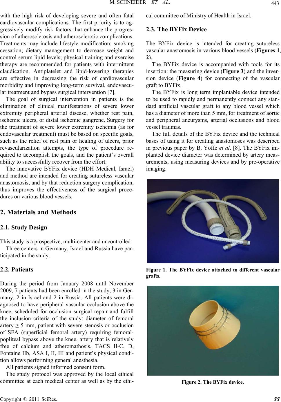 M. SCHNEIDER ET AL. 443 with the high risk of developing severe and often fatal cardiovascular complications. The first priority is to ag- gressively modify risk factors that enhance the progres- sion of atherosclerosis and atherosclerotic complications. Treatments may include lifestyle modification; smoking cessation; dietary management to decrease weight and control serum lipid levels; physical training and exercise therapy are recommended for patients with intermittent claudication. Antiplatelet and lipid-lowering therapies are effective in decreasing the risk of cardiovascular morbidity and improving long-term survival, endovascu- lar treatment and bypass surgical intervention [7]. The goal of surgical intervention in patients is the elimination of clinical manifestations of severe lower extremity peripheral arterial disease, whether rest pain, ischemic ulcers, or distal ischemic gangrene. Surgery for the treatment of severe lower extremity ischemia (as for endovascular treatment) must be based on specific goals, such as the relief of rest pain or healing of ulcers, prior revascularization attempts, the type of procedure re- quired to accomplish the goals, and the patient’s overall ability to successfully recover from the effort. The innovative BYFix device (HDH Medical, Israel) and method are intended for creating sutureless vascular anastomosis, and by that reduction surgery complication, thus improves the effectiveness of the surgical proce- dures on various blood vessels. 2. Materials and Methods 2.1. Study Design This study is a prospective, multi-center and uncontrolled. Three centers in Germany, Israel and Russia have par- ticipated in the study. 2.2. Patients During the period from January 2008 until November 2009, 7 patients had been enrolled in the study, 3 in Ger- many, 2 in Israel and 2 in Russia. All patients were di- agnosed to have peripheral vascular occlusion above the knee, scheduled for occlusion surgical repair and fulfill the inclusion criteria of the study: diameter of femoral artery ≥ 5 mm, patient with severe stenosis or occlusion of SFA (superficial femoral artery) requiring femoral- popliteal bypass above the knee, artery that is relatively free of calcium and atheromathosis, TACS II-C, D, Fontaine IIb, ASA I, II, III and patient’s physical condi- tion allows performing general anesthesia. All patients signed informed consent form. The study protocol was approved by the local ethical committee at each medical center as well as by the ethi- cal committee of Ministry of Health in Israel. 2.3. The BYFix Device The BYFix device is intended for creating sutureless vascular anastomosis in various blood vessels (Figures 1, 2). The BYFix device is accompanied with tools for its insertion: the measuring device (Figure 3) and the inver- sion device (Figure 4) for connecting of the vascular graft to BYFix. The BYFix is long term implantable device intended to be used to rapidly and permanently connect any stan- dard artificial vascular graft to any blood vessel which has a diameter of more than 5 mm, for treatment of aortic and peripheral aneurysms, arterial occlusions and blood vessel traumas. The full details of the BYFix device and the technical bases of using it for creating anastomoses was described in previous paper by B. Yoffe et al. [8]. The BYFix im- planted device diameter was determined by artery meas- urements, using measuring devices and by pre-operative imaging. Figure 1. The BYFix device attached to different vascular grafts. Figure 2. The BYFix device. Copyright © 2011 SciRes. SS 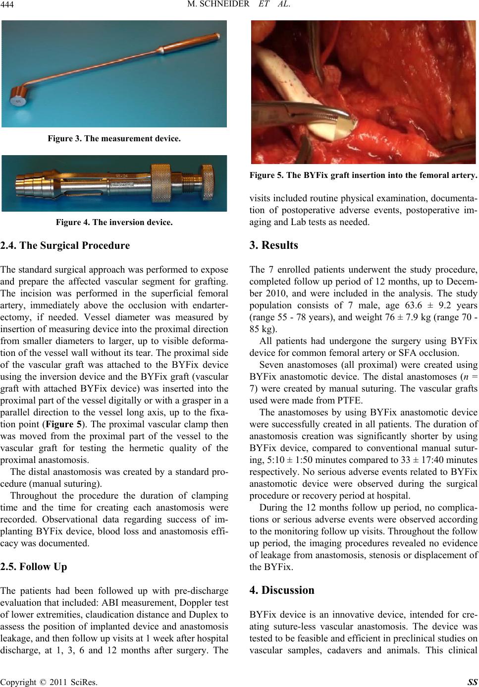 444 M. SCHNEIDER ET AL. Figure 3. The measurement device. Figure 4. The inversion device. 2.4. The Surgical Procedure The standard surgical approach was performed to expose and prepare the affected vascular segment for grafting. The incision was performed in the superficial femoral artery, immediately above the occlusion with endarter- ectomy, if needed. Vessel diameter was measured by insertion of measuring device into the proximal direction from smaller diameters to larger, up to visible deforma- tion of the vessel wall without its tear. The proximal side of the vascular graft was attached to the BYFix device using the inversion device and the BYFix graft (vascular graft with attached BYFix device) was inserted into the proximal part of the vessel digitally or with a grasper in a parallel direction to the vessel long axis, up to the fixa- tion point (Figure 5). The proximal vascular clamp then was moved from the proximal part of the vessel to the vascular graft for testing the hermetic quality of the proximal anastomosis. The distal anastomosis was created by a standard pro- cedure (manual suturing). Throughout the procedure the duration of clamping time and the time for creating each anastomosis were recorded. Observational data regarding success of im- planting BYFix device, blood loss and anastomosis effi- cacy was documented. 2.5. Follow Up The patients had been followed up with pre-discharge evaluation that included: ABI measurement, Doppler test of lower extremities, claudication distance and Duplex to assess the position of implanted device and anastomosis leakage, and then follow up visits at 1 week after hospital discharge, at 1, 3, 6 and 12 months after surgery. The Figure 5. The BYFix graft insertion into the femoral artery. visits included routine physical examination, documenta- tion of postoperative adverse events, postoperative im- aging and Lab tests as needed. 3. Results The 7 enrolled patients underwent the study procedure, completed follow up period of 12 months, up to Decem- ber 2010, and were included in the analysis. The study population consists of 7 male, age 63.6 ± 9.2 years (range 55 - 78 years), and weight 76 ± 7.9 kg (range 70 - 85 kg). All patients had undergone the surgery using BYFix device for common femoral artery or SFA occlusion. Seven anastomoses (all proximal) were created using BYFix anastomotic device. The distal anastomoses (n = 7) were created by manual suturing. The vascular grafts used were made from PTFE. The anastomoses by using BYFix anastomotic device were successfully created in all patients. The duration of anastomosis creation was significantly shorter by using BYFix device, compared to conventional manual sutur- ing, 5:10 ± 1:50 minutes compared to 33 ± 17:40 minutes respectively. No serious adverse events related to BYFix anastomotic device were observed during the surgical procedure or recovery period at hospital. During the 12 months follow up period, no complica- tions or serious adverse events were observed according to the monitoring follow up visits. Throughout the follow up period, the imaging procedures revealed no evidence of leakage from anastomosis, stenosis or displacement of the BYFix. 4. Discussion BYFix device is an innovative device, intended for cre- ating suture-less vascular anastomosis. The device was tested to be feasible and efficient in preclinical studies on vascular samples, cadavers and animals. This clinical Copyright © 2011 SciRes. SS 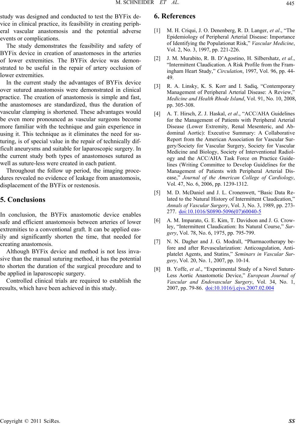 M. SCHNEIDER ET AL. Copyright © 2011 SciRes. SS 445 study was designed and conducted to test the BYFix de- vice in clinical practice, its feasibility in creating periph- eral vascular anastomosis and the potential adverse events or complications. The study demonstrates the feasibility and safety of BYFix device in creation of anastomoses in the arteries of lower extremities. The BYFix device was demon- strated to be useful in the repair of artery occlusion of lower extremities. In the current study the advantages of BYFix device over sutured anastomosis were demonstrated in clinical practice. The creation of anastomosis is simple and fast, the anastomoses are standardized, thus the duration of vascular clamping is shortened. These advantages would be even more pronounced as vascular surgeons become more familiar with the technique and gain experience in using it. This technique as it eliminates the need for su- turing, is of special value in the repair of technically dif- ficult aneurysms and suitable for laparoscopic surgery. In the current study both types of anastomoses sutured as well as suture-less were created in each patient. Throughout the follow up period, the imaging proce- dures revealed no evidence of leakage from anastomosis, displacement of the BYFix or restenosis. 5. Conclusions In conclusion, the BYFix anastomotic device enables safe and efficient anastomosis between arteries of lower extremities to a conventional graft. It can be applied eas- ily and significantly shorten the time, that needed for creating anastomosis. Although BYFix device and method is not less inva- sive than the manual suturing method, it has the potential to shorten the duration of the surgical procedure and to be applied in laparoscopic surgery. Controlled clinical trials are required to establish the results, which have been achieved in this study. 6. References [1] M. H. Criqui, J. O. Denenberg, R. D. Langer, et al., “The Epidemiology of Peripheral Arterial Disease: Importance of Identifying the Populationat Risk,” Vascular Medicine, Vol. 2, No. 3, 1997, pp. 221-226. [2] J. M. Murabito, R. B. D’Agostino, H. Silbershatz, et al., “Intermittent Claudication. A Risk Profile from the Fram- ingham Heart Study,” Circulation, 1997, Vol. 96, pp. 44- 49. [3] R. A. Linsky, K. S. Korr and I. Sadiq, “Contemporary Management of Peripheral Arterial Disease: A Review,” Medicine and Health Rhode Island, Vol. 91, No. 10, 2008, pp. 305-308. [4] A. T. Hirsch, Z. J. Haskal, et al., “ACC/AHA Guidelines for the Management of Patients with Peripheral Arterial Disease (Lower Extremity, Renal Mesenteric, and Ab- dominal Aortic): Executive Summary: A Collaborative Report from the American Association for Vascular Sur- gery/Society for Vascular Surgery, Society for Vascular Medicine and Biology, Society of Interventional Radiol- ogy and the ACC/AHA Task Force on Practice Guide- lines (Writing Committee to Develop Guidelines for the Management of Patients with Peripheral Arterial Dis- ease,” Journal of the American College of Cardiology, Vol. 47, No. 6, 2006, pp. 1239-1312. [5] M. D. McDaniel and J. L. Cronenwett, “Basic Data Re- lated to the Natural History of Intermittent Claudication,” Annals of Vascular Surgery, Vol. 3, No. 3, 1989, pp. 273- 277. doi:10.1016/S0890-5096(07)60040-5 [6] A. M. Imparato, G. E. Kim, T. Davidson and J. G. Crow- ley, “Intermittent Claudication: Its Natural Course,” Sur- gery, Vol. 78, No. 6, 1975, pp. 795-799. [7] N. N. Dagher and J. G. Modrall, “Pharmacotherapy be- fore and after Revascularization: Anticoagulation, Anti- platelet Agents, and Statins,” Seminars in Vascular Sur- gery, Vol. 20, No. 1, 2007, pp. 10-14. [8] B. Yoffe, et al., “Experimental Study of a Novel Suture- Less Aortic Anastomotic Device,” European Journal of Vascular and Endovascular Surgery, Vol. 34, No. 1, 2007, pp. 79-86. doi:10.1016/j.ejvs.2007.02.004 |