

Paper Menu >>
Journal Menu >>
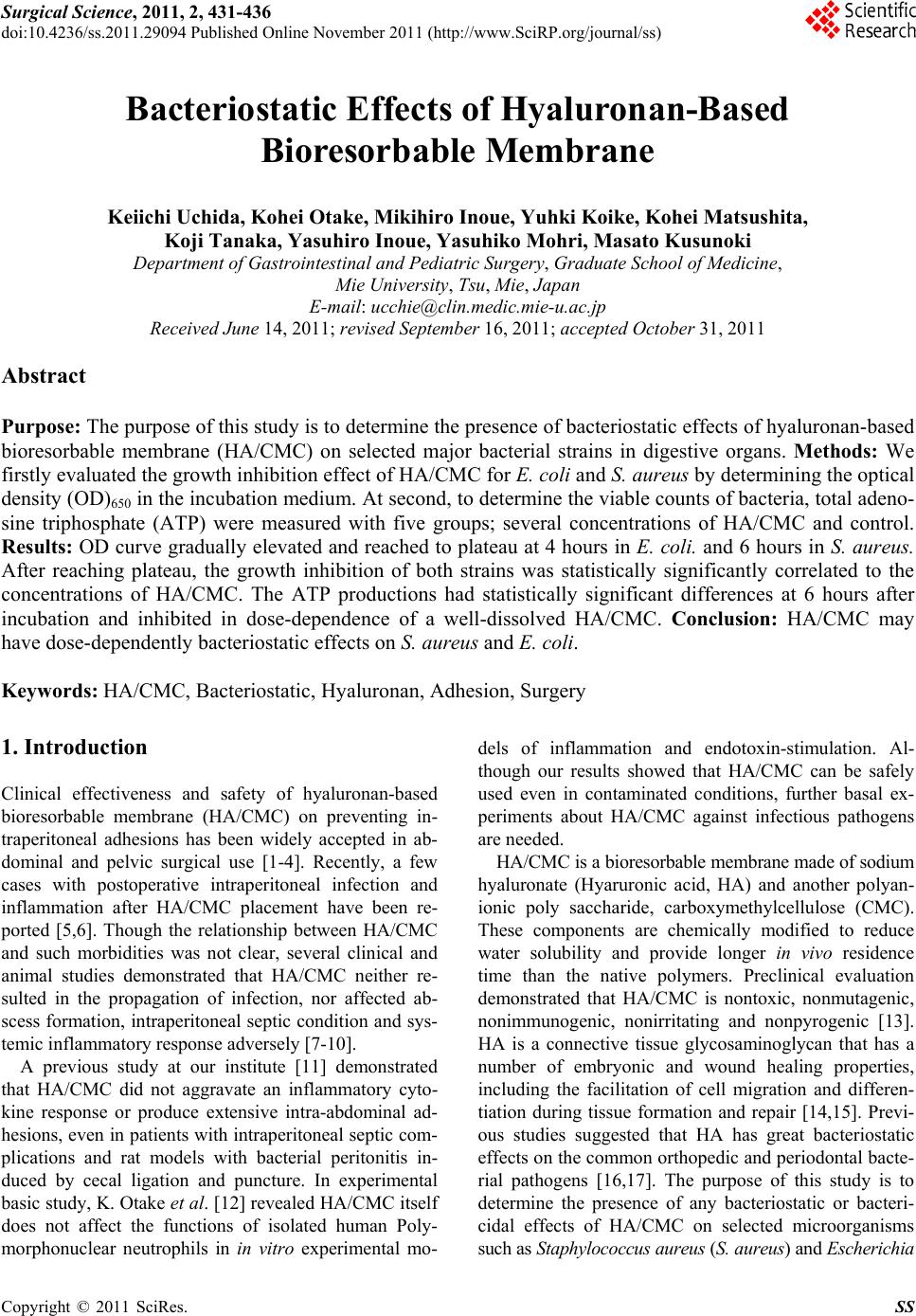 Surgical Science, 2011, 2, 431-436 doi:10.4236/ss.2011.29094 Published Online November 2011 (http://www.SciRP.org/journal/ss) Copyright © 2011 SciRes. SS Bacteriostatic Effects of Hyalurona n - Bas ed Bioresorbable Membrane Keiichi Uchida, Kohei Otake, Mikihiro Inoue, Yuhki Koike, Kohei Matsushita, Koji Tanaka, Yasuhiro Inoue, Yasuhiko Mohri, Masato Kusunoki Department of Gastrointestinal and Pediatric Surgery, Graduate School of Medicine, Mie University, Tsu, Mie, Japan E-mail: [email protected] Received June 14, 2011; revised September 16, 2011; accepted October 31, 2011 Abstract Purpose: The purpose of this study is to determine the presence of bacteriostatic effects of hyaluronan-based bioresorbable membrane (HA/CMC) on selected major bacterial strains in digestive organs. Methods: We firstly evaluated the growth inhibition effect of HA/CMC for E. coli and S. aureus by determining the optical density (OD)650 in the incubation medium. At second, to determine the viable counts of bacteria, total adeno- sine triphosphate (ATP) were measured with five groups; several concentrations of HA/CMC and control. Results: OD curve gradually elevated and reached to plateau at 4 hours in E. coli. and 6 hours in S. aureus. After reaching plateau, the growth inhibition of both strains was statistically significantly correlated to the concentrations of HA/CMC. The ATP productions had statistically significant differences at 6 hours after incubation and inhibited in dose-dependence of a well-dissolved HA/CMC. Conclusion: HA/CMC may have dose-dependently bacteriostatic effects on S. aureus and E. coli. Keywords: HA/CMC, Bacteriostatic, Hyaluronan, Adhesion, Surgery 1. Introduction Clinical effectiveness and safety of hyaluronan-based bioresorbable membrane (HA/CMC) on preventing in- traperitoneal adhesions has been widely accepted in ab- dominal and pelvic surgical use [1-4]. Recently, a few cases with postoperative intraperitoneal infection and inflammation after HA/CMC placement have been re- ported [5,6]. Though the relationship between HA/CMC and such morbidities was not clear, several clinical and animal studies demonstrated that HA/CMC neither re- sulted in the propagation of infection, nor affected ab- scess formation, intraperitoneal septic condition and sys- temic inflammatory response adversely [7-10]. A previous study at our institute [11] demonstrated that HA/CMC did not aggravate an inflammatory cyto- kine response or produce extensive intra-abdominal ad- hesions, even in patients with intraperitoneal septic com- plications and rat models with bacterial peritonitis in- duced by cecal ligation and puncture. In experimental basic study, K. Otake et al. [12] revealed HA/CMC itself does not affect the functions of isolated human Poly- morphonuclear neutrophils in in vitro experimental mo- dels of inflammation and endotoxin-stimulation. Al- though our results showed that HA/CMC can be safely used even in contaminated conditions, further basal ex- periments about HA/CMC against infectious pathogens are needed. HA/CMC is a bioresorbable membrane made of sodium hyaluronate (Hyaruronic acid, HA) and another polyan- ionic poly saccharide, carboxymethylcellulose (CMC). These components are chemically modified to reduce water solubility and provide longer in vivo residence time than the native polymers. Preclinical evaluation demonstrated that HA/CMC is nontoxic, nonmutagenic, nonimmunogenic, nonirritating and nonpyrogenic [13]. HA is a connective tissue glycosaminoglycan that has a number of embryonic and wound healing properties, including the facilitation of cell migration and differen- tiation during tissue formation and repair [14,15]. Previ- ous studies suggested that HA has great bacteriostatic effects on the common orthopedic and periodontal bacte- rial pathogens [16,17]. The purpose of this study is to determine the presence of any bacteriostatic or bacteri- cidal effects of HA/CMC on selected microorganisms such as Staphylococcus aureus (S. aureus) and Escherichia 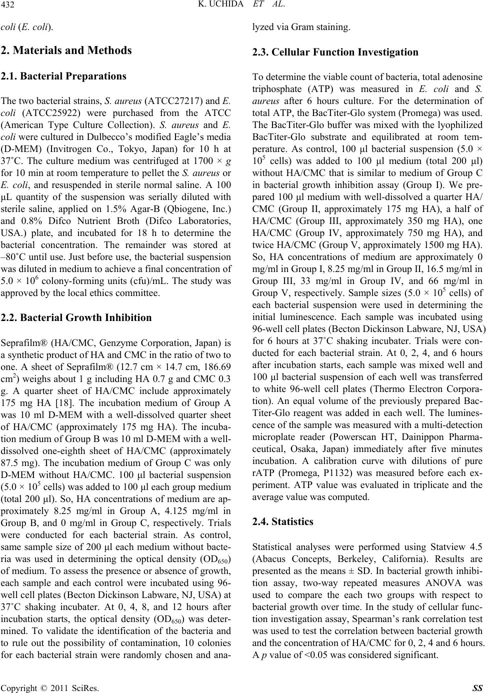 432 K. UCHIDA ET AL. coli (E. coli). 2. Materials and Methods 2.1. Bacterial Preparations The two bacterial strains, S. aureus (ATCC27217) and E. coli (ATCC25922) were purchased from the ATCC (American Type Culture Collection). S. aureus and E. coli were cultured in Dulbecco’s modified Eagle’s media (D-MEM) (Invitrogen Co., Tokyo, Japan) for 10 h at 37˚C. The culture medium was centrifuged at 1700 × g for 10 min at room temperature to pellet the S. aureus or E. coli, and resuspended in sterile normal saline. A 100 μL quantity of the suspension was serially diluted with sterile saline, applied on 1.5% Agar-B (Qbiogene, Inc.) and 0.8% Difco Nutrient Broth (Difco Laboratories, USA.) plate, and incubated for 18 h to determine the bacterial concentration. The remainder was stored at –80˚C until use. Just before use, the bacterial suspension was diluted in medium to achieve a final concentration of 5.0 × 10⁶ colony-forming units (cfu)/mL. The study was approved by the local ethics committee. 2.2. Bacterial Growth Inhibition Seprafilm® (HA/CMC, Genzyme Corporation, Japan) is a synthetic product of HA and CMC in the ratio of two to one. A sheet of Seprafilm® (12.7 cm × 14.7 cm, 186.69 cm2) weighs about 1 g including HA 0.7 g and CMC 0.3 g. A quarter sheet of HA/CMC include approximately 175 mg HA [18]. The incubation medium of Group A was 10 ml D-MEM with a well-dissolved quarter sheet of HA/CMC (approximately 175 mg HA). The incuba- tion medium of Group B was 10 ml D-MEM with a well- dissolved one-eighth sheet of HA/CMC (approximately 87.5 mg). The incubation medium of Group C was only D-MEM without HA/CMC. 100 μl bacterial suspension (5.0 × 10⁵ cells) was added to 100 μl each group medium (total 200 μl). So, HA concentrations of medium are ap- proximately 8.25 mg/ml in Group A, 4.125 mg/ml in Group B, and 0 mg/ml in Group C, respectively. Trials were conducted for each bacterial strain. As control, same sample size of 200 μl each medium without bacte- ria was used in determining the optical density (OD650) of medium. To assess the presence or absence of growth, each sample and each control were incubated using 96- well cell plates (Becton Dickinson Labware, NJ, USA) at 37˚C shaking incubater. At 0, 4, 8, and 12 hours after incubation starts, the optical density (OD650) was deter- mined. To validate the identification of the bacteria and to rule out the possibility of contamination, 10 colonies for each bacterial strain were randomly chosen and ana- lyzed via Gram staining. 2.3. Cellular Function Investigation To determine the viable count of bacteria, total adenosine triphosphate (ATP) was measured in E. coli and S. aureus after 6 hours culture. For the determination of total ATP, the BacTiter-Glo system (Promega) was used. The BacTiter-Glo buffer was mixed with the lyophilized BacTiter-Glo substrate and equilibrated at room tem- perature. As control, 100 μl bacterial suspension (5.0 × 10⁵ cells) was added to 100 μl medium (total 200 μl) without HA/CMC that is similar to medium of Group C in bacterial growth inhibition assay (Group I). We pre- pared 100 μl medium with well-dissolved a quarter HA/ CMC (Group II, approximately 175 mg HA), a half of HA/CMC (Group III, approximately 350 mg HA), one HA/CMC (Group IV, approximately 750 mg HA), and twice HA/CMC (Group V, approximately 1500 mg HA). So, HA concentrations of medium are approximately 0 mg/ml in Group I, 8.25 mg/ml in Group II, 16.5 mg/ml in Group III, 33 mg/ml in Group IV, and 66 mg/ml in Group V, respectively. Sample sizes (5.0 × 10⁵ cells) of each bacterial suspension were used in determining the initial luminescence. Each sample was incubated using 96-well cell plates (Becton Dickinson Labware, NJ, USA) for 6 hours at 37˚C shaking incubater. Trials were con- ducted for each bacterial strain. At 0, 2, 4, and 6 hours after incubation starts, each sample was mixed well and 100 μl bacterial suspension of each well was transferred to white 96-well cell plates (Thermo Electron Corpora- tion). An equal volume of the previously prepared Bac- Titer-Glo reagent was added in each well. The lumines- cence of the sample was measured with a multi-detection microplate reader (Powerscan HT, Dainippon Pharma- ceutical, Osaka, Japan) immediately after five minutes incubation. A calibration curve with dilutions of pure rATP (Promega, P1132) was measured before each ex- periment. ATP value was evaluated in triplicate and the average value was computed. 2.4. Statistics Statistical analyses were performed using Statview 4.5 (Abacus Concepts, Berkeley, California). Results are presented as the means ± SD. In bacterial growth inhibi- tion assay, two-way repeated measures ANOVA was used to compare the each two groups with respect to bacterial growth over time. In the study of cellular func- tion investigation assay, Spearman’s rank correlation test was used to test the correlation between bacterial growth and the concentration of HA/CMC for 0, 2, 4 and 6 hours. A p value of <0.05 was considered significant. Copyright © 2011 SciRes. SS 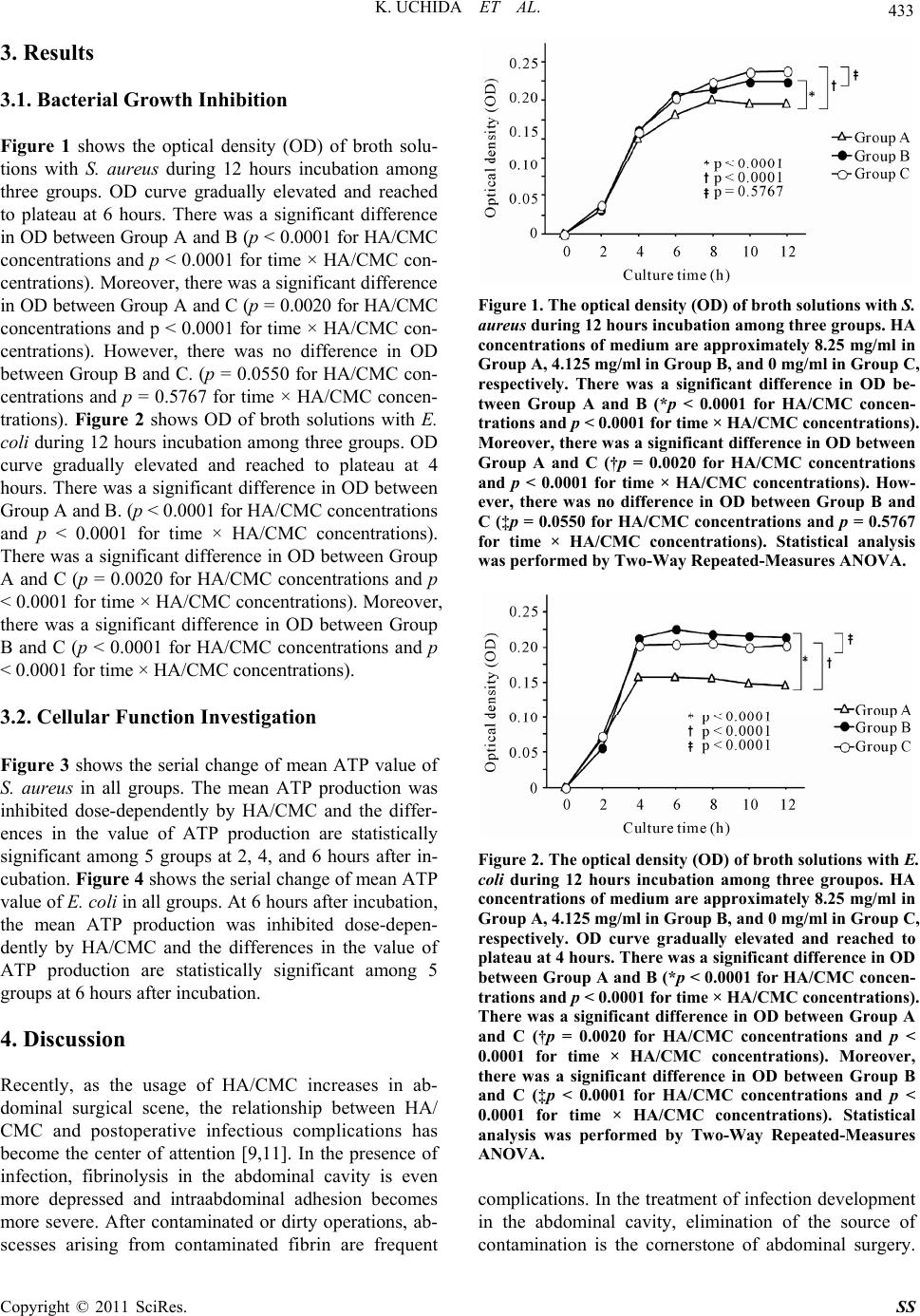 K. UCHIDA ET AL. 433 3. Results 3.1. Bacterial Growth Inhibition Figure 1 shows the optical density (OD) of broth solu- tions with S. aureus during 12 hours incubation among three groups. OD curve gradually elevated and reached to plateau at 6 hours. There was a significant difference in OD between Group A and B (p < 0.0001 for HA/CMC concentrations and p < 0.0001 for time × HA/CMC con- centrations). Moreover, there was a significant difference in OD between Group A and C (p = 0.0020 for HA/CMC concentrations and p < 0.0001 for time × HA/CMC con- centrations). However, there was no difference in OD between Group B and C. (p = 0.0550 for HA/CMC con- centrations and p = 0.5767 for time × HA/CMC concen- trations). Figure 2 shows OD of broth solutions with E. coli during 12 hours incubation among three groups. OD curve gradually elevated and reached to plateau at 4 hours. There was a significant difference in OD between Group A and B. (p < 0.0001 for HA/CMC concentrations and p < 0.0001 for time × HA/CMC concentrations). There was a significant difference in OD between Group A and C (p = 0.0020 for HA/CMC concentrations and p < 0.0001 for time × HA/CMC concentrations). Moreover, there was a significant difference in OD between Group B and C (p < 0.0001 for HA/CMC concentrations and p < 0.0001 for time × HA/CMC concentrations). 3.2. Cellular Function Investigation Figure 3 shows the serial change of mean ATP value of S. aureus in all groups. The mean ATP production was inhibited dose-dependently by HA/CMC and the differ- ences in the value of ATP production are statistically significant among 5 groups at 2, 4, and 6 hours after in- cubation. Figure 4 shows the serial change of mean ATP value of E. coli in all groups. At 6 hours after incubation, the mean ATP production was inhibited dose-depen- dently by HA/CMC and the differences in the value of ATP production are statistically significant among 5 groups at 6 hours after incubation. 4. Discussion Recently, as the usage of HA/CMC increases in ab- dominal surgical scene, the relationship between HA/ CMC and postoperative infectious complications has become the center of attention [9,11]. In the presence of infection, fibrinolysis in the abdominal cavity is even more depressed and intraabdominal adhesion becomes more severe. After contaminated or dirty operations, ab- scesses arising from contaminated fibrin are frequent Figure 1. The optical density (OD) of broth solutions with S. aureus during 12 hours incubation among three groups. HA concentrations of medium are approximately 8.25 mg/ml in Group A, 4.125 mg/ml in Group B, and 0 mg/ml in Group C, respectively. There was a significant difference in OD be- tween Group A and B (*p < 0.0001 for HA/CMC concen- trations and p < 0.0001 for time × HA/CMC concentrations). Moreover, there was a significant difference in OD between Group A and C (†p = 0.0020 for HA/CMC concentrations and p < 0.0001 for time × HA/CMC concentrations). How- ever, there was no difference in OD between Group B and C (‡p = 0.0550 for HA/CMC concentrations and p = 0.5767 for time × HA/CMC concentrations). Statistical analysis was performed by Two-Way Repeated-Measures ANOVA. Figure 2. The optical density (OD) of broth solutions with E. coli during 12 hours incubation among three groupos. HA concentrations of medium are approximately 8.25 mg/ml in Group A, 4.125 mg/ml in Group B, and 0 mg/ml in Group C, respectively. OD curve gradually elevated and reached to plateau at 4 hours. There was a significant difference in OD between Gr oup A and B (*p < 0.0001 for HA/CMC concen- trations and p < 0.0001 for time × HA/CMC concentrations). There was a significant difference in OD between Group A and C (†p = 0.0020 for HA/CMC concentrations and p < 0.0001 for time × HA/CMC concentrations). Moreover, there was a significant difference in OD between Group B and C (‡p < 0.0001 for HA/CMC concentrations and p < 0.0001 for time × HA/CMC concentrations). Statistical analysis was performed by Two-Way Repeated-Measures ANOVA. complications. In the treatment of infection development in the abdominal cavity, elimination of the source of contamination is the cornerstone of abdominal surgery. Copyright © 2011 SciRes. SS 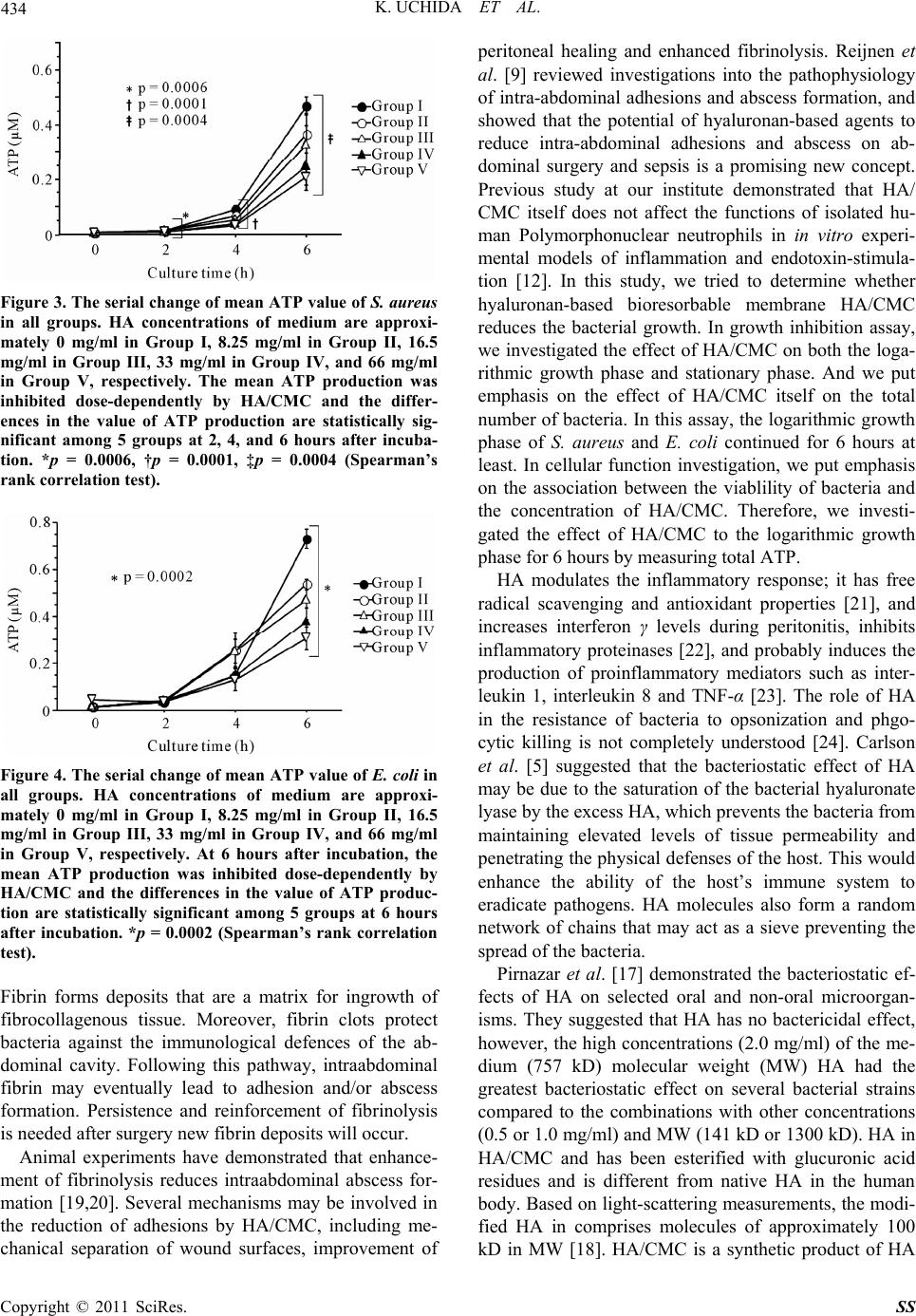 434 K. UCHIDA ET AL. Figure 3. The serial change of mean ATP value of S. aureus in all groups. HA concentrations of medium are approxi- mately 0 mg/ml in Group I, 8.25 mg/ml in Group II, 16.5 mg/ml in Group III, 33 mg/ml in Group IV, and 66 mg/ml in Group V, respectively. The mean ATP production was inhibited dose-dependently by HA/CMC and the differ- ences in the value of ATP production are statistically sig- nificant among 5 groups at 2, 4, and 6 hours after incuba- tion. *p = 0.0006, †p = 0.0001, ‡p = 0.0004 (Spearman’s rank correlation test). Figure 4. The serial change of mean ATP value of E. coli in all groups. HA concentrations of medium are approxi- mately 0 mg/ml in Group I, 8.25 mg/ml in Group II, 16.5 mg/ml in Group III, 33 mg/ml in Group IV, and 66 mg/ml in Group V, respectively. At 6 hours after incubation, the mean ATP production was inhibited dose-dependently by HA/CMC and the differences in the value of ATP produc- tion are statistically significant among 5 groups at 6 hours after incubation. *p = 0.0002 (Spearman’s rank correlation test). Fibrin forms deposits that are a matrix for ingrowth of fibrocollagenous tissue. Moreover, fibrin clots protect bacteria against the immunological defences of the ab- dominal cavity. Following this pathway, intraabdominal fibrin may eventually lead to adhesion and/or abscess formation. Persistence and reinforcement of fibrinolysis is needed after surgery new fibrin deposits will occur. Animal experiments have demonstrated that enhance- ment of fibrinolysis reduces intraabdominal abscess for- mation [19,20]. Several mechanisms may be involved in the reduction of adhesions by HA/CMC, including me- chanical separation of wound surfaces, improvement of peritoneal healing and enhanced fibrinolysis. Reijnen et al. [9] reviewed investigations into the pathophysiology of intra-abdominal adhesions and abscess formation, and showed that the potential of hyaluronan-based agents to reduce intra-abdominal adhesions and abscess on ab- dominal surgery and sepsis is a promising new concept. Previous study at our institute demonstrated that HA/ CMC itself does not affect the functions of isolated hu- man Polymorphonuclear neutrophils in in vitro experi- mental models of inflammation and endotoxin-stimula- tion [12]. In this study, we tried to determine whether hyaluronan-based bioresorbable membrane HA/CMC reduces the bacterial growth. In growth inhibition assay, we investigated the effect of HA/CMC on both the loga- rithmic growth phase and stationary phase. And we put emphasis on the effect of HA/CMC itself on the total number of bacteria. In this assay, the logarithmic growth phase of S. aureus and E. coli continued for 6 hours at least. In cellular function investigation, we put emphasis on the association between the viablility of bacteria and the concentration of HA/CMC. Therefore, we investi- gated the effect of HA/CMC to the logarithmic growth phase for 6 hours by measuring total ATP. HA modulates the inflammatory response; it has free radical scavenging and antioxidant properties [21], and increases interferon γ levels during peritonitis, inhibits inflammatory proteinases [22], and probably induces the production of proinflammatory mediators such as inter- leukin 1, interleukin 8 and TNF-α [23]. The role of HA in the resistance of bacteria to opsonization and phgo- cytic killing is not completely understood [24]. Carlson et al. [5] suggested that the bacteriostatic effect of HA may be due to the saturation of the bacterial hyaluronate lyase by the excess HA, which prevents the bacteria from maintaining elevated levels of tissue permeability and penetrating the physical defenses of the host. This would enhance the ability of the host’s immune system to eradicate pathogens. HA molecules also form a random network of chains that may act as a sieve preventing the spread of the bacteria. Pirnazar et al. [17] demonstrated the bacteriostatic ef- fects of HA on selected oral and non-oral microorgan- isms. They suggested that HA has no bactericidal effect, however, the high concentrations (2.0 mg/ml) of the me- dium (757 kD) molecular weight (MW) HA had the greatest bacteriostatic effect on several bacterial strains compared to the combinations with other concentrations (0.5 or 1.0 mg/ml) and MW (141 kD or 1300 kD). HA in HA/CMC and has been esterified with glucuronic acid residues and is different from native HA in the human body. Based on light-scattering measurements, the modi- fied HA in comprises molecules of approximately 100 kD in MW [18]. HA/CMC is a synthetic product of HA Copyright © 2011 SciRes. SS  K. UCHIDA ET AL. 435 and CMC in the ratio of two to one. A sheet of HA/CMC (12.7 cm × 14.7 cm, 186.69 cm2) weighs about 1 g in- cluding HA 0.7 g and CMC 0.3 g. A quarter sheet of HA/ CMC include approximately 175 mg HA. The concentra- tion of HA formulation used in Group A was 3.5 mg/ml and higher compared to Pirnazar’s study. This study clarified the higher concentrations of HA/CMC intro- duced, the greater bacteriostatic effects on S. aureus and E. coli using bacterial growth inhibition and cellular func- tion investigation. In conclusion, HA/CMC may have dose-dependently bacteriostatic effects on S. aureus and E. coli. Further investigations are needed about various bacterial strains which could be present in the abdominal cavity. 5. References [1] J. M. Becker, M. T. Dayton, V. W. Fazio, D. E. Beck, S. J. Stryker, S. D. Wexner, et al., “Prevention of Postoperative Abdominal Adhesions by a Sodium Hyaluronate-Based Bioresorbable Membrane: A Prospective, Randomized, Dou- ble-Blind Multicenter Study,” Journal of the American College of Surgeons, Vol. 183, No. 4, 1996, pp. 297-306. [2] Y. Mohri, K. Uchida, T. Araki, Y. Inoue, H. Tonouchi, C. Miki, et al., “Hyaluronic Acid-Carboxycellulose Mem- brane (Seprafilm®) Reduces Early Postoperative Small Bowel Obstruction in Gastrointestinal Surgery,” The American Surgeon, Vol. 71, No. 10, 2005, pp. 861-863. [3] M. Kusunoki, H. Ikeuchi, H. Yanagi, M. Noda, H. To- nouchi, Y. Mohri, et al., “Bioresorbable Hyaluronate-Car- boxymethylcellulose Membrane (Seprafilm) in Surgery for Rectal Carcinoma: A Prospective Randomized Clini- cal Trial,” Surgery Today, Vol. 35, No. 11, 2005, pp. 940-945. doi:10.1007/s00595-005-3061-0 [4] M. Inoue, K. Uchida, C. Miki and M. Kusunoki, “Effi- cacy of Seprafilm for Reducing Reoperative Risk in Pedia- tric Surgical Patients Undergoing Abdominal Surgery,” Journal of Pediatric Surgery, Vol. 40, No. 8, 2005, pp. 1301-1306. doi:10.1016/j.jpedsurg.2005.05.015 [5] P. J. Klingler, N. R. Floch, M. H. Seelig, S. A. Branton, J. T. Wolfe and P. P. Metzger, “Seprafilm-Induced Peritoneal Inflammation: A Previously Unknown Complication—Re- port of a Case,” Diseases of the Colon & Rectum, Vol. 42, No. 12, 1999, pp. 1639-1643. doi:10.1007/BF02236221 [6] F. H. Remzi, M. Oncel, J. M. Church, A. J. Senagore, C. P. Delaney and V. W. Fazio, “An Unusual Complication after Hyaluronate-Based Bioresorbable Membrane (Se- prafilm) Application,” The American Surgeon, Vol. 69, No. 4, 2003, pp. 356-357. [7] A. O. Tzianabos, R. L. Cisneros, J. Gershkovich, J. John- son, R. J. Miller, J. W. Burns, et al., “Effect of Surgical Adhesion Reduction Devices on the Propagation of Ex- perimental Intra-Abdominal Infection,” Archives of Sur- gery, Vol. 134, No. 11, 1999, pp. 1254-1259. doi:10.1001/archsurg.134.11.1254 [8] A. Tuzune, M. A. Kuzu, B. Akin, S. Karaca and S. Hazi- nedaroglu, “The Effect of Hyaluronan-Based Agents on Adhesion Formation in an Intra-Abdominal Sepsis Model,” Digestive Diseases and Sciences, Vol. 49, No. 6, 2004, pp. 1054-1061. doi:10.1023/B:DDAS.0000034571.30602.36 [9] M. M. Reijnen, J. F. Meis, V. A. Postma and H. van Goor, “Prevention of Intra-Abdominal Abscesses and adhesions Using a Hyaluronic Acid Solution in a Rat Peritonitis Model,” Archives of Surgery, Vol. 134, No. 9, 1999, pp. 997-1001. doi:10.1001/archsurg.134.9.997 [10] A. M. Ghellai, A. F. Stucchi, D. J. Lynch, K. C. Skinner, M. J. Colt and J. M. Becker, “Role of a Hyaluronate- Based Membrane in the Prevention of Peritonitis-Induced Adhesions,” Journal of Gastrointestinal Surgery, Vol. 4, No. 3, 2000, pp. 310-315. doi:10.1016/S1091-255X(00)80081-5 [11] K. Uchida, H. Urata, Y. Mohri, M. Inoue, C. Miki and M. Kusunoki, “Seprafilm® Does Not Aggravate Intraperito- neal Septic Conditions or Evoke Systemic Inflammatory Response,” Surgery Today, Vol. 35, No. 12, 2005, pp. 1054-1059. doi:10.1007/s00595-005-3085-5 [12] K. Otake, K. Uchida, S. Yoshiyama, M. Inoue, Y. Okita, H. Watanabe, et al., “Effects of a Hyaluronate-Carboxy- Methylcellulose Membrane (Seprafilm) on Human Po- lymorphonuclear Neutrophil Functions,” Journal of Sur- gical Research, Vol. 149, No. 2, 2008, pp. 243-249. doi:10.1016/j.jss.2008.01.011 [13] J. W. Burns, M. J. Colt, L. S. Burgees and K. C. Skinner, “Preclinical Evaluation of Seprafilm Bioresorbable Mem- brane,” European Journal of Surgery, No. 577, 1997, pp. 40-48. [14] B. P. Toole, In: D. E. Hay, Ed., Cell Biology of Extracellu- lar Matrix, Plenum Press, New York, 1991, pp. 305-341. [15] C. N. Bertolami, “Glycosaminoglycan Interactions in Early Wound Repair,” In: T. K. Hunt, R. B. Heppenstall, E. Pines, et al., Eds., Soft and Hard Tissue Repair, Biological and Clinical Aspects, Praeger Publishers, New York, 1984, pp. 67-97. [16] G. A. Carlson, J. L. Dragoo, B. Samimi, D. A. Bruckner, G. W. Bernard, M. Hedrick, et al., “Bacteriostatic Properties of Biomatrices against Common Orthopaedic Pathogens,” Biochemical and Biophysical Research Communications, Vol. 321, No. 2, 2004, pp. 472-478. doi:10.1016/j.bbrc.2004.06.165 [17] P. Pirnazar, L. Wolinsky, S. Nachnani, S. Haake, A. Pil- loni and G. W. Bernard, “Bacteriostatic Effects of Hya- luronic Acid,” Journal of Periodontology, Vol. 70, No. 4, 1999, pp. 370-374. doi:10.1902/jop.1999.70.4.370 [18] S. C. Hubbard and J. W. Burns, “Effects of a Hyaluronan- Based Membrane (Seprafilm) on Intraperitoneally Dis- seminated Human Colon Cancer Cell Growth in a Nude Mouse Model,” Diseases of the Colon & Rectum, Vol. 45, No. 3, 2002, pp. 334-344. doi:10.1007/s10350-004-6178-0 [19] H. van Goor, J. S. de Graaf, K. Kooi, W. J. Sluiter, V. J. Bom, J. van der Meer, et al., “Effect of Recombinant Tis- sue Plasminogen Activator on Intra-Abdominal Abscess Formation in Rats with Generalized Peritonitis,” Journal of the American College of Surgeons, Vol. 179, No. 4, Copyright © 2011 SciRes. SS  K. UCHIDA ET AL. Copyright © 2011 SciRes. SS 436 1994, pp. 407-411. [20] D. I. McRitchie, D. Cummings and O. D. Rotstein, “De- layed Administration of Tissue Plasminogen Activator Reduces Intra-Abdominal Abscess Formation,” Archives of Surgery, Vol. 124, No. 12, 1989, pp. 1406-1410. [21] D. Foschi, L. Castoldi, E. Radaelli, P. Abelli, G. Cal- derini, A. Rastrelli, et al., “Hyaluronic Acid Pevents Oxy- gen Free-Radical Damage to Granulation Tissue: A Study in Rats,” International Journal of Tissue Reactions, Vol. 12, No. 6, 1990, pp. 333-339. [22] M. Akatsuka, Y. Yamamoto, K. Tobetto, T. Yasui and T. Ando, “Suppressive Effects of Hyaluronic Acid on Elas- tase Release from Rat Peritoneal Leucocytes,” Journal of Pharmacy and Pharmacology, Vol. 45, No. 2, 1993, pp. 110-114. doi:10.1111/j.2042-7158.1993.tb03693.x [23] D. Hiro, A. Ito, K. Matsuta and Y. Mori, “Hyaluronic Acid Is an Endogenous Inducer of Interleukin-1 Produc- tion by Human Monocytes and Rabbit Macrophages,” Biochemical and Biophysical Research Communications, Vol. 140, No. 2, 1986, pp. 715-722. [24] A. E. Moses, M. R. Wessels, K. Zalcman, S. Alberti, S. Natanson-Yaron, T. Menes, et al., “Relative Contribu- tions of Hyaluronic Acid Capsule and M Protein to Viru- lence in a Mucoid Strain of the Group A Streptococcus,” Infection and Immunity, Vol. 65, No. 1, 1997, pp. 64-71. |