

Paper Menu >>
Journal Menu >>
 Advances in Biological Chemistry, 2011, 1, 58-64 doi:10.4236/abc.2011.13008 Published Online November 2011 (http://www.SciRP.org/journal/abc/ ABC ). Published Online November 2011 in SciRes. http://www.scirp.org/journal/ABC Reliability of enzyme assays in dried blood spots for diagnosis of 4 lysosomal storage disorders Romina Ceci, Pablo N. de Francesco, Juan M. Mucci, Lorena N. Cancelarich, Carlos A. Fossati, Paula A. Rozenfeld* LISIN, Departamento de Ciencias Biológicas, Facultad de Ciencias Exactas, Universidad Nacional de La Plata, La Plata, Argentina. Email: *[email protected] Received 3 August, 2011; revised 12 September, 2011; revised 23 September, 2011. ABSTRACT Introduction: Lysosomal storage disord ers ( LSD ) a re inherited diseases caused, in the majority of them, by the deficiency of lysosomal enzymatic activities. Ob- jectives: We aimed to analyze the usefulness of DBS samples for diagnosis of 4 LSDs, with the availability of a large quantity of patient samples. Design and methods: Blood samples from previously diagnosed patients with Fabry, Gaucher, Hunter, and Maro- teaux-Lamy syndromes and normal control indi- viduals, were collected and dispen-sed in filter paper, and used for enzymatic activity determination. Re- sults: Diagnosis of hemi/homo-zygous patients with Fabry, Hunter and Maroteaux-Lamy diseases using DBS samples showed ideal parameters of 100% sen- sitivity and specificity. DBS assay for Gaucher dis- ease would need a posterior confirmatory step. Con- clusions: Leukocyte measu-rement is the only reli- able way to diagnose Gaucher disease. For Hunter, Fabry and Maroteaux-Lamy disorders discrimina- tion between patients and controls seems adequate by DBS. Keywords: Lysosomal Storage Disorders; Diagnosis; Dried Blood Spots; Sensitivity; Enzymatic Activity; Re- liability 1. INTRODUCTION Lysosomal storage disorders (LSD) are severe inherited diseases caused, in the majority of them, by the defi- ciency of lysosomal enzymatic activities, resulting in the accumulation of specific substances, such as complex lipids, glycoproteins and glycosaminoglycans. Clinical manifestations are characterized by a spectrum of clini- cal manifestations, which may include organ dysfunction, neurological involvement and skeletal abnormalities [1]. Enzyme replacement therapy is now available for speci- fictreatment of 6 LSD, and is on development or in use in clinical trials for some others [2]. Early instauration of therapy, before irreversible organ damage, is needed for its better success. Therefore, early diagnosis is crucial. Diagnosis process generally starts by clinical suspi- cion by a physician [3]. However, due to age at onset of signs and symptoms is variable, and clinical manifesta- tions could be unspecific and multisystemic, LSD are generally underdiagnosed and/or there could be a sub- stantial delay between age at onset of manifestations and age at diagnosis. Diagnosis is confirmed by laboratory determination of the specific enzymatic activity in leukocytes or culture fibroblasts, using 4-methylumbelliferone conjugated ar- tificial substrate. The use of leukocytes as the enzyme source implies the isolation of these cells from a volume (10 mL) of liquid blood, requires a homogenisation step with a metal tip sonicator, and total protein measurement [4]. On the other hand, the use of fibroblasts requires a sample of skin biopsy, primary cell culture of skin fibro- blasts (avoiding the high risk of contamination), and to wait for the growing of fibroblasts to obtain enough cell counts [5]. Measurement of lysosomal enzymes activities in dried blood spots (DBS) on filter paper was introduced in 2001 [6] as a method with several advantages over test- ing in leukocytes/fibroblasts. First, it requires only a few drops of blood. Second, the main benefit is the possibili- ty to mail the DBS samples to specialized laboratories, making it possible to send samples to other cities, re- gions or countries for testing. This is especially impor- tant due to LSD laboratory diagnosis is currently under- gone in specialized reference laboratories. Therefore there are a few labs per country, or somewhere they are missing. Finally, this method allows microplate adapta- tion, permitting the simultaneously measurement of mul- tiple samples and automation. Potentially, it could also be applicable to newborn screening. Due to different techniques of blood sample collection could change the enzyme activity, specific lab recom-  R. Ceci et al. / Advances in Biological Chemistry 1 (2011) 58-64 59 mendations for collection should be follow [7], in terms of the specific filter paper and use or not of anticoagu- lant. The usefulness of DBS for LSD diagnosis is not uni- versally accepted by medical community. Generally, physicians recommend assaying enzymatic activity in leukocytes/fibroblasts in order to confirm an abnormal result in DBS [8]. However, to our knowledge, there is no study analyzing clinical parameters of efficacy of en- zymatic activity determination on DBS for diagnosis of the lysosomal storage disorders Fabry, Gaucher, Hunter (Mucopolysaccharidosis II) and Maroteaux-Lamy (Mu- copolysaccharidosis VI). For this reason, we aimed to analyze the usefulness of DBS samples for diagnosis of these 4 LSDs, with the availability of a large quantity of patient samples. 2. MATERIALS AND METHODS 2.1. Patients and Samples Samples from previously diagnosed patients with Fabry (n = 45), Gaucher (n = 30), Hunter (n = 85), and Maro- teaux-Lamy(n = 22) diseases and normal control indi- viduals (n = 155), were obtained for this study. All the samples were taken before instauration of any specific treatment. Samples from heterozygous Fabry (n = 59), Hunter (n = 10) and Gaucher (n = 33) individuals were also included. The protocol was approved by the scien- tific committee of AADELFA according to provisions of the Declaration of Helsinki in 1995. The nature and pur- pose of the study and its possible risks were explained to all volunteers. All the patients gave their informed con- sent previous to the participation in the study. Peripheral blood samples were collected by venopun- cture, no anticoagulant was added. Immediatly, 50ul drops were dispensed in circles of S & S nº903 filter paper cards (Schleier and Schuell, Dassel, Germany). The cards were allowed to dry for 4 hours at room tem- perature. In order to control sample quality a reference enzyme was assayed in all DBS samples prior to the specific enzyme test. The chosen reference enzyme was beta- galactosidase (BGL1, EC 3.1.2.23), and was carried out according to the method of Civallero et al. [9]. All the samples showed normal values on BGL1 assay (data not shown), confirming the good quality of the samples ana- lyzed in this work. 2.2. Enzymatic Activity Determination The determination of the enzymatic activities in DBS samples specific for each of the 4 diseases were under- gone by adaptation of reported methods (Table 1) stated in Table 1 in samples from patients and control indi- viduals. Alpha-galactosidase A (GLA): A 3 mm diameter cir- cle from DBS filter paper was placed into a well of a black microplate and 70 l of the reaction mixture con- taining 3.57 mol/l 4-methylumbelliferyl-α-D-galactopy- ranoside (Glycosynth, Cheshire, England) and 0.07 mol/l N-acetylgalactosamine (Toronto Research Chemicals Inc., North York, Ontario, Canada) in 0.15 mol/l acetate buffer pH = 4.5 was added. After an incubation for 8 h at 37˚C, the stop solution (180 l of 1 mol/l ethylenediamine, pH = 11.4) was added. Beta-glucosidase (BGLU): A 3-mm diameter circle from DBS filter paper was placed into a well of a black microplate and 150 l of the reaction mixture containing 4-methylumbelliferyl-β-D-glucopyranoside 10 mmol/l (G- lycosynth, Cheshire, England) and sodium taurocholate (Sigma, Saint Louis, MO, USA) 50 mmol/l in 0.54 mol/l phosphate-citrate buffer pH 5.5 was added. After an in- cubation for 24 h at 37˚C, the stop solution (150 l of 0.5 mol/l glycine-NaOH buffer pH = 10.3) was added. Iduronate-2-sulphatase (IDS): A 2-mm diameter circle from DBS filter paper was placed into an eppendorf and 50 l BSA 0.2% was added to elute the blood. 10 l of eluted blood in BSA was placed into a well of a black microplate and continued as the method reported by Vo z ny i et al. [11]. Briefly, 20 l of 4-Methylumbellifer- yl- -iduronato-2-sulfato 1.25 mM (Moscerdam substra- tes, Oegstgeest, the Netherlands) in 0.1 M sodium ace- tate pH = 5 containing 10mM lead acetate was added. After an incubation for 24 h at 37˚C, 20 l of 0.05 M cit- rate-phosphate pH = 4.5 buffer and 10 l of alfa-iduroni- dase purified from bovine testis (Moscerdam substrates, Oegstgeest, the Netherlands) were added. After an incu- bation for 24 h at 37˚C, the stop solution (150 l of 0.5 mol/l sodium carbonate/bicarbonate pH = 10.7 buffer) was added. Table 1. List of the enzymes, abbrevations and bibliographic references of the enzyme assays methods. Disease Enzyme Enzyme abbreviation Enzyme Classification number Reference Fabry Alpha-galactosidase A GLA EC 3.2.1.22 [6] Gaucher Beta-D-glucosidase BGLU EC 3.2.1.45 [10] Hunter Iduronate-2-sulfatase IDS EC 3.1.6.13 [11] Maroteaux Lamy Arylsulfatase B ASB EC 3.1.6.12 [8] C opyright © 2011 SciRes. ABC  R. Ceci et al. / Advances in Biological Chemistry 1 (2011) 58-64 60 Arylsulphatase B (ASB): A 2-mm diameter circle from DBS filter paper was placed into a well of a black microplate and 150 l of the reaction mixture containing 15 m mol/l lead acetate and 10 mmol/l 4-methylum- belliferyl-sulfate (Glycosynth, Cheshire, England) in 15 mmol/l acetate buffer pH = 5.0. After incubation for 24 h at 37˚C with agitation, the stop solution (150 ml of 0.085 mol/l glycine-NaOH buffer pH 10.5) was added. In all cases, the fluorescence of the product (excitation 365 nm; emission 450 nm) was measured on a Twinkle LB 970 fluorometer (Berthold Technologies, Bad Wild- bad, Germany). A standard curve of 4-methylumbelli- ferone (Sigma, Saint Louis, MO, USA) was used to ex- trapolate fluorescence counts to moles of enzymatic product. The enzymatic activity was expressed as mi- cromoles of 4-methylumbelliferone produced per liter of blood per hour. 2.3. Analitical Parameters Sensitivity, specificity, positive (PPV) and negative (NPV) predictive values and positive results proportion for each of the 4 enzymatic methods for diagnosis of Fabry, Gaucher, Hunter and Maroteaux-Lamy were calculated. 3. RESULTS Each sample was assayed in duplicate. One blank was included for each sample, consisted of addition of both substrate and stop solutions at the same time at the end of the incubation time. Appropriate incubation times for each enzyme were chosen, among the linearity curve, in order to obtain enough 4-MU to be detected by the fluorometer and in comparison to the counts of the blank. Intra-assay CVs were below 9% and inter-assay CVs below 15% for all the enzymes. Histogram of enzymatic activity of GLA, BGLU, IDS and ASB from patients and normal control individuals is shown in Figure 1. The analytical parameters calculated for each of the methods is shown in Table 2. 3.1. GLA Histogram of enzymatic activity of GLA from hemyzy- gous, heterozygous and normal control individuals is shown in Figure 1(a). Mean value of GLA from normal controls was 6.12 ± 2.28 moles/l·h, and the cut-off (mean - 2SD) was 1.56. As shown in Figure 1(a), there is no overlap between the values from normal controls C opyright © 2011 SciRes. ABC  R. Ceci et al. / Advances in Biological Chemistry 1 (2011) 58-64 61 Figure 1. Histograms and individuals values, respectively, of enzymatic activity determination in DBS for diagnosis of Fabry (1(a) and 1(b)), Gaucher (1(c) and 1(d)), Hunter (1(e) and 1(f)), and Maroteaux-Lamy (1(g) and 1(h)) diseases. Tabl e 2 . Analytical parameters for the enzymatic activity de- termination in DBS for diagnosis of Fabry, Gaucher, Hunter and Maroteaux-Lamy diseases. Fabry Gaucher Hunter Maroteaux Lamy Sensitivity 100 97 100 100 Specificity 100 100 100 100 VPP 100 100 100 100 VPN 100 99 100 100 Positive results (%) 100 99 100 100 and male patients, giving a sensitivity and specificity of 100%. Moreover both predictive values are 100% (Table 2). Values from heterozygous females with Fabry disease, showed a high variability, ranging from very low values to normal ones. 3.2. BGLU Histogram of values of BGLU enzymatic activity in pa- tients and normal controls revealed a minimal (but pre- sent) overlap between both groups (Figure 1(c)). Cut- off value of BGLU enzymatic activity is 1.87 μmoles/l.h. As can be seen in Figure 1(d), one confirmed Gaucher patient exhibited an enzymatic activity value coincident with the cut-off value of this enzymatic assay. This result reduces the sensitivity of this method to 97%. However, the specificity was 100%. We carried out a parallel analysis of BGLU in leuko- cytes and DBS of samples from patients belonging to the 3 groups. The results are shown in Figure 2. Samples from a few normal controls and heterozygotes on DBS revealed enzyme values close to or at the cut-off value; however, the respective value in leukocyte is above its cut-off. Moreover, a homozygous patient with a DBS value close to cut-off, showed very low enzymatic activ- ity in leukocyte homogenates. 3.3. IDS IDS enzymatic activity values obtained from Hunter pa- tients and normal individuals showed no overlap between both groups, with a good separation (Figures 1(e) and (f)). All analytical parameters of this method were 100%. C opyright © 2011 SciRes. ABC 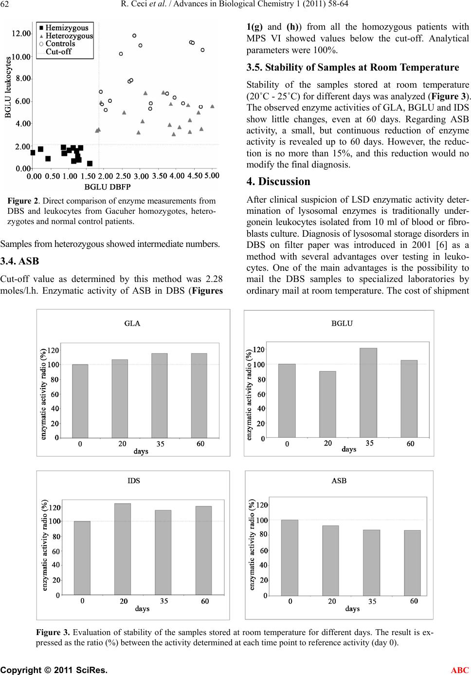 R. Ceci et al. / Advances in Biological Chemistry 1 (2011) 58-64 62 Figure 2. Direct comparison of enzyme measurements from DBS and leukocytes from Gacuher homozygotes, hetero- zygotes and normal control patients. Samples from heterozygous showed intermediate numbers. 3.4. ASB Cut-off value as determined by this method was 2.28 moles/l.h. Enzymatic activity of ASB in DBS (Figures 1(g) and (h)) from all the homozygous patients with MPS VI showed values below the cut-off. Analytical parameters were 100%. 3.5. Stability of Samples at Room Temperature Stability of the samples stored at room temperature (20˚C - 25˚C) for different days was analyzed (Figure 3). The observed enzyme activities of GLA, BGLU and IDS show little changes, even at 60 days. Regarding ASB activity, a small, but continuous reduction of enzyme activity is revealed up to 60 days. However, the reduc- tion is no more than 15%, and this reduction would no modify the final diagnosis. 4. Discussion After clinical suspicion of LSD enzymatic activity deter- mination of lysosomal enzymes is traditionally under- gonein leukocytes isolated from 10 ml of blood or fibro- blasts culture. Diagnosis of lysosomal storage disorders in DBS on filter paper was introduced in 2001 [6] as a method with several advantages over testing in leuko- cytes. One of the main advantages is the possibility to mail the DBS samples to specialized laboratories by ordinary mail at room temperature. The cost of shipment Figure 3. Evaluation of stability of the samples stored at room temperature for different days. The result is ex- pressed as the ratio (%) between the activity determined at each time point to reference activity (day 0). C opyright © 2011 SciRes. ABC 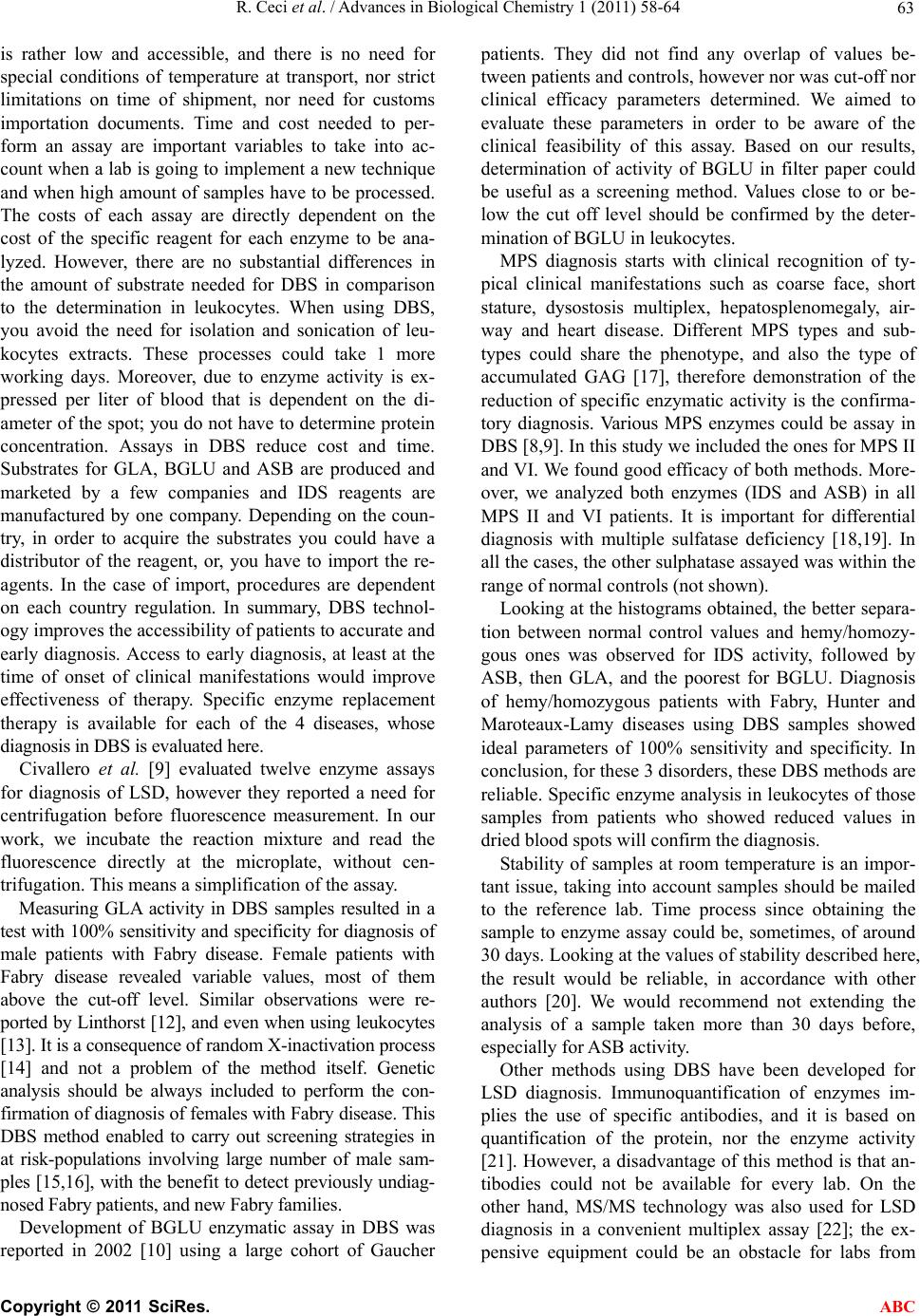 R. Ceci et al. / Advances in Biological Chemistry 1 (2011) 58-64 63 is rather low and accessible, and there is no need for special conditions of temperature at transport, nor strict limitations on time of shipment, nor need for customs importation documents. Time and cost needed to per- form an assay are important variables to take into ac- count when a lab is going to implement a new technique and when high amount of samples have to be processed. The costs of each assay are directly dependent on the cost of the specific reagent for each enzyme to be ana- lyzed. However, there are no substantial differences in the amount of substrate needed for DBS in comparison to the determination in leukocytes. When using DBS, you avoid the need for isolation and sonication of leu- kocytes extracts. These processes could take 1 more working days. Moreover, due to enzyme activity is ex- pressed per liter of blood that is dependent on the di- ameter of the spot; you do not have to determine protein concentration. Assays in DBS reduce cost and time. Substrates for GLA, BGLU and ASB are produced and marketed by a few companies and IDS reagents are manufactured by one company. Depending on the coun- try, in order to acquire the substrates you could have a distributor of the reagent, or, you have to import the re- agents. In the case of import, procedures are dependent on each country regulation. In summary, DBS technol- ogy improves the accessibility of patients to accurate and early diagnosis. Access to early diagnosis, at least at the time of onset of clinical manifestations would improve effectiveness of therapy. Specific enzyme replacement therapy is available for each of the 4 diseases, whose diagnosis in DBS is evaluated here. Civallero et al. [9] evaluated twelve enzyme assays for diagnosis of LSD, however they reported a need for centrifugation before fluorescence measurement. In our work, we incubate the reaction mixture and read the fluorescence directly at the microplate, without cen- trifugation. This means a simplification of the assay. Measuring GLA activity in DBS samples resulted in a test with 100% sensitivity and specificity for diagnosis of male patients with Fabry disease. Female patients with Fabry disease revealed variable values, most of them above the cut-off level. Similar observations were re- ported by Linthorst [12], and even when using leukocytes [13]. It is a consequence of random X-inactivation process [14] and not a problem of the method itself. Genetic analysis should be always included to perform the con- firmation of diagnosis of females with Fabry disease. This DBS method enabled to carry out screening strategies in at risk-populations involving large number of male sam- ples [15,16], with the benefit to detect previously undiag- nosed Fabry patients, and new Fabry families. Development of BGLU enzymatic assay in DBS was reported in 2002 [10] using a large cohort of Gaucher patients. They did not find any overlap of values be- tween patients and controls, however nor was cut-off nor clinical efficacy parameters determined. We aimed to evaluate these parameters in order to be aware of the clinical feasibility of this assay. Based on our results, determination of activity of BGLU in filter paper could be useful as a screening method. Values close to or be- low the cut off level should be confirmed by the deter- mination of BGLU in leukocytes. MPS diagnosis starts with clinical recognition of ty- pical clinical manifestations such as coarse face, short stature, dysostosis multiplex, hepatosplenomegaly, air- way and heart disease. Different MPS types and sub- types could share the phenotype, and also the type of accumulated GAG [17], therefore demonstration of the reduction of specific enzymatic activity is the confirma- tory diagnosis. Various MPS enzymes could be assay in DBS [8,9]. In this study we included the ones for MPS II and VI. We found good efficacy of both methods. More- over, we analyzed both enzymes (IDS and ASB) in all MPS II and VI patients. It is important for differential diagnosis with multiple sulfatase deficiency [18,19]. In all the cases, the other sulphatase assayed was within the range of normal controls (not shown). Looking at the histograms obtained, the better separa- tion between normal control values and hemy/homozy- gous ones was observed for IDS activity, followed by ASB, then GLA, and the poorest for BGLU. Diagnosis of hemy/homozygous patients with Fabry, Hunter and Maroteaux-Lamy diseases using DBS samples showed ideal parameters of 100% sensitivity and specificity. In conclusion, for these 3 disorders, these DBS methods are reliable. Specific enzyme analysis in leukocytes of those samples from patients who showed reduced values in dried blood spots will confirm the diagnosis. Stability of samples at room temperature is an impor- tant issue, taking into account samples should be mailed to the reference lab. Time process since obtaining the sample to enzyme assay could be, sometimes, of around 30 days. Looking at the values of stability described here, the result would be reliable, in accordance with other authors [20]. We would recommend not extending the analysis of a sample taken more than 30 days before, especially for ASB activity. Other methods using DBS have been developed for LSD diagnosis. Immunoquantification of enzymes im- plies the use of specific antibodies, and it is based on quantification of the protein, nor the enzyme activity [21]. However, a disadvantage of this method is that an- tibodies could not be available for every lab. On the other hand, MS/MS technology was also used for LSD diagnosis in a convenient multiplex assay [22]; the ex- pensive equipment could be an obstacle for labs from C opyright © 2011 SciRes. ABC 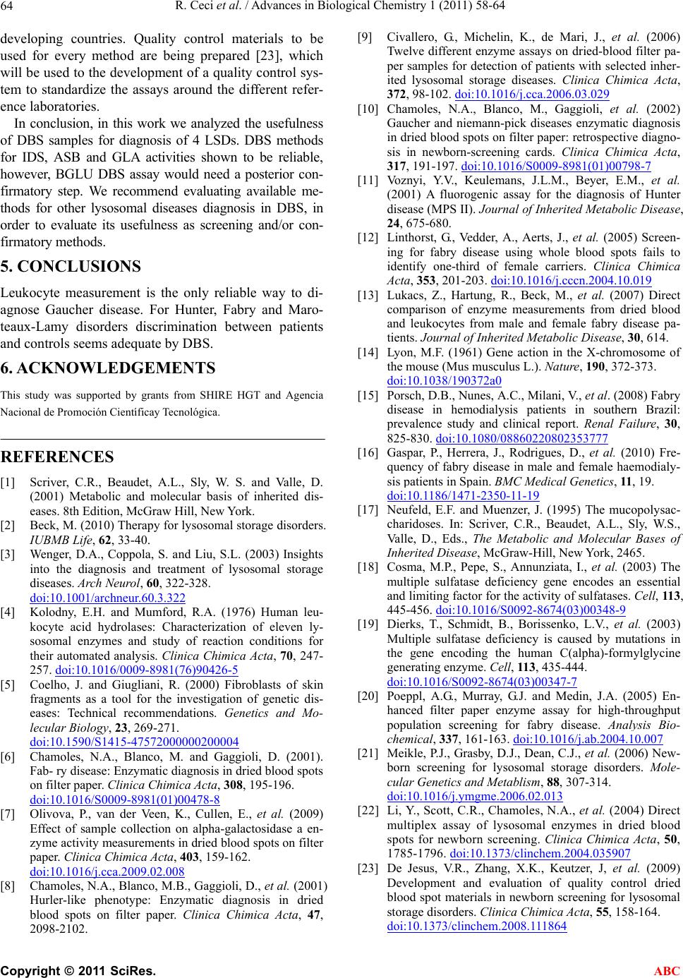 R. Ceci et al. / Advances in Biological Chemistry 1 (2011) 58-64 64 developing countries. Quality control materials to be used for every method are being prepared [23], which will be used to the development of a quality control sys- tem to standardize the assays around the different refer- ence laboratories. In conclusion, in this work we analyzed the usefulness of DBS samples for diagnosis of 4 LSDs. DBS methods for IDS, ASB and GLA activities shown to be reliable, however, BGLU DBS assay would need a posterior con- firmatory step. We recommend evaluating available me- thods for other lysosomal diseases diagnosis in DBS, in order to evaluate its usefulness as screening and/or con- firmatory methods. 5. CONCLUSIONS Leukocyte measurement is the only reliable way to di- agnose Gaucher disease. For Hunter, Fabry and Maro- teaux-Lamy disorders discrimination between patients and controls seems adequate by DBS. 6. ACKNOWLEDGEMENTS This study was supported by grants from SHIRE HGT and Agencia Nacional de Promoción Científicay Tecnológica. REFERENCES [1] Scriver, C.R., Beaudet, A.L., Sly, W. S. and Valle, D. (2001) Metabolic and molecular basis of inherited dis- eases. 8th Edition, McGraw Hill, New York. [2] Beck, M. (2010) Therapy for lysosomal storage disorders. IUBMB Life, 62, 33-40. [3] Wenger, D.A., Coppola, S. and Liu, S.L. (2003) Insights into the diagnosis and treatment of lysosomal storage diseases. Arch Neurol, 60, 322-328. doi:10.1001/archneur.60.3.322 [4] Kolodny, E.H. and Mumford, R.A. (1976) Human leu- kocyte acid hydrolases: Characterization of eleven ly- sosomal enzymes and study of reaction conditions for their automated analysis. Clinica Chimica Acta, 70, 247- 257. doi:10.1016/0009-8981(76)90426-5 [5] Coelho, J. and Giugliani, R. (2000) Fibroblasts of skin fragments as a tool for the investigation of genetic dis- eases: Technical recommendations. Genetics and Mo- lecular Biology, 23, 269-271. doi:10.1590/S1415-47572000000200004 [6] Chamoles, N.A., Blanco, M. and Gaggioli, D. (2001). Fab- ry disease: Enzymatic diagnosis in dried blood spots on filter paper. Clinica Chimica Acta, 308, 195-196. doi:10.1016/S0009-8981(01)00478-8 [7] Olivova, P., van der Veen, K., Cullen, E., et al. (2009) Effect of sample collection on alpha-galactosidase a en- zyme activity measurements in dried blood spots on filter paper. Clinica Chimica Acta, 403, 159-162. doi:10.1016/j.cca.2009.02.008 [8] Chamoles, N.A., Blanco, M.B., Gaggioli, D., et al. (2001) Hurler-like phenotype: Enzymatic diagnosis in dried blood spots on filter paper. Clinica Chimica Acta, 47, 2098-2102. [9] Civallero, G., Michelin, K., de Mari, J., et al. (2006) Twelve different enzyme assays on dried-blood filter pa- per samples for detection of patients with selected inher- ited lysosomal storage diseases. Clinica Chimica Acta, 372, 98-102. doi:10.1016/j.cca.2006.03.029 [10] Chamoles, N.A., Blanco, M., Gaggioli, et al. (2002) Gaucher and niemann-pick diseases enzymatic diagnosis in dried blood spots on filter paper: retrospective diagno- sis in newborn-screening cards. Clinica Chimica Acta, 317, 191-197. doi:10.1016/S0009-8981(01)00798-7 [11] Voznyi, Y.V., Keulemans, J.L.M., Beyer, E.M., et al. (2001) A fluorogenic assay for the diagnosis of Hunter disease (MPS II). Journal of Inherited Metabolic Disease, 24, 675-680. [12] Linthorst, G., Vedder, A., Aerts, J., et al. (2005) Screen- ing for fabry disease using whole blood spots fails to identify one-third of female carriers. Clinica Chimica Acta, 353, 201-203. doi:10.1016/j.cccn.2004.10.019 [13] Lukacs, Z., Hartung, R., Beck, M., et al. (2007) Direct comparison of enzyme measurements from dried blood and leukocytes from male and female fabry disease pa- tients. Journal of Inherited Metabolic Disease, 30, 614. [14] Lyon, M.F. (1961) Gene action in the X-chromosome of the mouse (Mus musculus L.). Nature, 190, 372-373. doi:10.1038/190372a0 [15] Porsch, D.B., Nunes, A.C., Milani, V., et al. (2008) Fabry disease in hemodialysis patients in southern Brazil: prevalence study and clinical report. Renal Failure, 30, 825-830. doi:10.1080/08860220802353777 [16] Gaspar, P., Herrera, J., Rodrigues, D., et al. (2010) Fre- quency of fabry disease in male and female haemodialy- sis patients in Spain. BMC Medical Genetics, 11, 19. doi:10.1186/1471-2350-11-19 [17] Neufeld, E.F. and Muenzer, J. (1995) The mucopolysac- charidoses. In: Scriver, C.R., Beaudet, A.L., Sly, W.S., Valle, D., Eds., The Metabolic and Molecular Bases of Inherited Disease, McGraw-Hill, New York, 2465. [18] Cosma, M.P., Pepe, S., Annunziata, I., et al. (2003) The multiple sulfatase deficiency gene encodes an essential and limiting factor for the activity of sulfatases. Cell, 11 3, 445-456. doi:10.1016/S0092-8674(03)00348-9 [19] Dierks, T., Schmidt, B., Borissenko, L.V., et al. (2003) Multiple sulfatase deficiency is caused by mutations in the gene encoding the human C(alpha)-formylglycine generating enzyme. Cell, 113, 435-444. doi:10.1016/S0092-8674(03)00347-7 [20] Poeppl, A.G., Murray, G.J. and Medin, J.A. (2005) En- hanced filter paper enzyme assay for high-throughput population screening for fabry disease. Analysis Bio- chemical, 337, 161-163. doi:10.1016/j.ab.2004.10.007 [21] Meikle, P.J., Grasby, D.J., Dean, C.J., et al. (2006) New- born screening for lysosomal storage disorders. Mole- cular Genetics and Metablism, 88, 307-314. doi:10.1016/j.ymgme.2006.02.013 [22] Li, Y., Scott, C.R., Chamoles, N.A., et al. (2004) Direct multiplex assay of lysosomal enzymes in dried blood spots for newborn screening. Clinica Chimica Acta, 50, 1785-1796. doi:10.1373/clinchem.2004.035907 [23] De Jesus, V.R., Zhang, X.K., Keutzer, J, et al. (2009) Development and evaluation of quality control dried blood spot materials in newborn screening for lysosomal storage disorders. Clinica Chimica Acta, 55, 158-164. doi:10.1373/clinchem.2008.111864 C opyright © 2011 SciRes. ABC |