

Paper Menu >>
Journal Menu >>
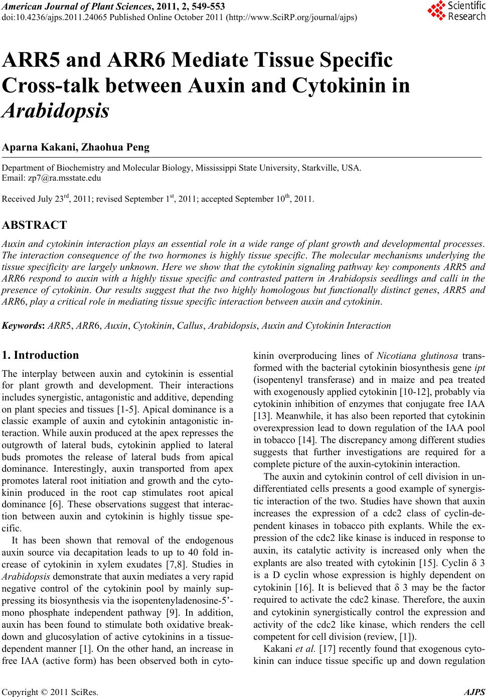 American Journal of Plant Sciences, 2011, 2, 549-553 doi:10.4236/ajps.2011.24065 Published Online October 2011 (http://www.SciRP.org/journal/ajps) Copyright © 2011 SciRes. AJPS 549 ARR5 and ARR6 Mediate Tissue Specific Cross-talk between Auxin and Cytokinin in Arabidopsis Aparna Kakani, Zhaohua Peng Department of Biochemistry and Molecular Biology, Mississippi State University, Starkville, USA. Email: [email protected] Received July 23rd, 2011; revised September 1st, 2011; accepted September 10th, 2011. ABSTRACT Auxin and cytokinin interaction plays an essential role in a wide range of plant growth and developmental processes. The interaction consequence of the two hormones is highly tissue specific. The molecular mechanisms underlying the tissue specificity are largely unknown. Here we show that the cytokinin signaling pathway key components ARR5 and ARR6 respond to auxin with a highly tissue specific and contrasted pattern in Arabidopsis seedlings and calli in the presence of cytokinin. Our results suggest that the two highly homologous but functionally distinct genes, ARR5 and ARR6, play a critical role in mediating tissue sp ecific interaction between auxin and cytokinin. Keywords: ARR5, ARR6, Auxin, Cytokinin, Callus, Arabidopsis, Auxin and Cyto kinin Interaction 1. Introduction The interplay between auxin and cytokinin is essential for plant growth and development. Their interactions includes synergistic, antagonistic and additive, depending on plant species and tissues [1-5]. Apical dominance is a classic example of auxin and cytokinin antagonistic in- teraction. While auxin produced at the apex represses the outgrowth of lateral buds, cytokinin applied to lateral buds promotes the release of lateral buds from apical dominance. Interestingly, auxin transported from apex promotes lateral root initiation and growth and the cyto- kinin produced in the root cap stimulates root apical dominance [6]. These observations suggest that interac- tion between auxin and cytokinin is highly tissue spe- cific. It has been shown that removal of the endogenous auxin source via decapitation leads to up to 40 fold in- crease of cytokinin in xylem exudates [7,8]. Studies in Arabidopsis demonstrate that auxin mediates a very rapid negative control of the cytokinin pool by mainly sup- pressing its biosynthesis via the isopentenyladenosine-5’- mono phosphate independent pathway [9]. In addition, auxin has been found to stimulate both oxidative break- down and glucosylation of active cytokinins in a tissue- dependent manner [1]. On the other hand, an increase in free IAA (active form) has been observed both in cyto- kinin overproducing lines of Nicotiana glutinosa trans- formed with the bacterial cytokinin biosynthesis gene ipt (isopentenyl transferase) and in maize and pea treated with exogenously applied cytokinin [10-12], probably via cytokinin inhibition of enzymes that conjugate free IAA [13]. Meanwhile, it has also been reported that cytokinin overexpression lead to down regulation of the IAA pool in tobacco [14]. The discrepancy among different studies suggests that further investigations are required for a complete picture of the auxin-cytokinin interaction. The auxin and cytokinin control of cell division in un- differentiated cells presents a good example of synergis- tic interaction of the two. Studies have shown that auxin increases the expression of a cdc2 class of cyclin-de- pendent kinases in tobacco pith explants. While the ex- pression of the cdc2 like kinase is induced in response to auxin, its catalytic activity is increased only when the explants are also treated with cytokinin [15]. Cyclin δ 3 is a D cyclin whose expression is highly dependent on cytokinin [16]. It is believed that δ 3 may be the factor required to activate the cdc2 kinase. Therefore, the auxin and cytokinin synergistically control the expression and activity of the cdc2 like kinase, which renders the cell competent for cell division (review, [1]). Kakani et al. [17] recently found that exogenous cyto- kinin can induce tissue specific up and down regulation  ARR5 and ARR6 Mediate Tissue Specific Cross-talk between Auxin and Cytokinin in Arabidopsis 550 of auxin in Arabidopsis. While auxin levels are reduced by exogenous cytokinin in cotyledons, auxin is elevated by exogenous cytokinin in roots and calli. More interest- ingly, they found that AUX1 plays a critical role in medi- ating cytokinin stimulated auxin accumulation in young roots and calli. Müller and Sheen reported that cytokinin and auxin interact antagonistically in root stem-cell specification in early embryogenesis [18]. Auxin antago- nizes cytokinin signaling by directly activating the rep- ressors of cytokinin signaling, ARR7 and ARR15. How- ever, how cytokinin and auxin interact during shoot stem cell specification was not reported. Both the auxin and cytokinin signaling pathways in- volve highly complicated networks that contain a large number of genes. The cytokinin signaling is perceived via a phosphorelay that is similar to the two-component systems used by bacteria for sensing and responding to environmental stimuli (recent review [18,19]). The path- way involves hybrid histidine protein kinases (AHK2, AHK3 and CRE1/WOL/AHK4) as receptors, histine phophotransfer proteins (AHPs), and nuclear response regulators (type A-ARRs and type B-ARRs). There are ten type-A ARR proteins (ARR3-ARR9 and ARR15- ARR17) and 11 type-B ARR proteins (ARR1, ARR2, ARR10-ARR14 and ARR18-ARR21) in Arabidopsis. The type-A ARRs were originally identified as cytokinin induced genes (review, [19]). At least eight of the ten type-A ARRs are negative regulators of cytokinin sig- naling with overlapping function. Both gene redundancy and tissue-specific roles have been observed among type- A ARRs in cytokinin response [20-22]. ARR5 and ARR6 share highest homology with each other compared with other members in the same gene family [21]. ARR5 ex- pression was found in the root and shoot meristems in the absence of exogenous cytokinin [23]. In the presence of exogenous cytokinin, the ARR5:GUS expression region was enlarged to include tissues around the shoot meris- tematic region in the shoot and all tissues in the roots, from the hypocotyl-root junction through the root tip. ARR6:GUS expression was detected in the shoot meris- tematic region and cotyledon vasculature in young seed- lings [21]. Cytokinin treatment resulted in overall higher level expression of ARR6:GUS, including tissues in hy- pocotyl and root except the root tip [21]. The arr5 knock- out mutant has an altered rosette morphology, which dis- appeared in arr5 arr6 double mutant, indicating that ARR5 and ARR6 have opposite functions [21]. Although the essential role of the interplay between auxin and cytokinin in plant growth and development has been well documented, little is known of the underlying molecular mechanisms. Most of the reported studies lim- ited to how the auxin and cytokinin levels modulated each other and in meristem tissue development. In this study, we report that the cytokinin induction of ARR5 and ARR6 expression is subjected to the regulation of auxin in a tissue and gene specific manner. The distinct auxin regulatory patterns of these two highly homolo- gous genes provide novel insights into the mechanisms underlying auxin and cytokinin interactions, especially the tissue specific interactions of these two hormones. 2. Materials and Methods 2.1. Arabidopsis Growth and Callus Induction Arabidopsis seeds were sterilized and placed on Gam- borg’s B5 medium (pH 5.7) supplemented with 1% su- crose, 0.9% agar, and 2,4-D (2,4-Dichlorophenoxyacetic acid) and KT (Kinetin) as indicated in the text. After 5 days of cold treatment at 4˚C, the Petri dishes were transferred to a 22˚C growth chamber for seed germina- tion or callus induction under 16 hrs of light and 8 hrs of dark. The light intensity was 150 mol m–2sec–2 for ger- mination and 50 mol m–2sec–2 for callus induction. 2.2. ARR5:GUS and ARR6:GUS Transgenic Lines The Arabidopsis (Columbia) seeds of ARR5:GUS and ARR6:GUS lines were ordered from Arabidopsis Bio- logical Research Center (ABRC) at the Ohio State Uni- versity [21]. 2.3. Histochemical Analyses of GUS Activities The histochemical stain of GUS was carried out as re- ported by Sessions et al. [24] without sectioning. Briefly, plant materials were stained in GUS staining solution (100 mM Sodium Phosphate at pH 7.0, 10 mM EDTA, 0.1% Triton X-100, 1 mM potassium ferricyanide, 1 mM potassium ferrocyanide and 1 mg/mL of X-Gluc [Gold Bio Technology, Inc.]). The samples were incubated at 37˚C overnight after being placed under a vacuum for 10 min in a desiccator. The staining solution was removed and the tissues were cleaned by incubating with several changes of 70% ethanol. The GUS images are acquired using a Zeiss Stemi SV11 (Apo) light Microscope. 3. Results and Discussions 3.1. Auxin Modulates ARR5 and ARR6 Expression with High Tissue Specificity and a Contrast Pattern In our previous studies, we found that cytokinin can in- duce auxin redistribution in seedlings and calli and AUX1 plays an essential role in mediating cytokinin in- duced auxin redistribution [17]. To examine how the cytokinin signaling pathways interact with auxin, we examined the expression of cytokinin pathway genes in response to exogenously applied auxin. ARR5 and ARR6 Copyright © 2011 SciRes. AJPS  ARR5 and ARR6 Mediate Tissue Specific Cross-talk between Auxin and Cytokinin in Arabidopsis 551 share highest sequence homology in the Arabidopsis ge- nome but may have opposite functions. We found that ARR5 expression was in the root and shoot meristems of wild-type seedlings as reported [23] (Figure 1(a)). In the presence of 0.2 mg/l KT, the ARR5:GUS expression was highly induced in roots, from the hypocotyl-root junction through the root tip, and regions surrounding the shoot apical meristem (Figure 1(b)). When auxin (2,4-D) was also applied, the ARR5:GUS expression in roots were strongly enhanced from the hypocotyl-root junction to root tip (Figure 1(c)-(f)). In contrast, ARR5:GUS ex- pression in cotyledons, shoot apical meristem, and hy- pocotyls were suppressed. When auxin concentration increased to 2.0 mg/l, the ARR5:GUS expression was completely shut off in coty- ledons and shoot apical meristem regions. But the gene expression in roots was very high. Interestingly, auxin regulation of ARR6 expression followed a contrast pattern. In wild-type seedlings, ARR6: GUS expression was detected in the shoot apical meris- tematic region and cotyledon vasculature (Figure 2(a)) [21]. Cytokinin treatment elevated the expression of ARR6:GUS, particularly in hypocotyls and roots as re- ported (Figure 2(b)). When low auxin (0.05 mg/l and 0.25 mg/l 2,4-D) was applied, ARR6:GUS expression was strongly induced in cotyledons, shoot apical meris- tem region, and hypocotyls. On the other hand, its ex- pression in roots was suppressed starting from the elon- gation zone. With the increase of 2,4-D, the suppressed region extended to hypocotyl region and root tip in both directions. When 2,4-D reached 2.0 mg/l, ARR6:GUS Figure 1. Histochemical assay of ARR5:GUS expression in response to cytokinin and auxin induction. The seedlings were germinated in B5 media supplemente d with KT and 2, 4-D in concentrations as indicated below. The seedlings were five days old. (a) Wild-type seedling; (b) Seedling grown in medium with 0.2 mg/l KT; (c) Seedling grown in medium with 0.2 mg/l KT and 0.05 mg/l 2,4-D. (d) Seedling grown in medium with 0.2 mg/l KT and 0.25 mg/l 2,4-D; (e) Seedling grown in medium with 0.2 mg/l KT and 1.0 mg/l 2, 4-D; (f) Seedling grown in medium with 0.2 mg/l KT and 2.0 mg/l 2,4-D. Figure 2. Histochemical assay of ARR6:GUS expression in response to cytokinin and auxin induction. The seedlings were germinated in B5 media supplemente d with KT and 2, 4-D in concentrations as indicated below. The seedlings were five days old. (a) Wild-type seedling; (b) Seedling grown in medium with 0.2 mg/l KT; (c) Seedling grown in medium with 0.2 mg/l KT and 0.05 mg/l 2,4-D. (d) Seedling grown in medium with 0.2 mg/l KT and 0.25 mg/l 2,4-D; (e) Seedling grown in medium with 0.2 mg/l KT and 1.0 mg/l 2, 4-D; (f) Seedling grown in medium with 0.2 mg/l KT and 2.0 mg/l 2,4-D. was expressed only in cotyledons. The expression in other tissues was almost completely suppressed. When the ARR6:GUS and ARR5:GUS expression patterns were compared (compare Figure 1 with Figure 2), it was clear that the expression of ARR5:GUS and ARR6:GUS followed a reversed pattern with the increase of auxin, suggesting a tissue specific role of these two genes in the interplay between auxin and cytokinin. While examining the mutant phenotype in Arabidopsis [21], it was observed that the reduced rosette size of ARR5 mutant was not enhanced by the mutation of ARR6. In- stead, the phenotype disappeared in the double mutant, indicating a different function of these two genes al- though these two genes have a high level of spatial over- lapping in gene expression in cotyledons and hypocotyls in wildtype seedlings. Given that ARR5 and ARR6 nega- tively regulate cytokinin response, our observations sug- gest that auxin suppress the cytokinin stimuli in plants via modulation of the negative regulators in the cytokinin pathway. The tissue specificity of auxin and cytokinin cross-talk is achieved via differential regulation of the highly homologous genes with different functions. 3.2. Auxin Regulation of ARR5 and ARR6 Expression in Calli To understand auxin and cytokinin interaction in calli, we examined the expression of ARR5:GUS and ARR6: GUS in calli induced from germinating seeds with dif- ferent auxin/cytokinin ratios and culturing time. ARR5: GUS was highly expressed in calli (Figure 3). Some cells, derived from cotyledons, barely expressed ARR5 Copyright © 2011 SciRes. AJPS  ARR5 and ARR6 Mediate Tissue Specific Cross-talk between Auxin and Cytokinin in Arabidopsis 552 Figure 3. Histochemical assay of ARR5:GUS expression in calli. The calli were induced in B5 media supplemented with KT and 2.4-D in concentrations as indicated below. (a) Four weeks old calli induced by 0.2 mg/l KT and 0.05 mg/l 2.4-D; (b) Four weeks old calli induced by 0.2 mg/l KT and 0.25 mg/l 2.4-D; (c) Four weeks old calli induced by 0.2 mg/l KT and 1.0 mg/l 2.4-D; (d) Four weeks old calli induced by 0.2 mg/l KT and 2.0 mg/l 2.4-D; (e) Six weeks old calli induced by 0.2 mg/l KT and 2.0 mg/l 2.4-D; (f) Eight weeks old calli induced by 0.2 mg/l KT and 2.0 mg/l 2.4-D. initially when auxin was high. ARR5:GUS became highly expressed across the entire callus after eight weeks of culture. In contrast, the expression of ARR6: GUS in calli was low (Figure 4). Although small portion of the tissues, which derived from cotyledons, expressed ARR6:GUS initially, the GUS expression substantially decreased with time. After eight weeks, the ARR6:GUS expression was barely detected in the calli. Our results suggest that ARR5 is highly expressed in calli but ARR6 is silenced in calli. Figure 4. Histochemical assay of ARR6:GUS expression in calli. The calli were induced in B5 media supplemented with KT and 2.4-D in concentrations as indicated below. (a) Four weeks old calli induced by 0.2 mg/l KT and 0.05 mg/l 2.4-D; (b) Four weeks old calli induced by 0.2 mg/l KT and 0.25 mg/l 2.4-D; (c) Four weeks old calli induced by 0.2 mg/l KT and 1.0 mg/l 2.4-D; (d) Four weeks old calli induced by 0.2 mg/l KT and 2.0 mg/l 2.4-D; (e) Six weeks old calli induced by 0.2 mg/l KT and 2.0 mg/l 2.4-D; (f) Eight weeks old calli induced by 0.2 mg/l KT and 2.0 mg/l 2.4-D. 4. Conclusions The interaction of auxin and cytokinin is essential to plant growth and development. The molecular mecha- nisms underlying the interaction, particularly the tissue specific response, are still poorly understood. Our results reported here suggest that auxin interacts with the cyto- kinin pathway by directly regulating the expression of negative regulators ARR5 and ARR6 in the cytokinin signaling pathway. The tissue specific response is a- chieved by differential regulation of homologous genes with distinct functions. 5. Acknowledgements We are grateful to Dr. Scott Willard, Dr. Jiaxu Li, Dr. Din-Pow Ma, and Ms Hana Mujahid for a critical reading of this manuscript. This research was approved for pub- lication as Journal Article J-10944 of the MAFES, Mis- sissippi State University. REFERENCES [1] C. Coenen and T. L. Lomax, “Auxin Cytokinin Interac- tions in Higher Plants: Old Problems and New Tools,” Trends in Plant Science, Vol. 2, 1997, pp. 351-356. doi:10.1016/S1360-1385(97)84623-7 [2] K. Hartig and E. Beck, “Crosstalk between Auxin, Cyto- kinins and Sugars in the Plant Cell Cycle,” Plant Biology (Stuttg), Vol. 8, No. 3, 2006, pp. 389-396. doi:10.1055/s-2006-923797 [3] R. Swarup, G. Parry, N. Graham, T. Allen and M. Ben- nett, “Auxin Cross-talk: Integration of Signaling Path- ways to Control Plant Development,” Plant Molecular Biology, Vol. 49, No. 3-4, 2002, pp. 411-426. doi:10.1023/A:1015250929138 [4] Y. -H. Su, Y. -B. Liu and X. -S. Zhang, “Auxin―Cyto- kinin Interaction Regulates Meristem Development,” Molecular Plant, 2011. [5] S. Perilli, L. Moubayidin and S. Sabatini, “The Molecular Basis of Cytokinin Function,” Current Opinion in Plant Biology, Vol. 13, No. 1, 2010, pp. 21-26. doi:10.1016/j.pbi.2009.09.018 [6] R. Aloni, E. Aloni, M. Langhans and C. I. Ullrich, “Role of Cytokinin and Auxin in Shaping Root Architecture: Regulating Vascular Differentiation, Lateral Root Initia- tion,” Root Apical Dominance and Root Gravitropism Annals of Botany, Vol. 97, 2006, pp. 883-893. [7] F. Bangerth, “Response of Cytokinin Concentration in the Xylem Exudates of Bean (Phaseolus vulgaris L.) Plants to Decapitation and Auxin Treatment, and Relationship to Apical Dominance,” Planta, Vol. 194, No. 4, 1994, pp. 439-442. doi:10.1007/BF00197546 [8] C. J. Li, E. Guevara, J. Herrera and F. Bangerth, “Effect of Apex Excision and Replacement by 1-Naphthy-Lacetic Acid on Cytokinin Concentration and Apical Dominance in Pea Plants,” Physiologia Plantarum, Vol. 94, No. 3, 1995, pp. 465-469. Copyright © 2011 SciRes. AJPS  ARR5 and ARR6 Mediate Tissue Specific Cross-talk between Auxin and Cytokinin in Arabidopsis Copyright © 2011 SciRes. AJPS 553 doi:10.1111/j.1399-3054.1995.tb00955.x [9] A. Nordstrom, P. Tarkowski, D. Tarkowska, R. Norbaek, C. Astot, K. Dolezal and G. Sandberg, “Auxin Regulation of Cytokinin Biosynthesis in Arabidopsis Thaliana: A Factor of Potential Importance for Auxin-cyto- kinin-Regulated Development,” Proceedings of the Na- tional Academy of Sciences of the United States of Amer- ica, Vol. 101, No. 21, 2004, pp. 8039-8044. doi:10.1073/pnas.0402504101 [10] G. Bertell and L. Eliasson, “Cytokinin Effects on Root-Growth and Possible Interactions with Ethylene and Indole-3-Acetic Acid,” Physiologia Plantarum, Vol. 84, No. 2, 1992, pp. 255-261. doi:10.1111/j.1399-3054.1992.tb04662.x [11] A. N. Binns, J. Labriola and R. C. Black, “Initiation of Auxin Autonomy in Nicotiana Glutinosa Cells by the Cytokinin-Biosysnthesis Gene from Agrobacterium Tumifaciens,” Planta, Vol. 171, No. 4, 1987, pp. 539-548. doi:10.1007/BF00392304 [12] M. Bourquin and P. E. Pilet, “Effect of Zeatin on the Growth and Indolyl-3-Acetic Acid and Abscisic Acid Levels in Maize Roots,” Physiologia Plantarum, Vol. 80, No. 3, 1990, pp. 342-349. doi:10.1111/j.1399-3054.1990.tb00051.x [13] W. K. Yip and S. F. Yang, “Effect of Thidiazuron, a Cyto kinin-Active Urea Derivative, in Cytokinin-Dependent Ethylene Production Systems,” Plant Physiology, Vol. 80, No. 2, 1986, pp. 515-519. doi:10.1104/pp.80.2.515 [14] S. Eklof, C. Astot, J. Blackwell, T. Moritz, O. Olsson and G. Sandberg, “Auxin-Cytokinin Interactions in Wildtype and Transgenic Tobacco,” Plant and Cell Physiology, Vol. 38, 1997, pp. 225-235. [15] P. C. L. John, K. Zhang, C. Dong, L. Diederich and F. Wightman, “P34 (CDC2) Related Proteins in Control of Cell Cycle Progression, the Switch between Division and Differentiation in Tissue Development and Stimulation of Division by Auxin and Cytokinin,” Australian Journal of Plant Physiology, Vol. 20, No. 5, 1993, pp. 503-526. doi:10.1071/PP9930503 [16] R. Soni, J. P. Carmichael, Z. H. Shah and J. A. H. Murray, “A Family of Cyclin D-Homologs from Plants Differen- tially Controlled by Growth Regulators and Containing the Conserved Retinoblastoma Protein Interaction Motif,” Plant Cell, Vol. 7, 1995, pp. 85-103. [17] A. Kakani, G. S. Li and Z. Peng, “Role of AUX1 in the Control of Organ Identity during in vitro Organogenesis and in Mediating Tissue Specific Auxin and Cytokinin Interaction in Arabidopsis,” Planta, Vol. 229, No. 3, 2009, pp. 645-657. doi:10.1007/s00425-008-0846-6 [18] B. Muller and J. Sheen, “Cytokinin and Auxin Interaction in Root Stem-Cell Specification during Early Embryo- genesis,” Nature, Vol. 453, 2008, pp. 1094-1097. doi:10.1038/nature06943 [19] J. P. C. To and J. J. Kieber, “Cytokinin Signaling: Two-Components and More,” Trends in Plant Science, Vol. 13, 2008, pp. 85-92. doi:10.1016/j.tplants.2007.11.005 [20] P. A. Salome, J. P. C. To, J. J. Kieber and C. R. McClung, “Arabidopsis Response Regulators ARR3 and ARR4 Play Cytokinin-Independent Roles in the Control of Circadian Period,” Plant Cell, Vol. 18, No. 1, 2006, pp. 55-69. doi:10.1105/tpc.105.037994 [21] J. P. C. To, et al., “Type-A Arabidopsis Response Regu- lators Are Partially Redundant Negative Regulators of Cytokinin Signaling,” Plant Cell, Vol. 16, No. 3, 2004, pp. 658-671. doi:10.1105/tpc.018978 [22] A. Leibfried, J. P. C. To, W. Busch, S. Stehling, A. Kehle, M. Demar, J. J. Kieber and J. U. Lohmann, “WUSCHEL Controls Meristem Function by Direct Regulation of Cy- tokinin-Inducible Response Regulators,” Nature, Vol. 438, 2005, pp. 1172-1175. doi:10.1038/nature04270 [23] I. B. D. Agostino, J. Deruere and J. J. Kieber, “Charac- terization of the Response of the Arabidopsis Response Regulator Gene Family to Cytokinin,” Plant Physiology, Vol. 124, No. 4, 2000, pp. 1706-1717. doi:10.1104/pp.124.4.1706 [24] A. Sessions, D. Weigel and M. F. Yanofsky, “The Arabidopsis Thaliana Meristem Layer 1 Promoter Speci- fies Epidermal Expression in Meristems and Young Pri- mordia,” Plant Journal, Vol. 20, No. 2, 1999, pp. 259-263. doi:10.1046/j.1365-313x.1999.00594.x |