

Paper Menu >>
Journal Menu >>
 Open Journal of Urology, 2011, 1, 86-88 doi:10.4236/oju.2011.14018 Published Online November 2011 (http://www.SciRP.org/journal/oju) Copyright © 2011 SciRes. OJU Xanthogranulomatous Cystitis Presenting as Urinary Incontinence and a Vesicovaginal Fistula: A Case Report and Review of the Literature Shivon N. Abdullah, Adamantios M. Mellis, Corinne L. Puzio, Daniel J. Culkin* Department of Urology, University of Oklah om a Health Sciences Cent er , Okl a ho m a Ci t y, USA E-mail: *[email protected] Received June 7, 2011; revised July 15, 2011; ac cepted Ju ly 28, 2011 Abstract Anthogranulomatous cystitis (XC) is an inflammatory condition of the urinary bladder that is benign and rarely seen. XC is a unique disease in which there have been only 26 cases described in the literature, in- cluding our case. Patients with XC often present with lower urinary tract symptoms, hematuria, abdominal pain, or abdominal mass. We present the unusual case of an 81 year old female who presents with urinary incontinence, which was later diagnosed as a vesicovaginal fistula. After describing the treatment of this pa- tient, a review of the literature is detailed including presentation, possible etiologies, and treatment of XC. Keywords: Xanthogranulomatous Cystitis, Inflammation, Bladder, Vesicovaginal Fistula, Urinary Incontinence 1. Introduction Xanthogranulomatous cystitis is an uncommon but be- nign inflammatory condition of the bladder that is chronic in duration for which the etiology is unknown [1]. XC was first described by Ota in 1933[2]. Histologically, the disease is characterized by multinucleated giant cells and chronic inflammation. The characteristic cell is the lipid-laden macrophages, known as “foamy histiocytes,” which stain strongly for CD 68. Although the kidney is the most common site of xanthogranulomatous disease, there are currently twenty-five previously described cases of this disease reported in the bladder [3]. Most of these cases involve urachal remnants with disease most often isolated to the dome of the bladder [4]. We add our case of XC in an 81 year old female with the unusual initial presentation of urinary incontinence and vesico- vaginal fistula (VVF) and provide a brief review of the literature 2. Case Report An 81 year old Caucasian female of German descent was referred to clinic with primary complaint of continuous urinary incontinence which had persisted for several months and believed to be secondary to stress urinary incontinence. The patient also had symptoms of urinary frequency and intermittent gross hematuria. However, the patient denied having any urinary tract infections. She also reported a significant unintentional weight loss in the past year. Past medical history included hyperten- sion, diabetes mellitus type II, as well as dyslipidemia. On physical examination, the patient was found to have a large tumor involving the anterior vaginal wall with pooling of urine in the vaginal vault, suspicious for a vesicovaginal fistula. Laboratory analysis revealed nor- mal renal function, absen ce of hematolo gic abnormalities, including eosinophilia. Urinalysis and urine culture fai- led to show to urinary tract infection. Con trast computed tomography with urogram phase revealed a midline pos- terior bladder wall lesion and a 4 mm communication between the posterior bladder wall and the anterior vagi- nal wall (Figures 1 and 2). Cystoscopy was performed identifying significant amount of bullous edema as well as a bulky solid lobu- lated mass approximately 2 cm in size in the area of the left posterior bladder neck extending to the trigone of the bladder. A midline vesicovaginal fistula was identified in the posterior bladder wall. The lateral walls of the blad- der, as well as the dome of the bladder were free of masses or inflammation. Due to concern for malignancy, endoscopic biopsy and resection were performed of the 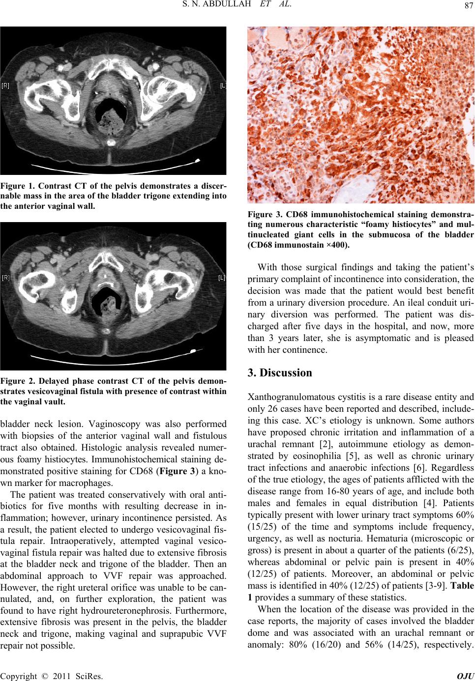 87 S. N. ABDULLAH ET AL. Figure 1. Contrast CT of the pelvis demonstrates a discer- nable mass in the area of the bladder trigone extending into the anterior vaginal wall. Figure 2. Delayed phase contrast CT of the pelvis demon- strates vesicovaginal fistula with presence of contrast within the vaginal vault. bladder neck lesion. Vaginoscopy was also performed with biopsies of the anterior vaginal wall and fistulous tract also obtained. Histologic analysis revealed numer- ous foamy histiocytes. Immunohistochemical staining de- monstrated positive staining for CD68 (Figure 3) a kno- wn marker fo r macrophages. The patient was treated conservatively with oral anti- biotics for five months with resulting decrease in in- flammation; however, urinary incontinence persisted. As a result, the patient elected to undergo vesicovaginal fis- tula repair. Intraoperatively, attempted vaginal vesico- vaginal fistula repair was halted due to extensive fibrosis at the bladder neck and trigone of the bladder. Then an abdominal approach to VVF repair was approached. However, the right ureteral orifice was unable to be can- nulated, and, on further exploration, the patient was found to have right hydroureteronephrosis. Furthermore, extensive fibrosis was present in the pelvis, the bladder neck and trigone, making vaginal and suprapubic VVF repair not possible. Figure 3. CD68 immunohistochemical staining demonstra- ting numerous characteristic “foamy histiocytes” and mul- tinucleated giant cells in the submucosa of the bladder (CD68 immunostain ×400). With those surgical findings and taking the patient’s primary complaint of incontinence into consideration, the decision was made that the patient would best benefit from a urinary diversion procedure. An ileal conduit uri- nary diversion was performed. The patient was dis- charged after five days in the hospital, and now, more than 3 years later, she is asymptomatic and is pleased with her continence. 3. Discussion Xanthogranulomatous cystitis is a rare disease entity and only 26 cases have been reported and described, include- ing this case. XC’s etiology is unknown. Some authors have proposed chronic irritation and inflammation of a urachal remnant [2], autoimmune etiology as demon- strated by eosinophilia [5], as well as chronic urinary tract infections and anaerobic infections [6]. Regardless of the true etiology, the ag es of patients afflicted with the disease range from 16-80 years of age, and include both males and females in equal distribution [4]. Patients typically present with lower urinary tract symptoms 60% (15/25) of the time and symptoms include frequency, urgency, as well as nocturia. Hematuria (microscopic or gross) is present in ab out a quarter of the patients (6 /25), whereas abdominal or pelvic pain is present in 40% (12/25) of patients. Moreover, an abdominal or pelvic mass is identified in 40% (1 2/25) of patien ts [3-9]. Table 1 provides a summary of these statistics. When the location of the disease was provided in the case reports, the majority of cases involved the bladder dome and was associated with an urachal remnant or anomaly: 80% (16/20) and 56% (14/25), respectively. Copyright © 2011 SciRes. OJU 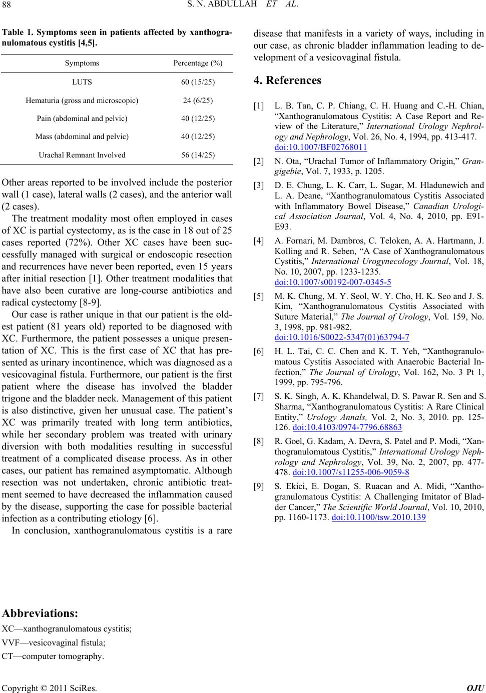 S. N. ABDULLAH ET AL. Copyright © 2011 SciRes. OJU 88 Table 1. Symptoms seen in patients affected by xanthogra- nulomatous cystitis [4,5]. Symptoms Percentage (%) LUTS 60 (15/25) Hematuria (gr oss a nd microscopic) 24 (6/25) Pain (abdominal and pelvic) 40 (12/25) Mass (abdominal and pelvic) 40 (12/25) Urachal Remnant Involved 56 (14/25) Other areas reported to be involved include the posterior wall (1 case), lateral walls (2 cases), and the anterior wall (2 cases). The treatment modality most often employed in cases of XC is partial cystectomy, as is the case in 18 out of 25 cases reported (72%). Other XC cases have been suc- cessfully managed with surgical or endoscopic resection and recurrences have never been reported, even 15 years after initial resection [1]. Other treatment modalities that have also been curative are long-course antibiotics and radical cystectomy [8-9]. Our case is rather unique in that our patient is the old- est patient (81 years old) reported to be diagnosed with XC. Furthermore, the patient possesses a unique presen- tation of XC. This is the first case of XC that has pre- sented as urinary incontinence, which was diagnosed as a vesicovaginal fistula. Furthermore, our patient is the first patient where the disease has involved the bladder trigone and the bladder neck. Management of this patient is also distinctive, given her unusual case. The patient’s XC was primarily treated with long term antibiotics, while her secondary problem was treated with urinary diversion with both modalities resulting in successful treatment of a complicated disease process. As in other cases, our patient has remained asymptomatic. Although resection was not undertaken, chronic antibiotic treat- ment seemed to have decreased the inflammation caused by the disease, supporting the case for possible bacterial infection as a contributing etiology [6]. In conclusion, xanthogranulomatous cystitis is a rare disease that manifests in a variety of ways, including in our case, as chronic bladder inflammation leading to de- velopment of a vesicovaginal fistula. 4. References [1] L. B. Tan, C. P. Chiang, C. H. Huang and C.-H. Chian, “Xanthogranulomatous Cystitis: A Case Report and Re- view of the Literature,” International Urology Nephrol- ogy and Nephrology, Vol. 26, No. 4, 1994, pp. 413-417. doi:10.1007/BF02768011 [2] N. Ota, “Urachal Tumor of Inflammatory Origin,” Gran- gigebie, Vol. 7, 1933, p. 1205. [3] D. E. Chung, L. K. Carr, L. Sugar, M. Hladunewich and L. A. Deane, “Xanthogranulomatous Cystitis Associated with Inflammatory Bowel Disease,” Canadian Urologi- cal Association Journal, Vol. 4, No. 4, 2010, pp. E91- E93. [4] A. Fornari, M. Dambros, C. Teloken, A. A. Hartmann, J. Kolling and R. Seben, “A Case of Xanthogranulomatous Cystitis,” International Urogynecology Journal, Vol. 18, No. 10, 2007, pp. 1233-1235. doi:10.1007/s00192-007-0345-5 [5] M. K. Chung, M. Y. Seol, W. Y. Cho, H. K. Seo and J. S. Kim, “Xanthogranulomatous Cystitis Associated with Suture Material,” The Journal of Urology, Vol. 159, No. 3, 1998, pp. 981-982. doi:10.1016/S0022-5347(01)63794-7 [6] H. L. Tai, C. C. Chen and K. T. Yeh, “Xanthogranulo- matous Cystitis Associated with Anaerobic Bacterial In- fection,” The Journal of Urology, Vol. 162, No. 3 Pt 1, 1999, pp. 795-796. [7] S. K. Singh, A. K. Khandelwal, D. S. Pawar R. Sen and S. Sharma, “Xanthogranulomatous Cystitis: A Rare Clinical Entity,” Urology Annals, Vol. 2, No. 3, 2010. pp. 125- 126. doi:10.4103/0974-7796.68863 [8] R. Goel, G. Kadam, A. Devra, S. Patel and P. Modi, “Xan- thogranulomatous Cystitis,” International Urology Neph- rology and Nephrology, Vol. 39, No. 2, 2007, pp. 477- 478. doi:10.1007/s11255-006-9059-8 [9] S. Ekici, E. Dogan, S. Ruacan and A. Midi, “Xantho- granulomatous Cystitis: A Challenging Imitator of Blad- der Cancer,” The Scientific World Journal, Vol. 10, 2010, pp. 1160-1173. doi:10.1100/tsw.2010.139 Abbreviations: XC—xanthogranulomatous cystitis; VVF—vesicovaginal fistula; CT—computer tomography. |