

Paper Menu >>
Journal Menu >>
 Advances in Chemical Engi neering and Science , 2011, 1, 299-304 doi:10.4236/aces.2011.14041 Published Online October 2011 (http://www.SciRP.org/journal/aces) Copyright © 2011 SciRes. ACES Preparation of Highly Concentrated Silver Nanoparticles in Reverse Micelles of Sucrose Fatty Acid Esters through Solid-Liquid Extraction Method Hidetaka Noritomi*, Yoshihiro Umezawa, Saori Miyagawa, Satoru Kato Department of Applied Chemistry, Tokyo Metropolitan University, Tokyo, Japan E-mail: *[email protected] Recieved August 12, 2011; revised September 16, 2011; accepted September 20, 2011 Abstract Silver nanoparticles were synthesized in reverse micelles consisting of sucrose fatty acid esters by dissolving reactant powder in the water pool of reverse micelles through the solid-liquid extraction. Silver nanoparticles having various sizes and shapes were obtained at high concentration. The size of silver nanoparticles was controlled by reaction temperature. Moreover, the size of silver nanoparticles was dependent upon the aver- age esterification degree of sucrose fatty acid esters forming reverse micelles. The wavelength in the peaks, which corresponded upon the localized surface plasmon resonance of resultant silver nanoparticles, was cor- related with their sizes. Keywords: Reverse Micelle, Silver Nanoparticle, Size Control, Solid-Liquid Extraction, Sucrose Fatty Acid Ester 1. Introduction In recent years, the production and applications of metal nanoparticles have rapidly been increased in the field of nanotechnology, since their novel physical and chemical properties are not only different from those of bulk sub- stances due to their extremely small size and large spe- cific surface area, but also metal nanoparticles exhibit specific colors due to localized surface plasmon reso- nance (LSPR) corresponding to the wavelength of light that induces the largest electromagnetic field on the nanopartices, when metal nanoparticles are impinged by a beam of light [1]. Especially, the applications of silver nanoparticles have widely been investigated, since they exhibit some profitable properties such as catalysis [2], antibacterial agent [3], nanoparticle colorant [4], nano- paste for electrical circuits and electrodes [5], and sub- strates for surface-enhanced Raman scattering [6]. The colors of silver nanoparticles result from changes of LSPR induced by their size and shape. In order to produce metal nanoparticles having suit- able sizes and/or shapes, many physical or chemical techniques such as coprecipitation, gas-evaporation, sol- gel method, and sputtering have been developed so far [1]. Additionally, more attention has been paid on the preparation of metal nanoparticles in reverse micelles [7]. Reverse micelles are thermodynamically stable nanome- ter-sized aggregates of surfactant molecules dispersed in a hydrophobic organic phase like octane, and can form w/o type microemulsions containing a small amount of water in their centers. The water phase in w/o type mi- croemulsions is called water pool. Metal nanoparticles are synthesized in the water pool, in which reactants are dissolved. As the preparation of metal nanoparticles us- ing reverse micelles does not require a special apparatus and extreme conditions of temperature and pressure, it is comparatively easy to expand the scale of reverse micel- lar reaction system. However, the productivity of metal nanoparticles per volume of reverse micellar system is limited due to low overall concentration of reactants in the reverse micellar system, since the volumetric ratio of water phase playing a role as the dissolution of reactants and the reaction field to the bulk organic phase is too small [1]. In order to solubilize reactants in reverse micelles, three solubilization methods are utilized: injection me- thod; liquid-liquid extraction method; solid-liquid ex- traction method [8]. First, the injection method is carried out by injecting a few microliters of the concentrated stock solution of reactants into the hydrocarbon solution  300 H. NORITOMI ET AL. of surfactants. This method is most commonly used to prepare metal nanoparticles in reverse micelles. Second, the liquid-liquid extraction method is carried out by transferring solutes dissolved in an aqueous phase from the aqueous phase into the hydrocarbon phase of surface- tants. This method is relatively slow, and the concentra- tion of reactants in the hydrocarbon phase is limited due to the distribution between aqueous and reverse micellar organic phases in thermodynamic equ ilibrium. Third, the solid-liquid extraction method is carried out by mixing solid reactants with the reverse micellar solution con- taining already a certain amount of water. This method can dissolve reactants until their dissolution reaches saturation in water pool. Moreover, when using the solid-liquid extraction instead of the conventional inject- tion method to solubilize reactants into reverse micelles, the following benefits are expected. First, the productive- ity of metal nanoparticles is markedly promoted, since reactants consumed for the formation of metal nanoparti- cles are successively supplied from solid reactants al- ready added in large excess, compared to the solubility of the water pool of reverse micelles. Second, only a small amount of water is needed for solid-liquid extrac- tion. Consequently, the aggregation of resultant nanopar- ticles is inhibited, since the lower water content is kept in reverse micellar system, the more stably nanoparticles are dispersed [9]. However, there have not been any re- ports about the preparation of nanoparticles in reverse micelles using the solid -liquid extraction method. In our previous work, we have reported that silver nanoparticles are prepared in reverse micelles of sucrose fatty acid esters by using the conventional injection method [10]. Sucrose fatty acid esters are commercial food grade nonionic surfactants, and are biodegradable and nonhazardous to the environment [11]. In the present work, we examined the preparation of silver nanoparti- cles in reverse micelles of sucrose fatty acid esters by using solid-liquid extraction method to address how the preparation conditions such as the reaction temperature and the composition of surfactants affect the size and shape of silver nanoparticles. 2. Experimental Silver nitrate and sodium borohydride were the guaran- teed reagents of Kanto Chemicals (Tokyo, Japan). DK- SS (sucrose fatty acids of 99 wt% monoesters and 1 wt% di- and triesters, fatty acid constituent consisting of 60 wt% stearic acid and 40 wt% palmitic acid) and DK-F- 20W (sucrose fatty acids of 11wt% monoesters, 36wt% di- and triesters, and 53 wt% more than tetraesters, fatty acid constituent con sisting of 70 wt% stearic acid and 30 wt% palmitic acid) were supplied from Dai-Ichi Kogyo Seiyaku (Kyoto, Japan). The surfactant was used without further purification. Isooctane and n-butanol were from Kanto Chemicals (Tokyo, Japan), and were of analytical grade. The reverse micellar solutions were prepared by add- ing the required amounts of sucrose fatty acid esters and a small amount of water into the solution of n-butanol/ isooctane (3:7 (v/v)), and then were used for the prepa- ration of nanoparticles within a few minutes. The preparation of nanoparticles was carried out by adding a certain amounts of AgNO3 powder and sodium borohydride powder into the reverse micellar solution. Since the preliminary study indicated that the synthe- sis of particles finished at 25˚C after 3 h, and no residual solid reactants existed in the reaction medium, the sam- ples for various analyses were taken after 3 h. In the pre- sent work, the concentrations of sucrose fatty acid esters and water based on the overall volume of reverse micel- lar solution were fixed at 50 g/L and 60 mM, respect- tively. As a typical condition, the overall concentrations of AgNO3 and sodium borohydride were 0.1 and 1.0 M, respectively, and the temperature was fixed at 25˚C. The TEM micrograph was obtained using a JEOL JEM-2000FX electron microscope operating at 200 kV. The sample for TEM was prepared by dilutin g a resultan t colloidal solution one hundred-fold with n-butanol and placing a drop of colloidal solution onto the standard carbon-coated copper grids and drying it under vacuum. The UV-vis spectra of the reverse micellar solutions containing nanoparticles were measured by UV/vis spec- trophotometer (Ubest-55, Japan Spectroscopic Co. Ltd.) with a 10 mm quartz cell. 3. Results and Discussion 3.1. Productivity of Silvar Nanoparticles through Solid-Liquid Extraction Method In order to successively supply reactants consumed for the formation of silver nanoparticles to the water poo ls of reverse micelles, which are reaction fields, and produce silver nanoparticles at high concentration, we have em- ployed solid-liquid extraction method instead of the conventional injection method [8]. The colloidal solution of silver nanoparticles prepared by solid-liqu id ex traction method was much concentrated, compared to that pre- pared by the injection method examined in our previous method [10]. The concentration of silver nanoparticles obtained by the present system was about one hundred and thirty times larger than that obtained by the injection method. After diluting the resultant colloidal solution one hundred-fold with n-butanol, the measurement of UV-visible absorption spectrum was carried out. The Copyright © 2011 SciRes. ACES  H. NORITOMI ET AL. 301 color of the solution after dilution was yello w, similar to the case of injection method, and, as shown in Figure 1, its UV-visible absorption spectrum exhibited the peak around 403 nm, which corresponds upon the LSPR of silver nanoparticles [12]. Figure 2 shows the relationship between surfactant concentration and the absorbance at 403 nm of colloidal solutions of silver na noparticles pre- liminarily diluted one handred-fold with n-butanol. Any absorbance at 403 nm was not observed without surfac- tant. On the other hand, the absorbance at 403 nm in- creased above 20 g/L of surfactant concentration. We have reported that the apparent critical micelle concen- tration in sucrose fatty acid/n-butanol/isooctane system was observed around 10 g/L [13]. Consequently, it is suggested that the preparation of silver nanoparticles proceeds as follows. First, empty reverse micelles con- taining a small amount of water approach solid reactants added in large excess, compared to the solubility of the water pool of reverse micelles. When the water pools of reverse micelles contact solid reactants, reactants are solubilized into the water pools. Then, silver nanoparti- cles are synthesized from reactants solubilized in reverse micelles. Resultant silver nanoparticles are coated by surfactants, and are dispersed in organic solvents. Emp- Figure 1. Absorption spectrum of silver nanoparticles pre- pared by solid-liquid extraction method. Figure 2. Effect of surfactant concentration on absorbance at 403 nm of colloidal solution dispersing silver nanoparti- cles prepared by solid-liquid extraction method: [AgNO3]ov = 0.1 M; [NaBH4]ov = 1.0 M; n-butanol/isooctane containing 50 g/L DK-SS and 60 mM H2O; reaction time = 3 h; reac- tion temperature = 25˚C. tied reverse micelles approach solid reactants again. Thus, the production of silver nanoparticles is promoted by repeating the process composed of the extraction of reactants, the synthesis of silver nanoparticles, and the dispersion of resultant silver nanoparticles. 3.2. Dependence of Reaction Temperature upon Size of Silver Nanopartices Figure 3 shows the typical transmission electron micro- graphs and size distributions of silver nanoparticles when silver nanoparticles were prepared in the reverse micellar system of DK-SS for 3 h at different reaction tempera- tures. The TEM images showed that the obtained silver nanoparticles displayed a wide variety of shapes. The size of resultant silver nanoparticles was almost similar to that prepared in DK-SS or AOT (sodium bis(2-ethyl- hexyl) sulfosuccinate) reverse micelles by the conven- tional injection method [10,14,15]. The size of silver na- noparticles was strongly dependent upon reaction tem- perature. Figure 4 shows the plots of the mean diameter of silver nanoparticles against reaction temperature. As seen in the figure, the mean diameter of silver nanoparti- cles gradually increased with an increase in reaction temperature. The line in the figure represents the fitting curve with the correlation coefficient of 0.99 as 0.079 6.2dt (1) where d is the mean diameter of silver nanoparticles, and t is the reaction temperature. This tendency was similar to the case using the conventional injection method [10]. The solubilization of reactants, the exchange of reactants between reverse micelles, the reduction reaction, and the growth of nanoparticles increase with increasing the re- action temperature [16-19]. Figure 5 shows the plots of the mean diameter of sil- ver nanoparticles obtained at different reaction tempera- ture against the wavelength in the peak of UV-visible absorption spectrum due to the LSPR of silver nanopar- ticles. The standard deviation concerning the size distri- bution of silver nanoparticles depended upon reaction temperature. The wavelength in the peaks increases with an increase in the mean diameter of silver nanoparticles. The line in the figure represents th e fitting curve with the correlation coefficient of 0.99 as λ = 8.2d + 340 (2) where λ is the wavelength of peaks, and d is the mean diameter of silver nanoparticles. The position of the plasmon adsorption peak depends upon the particle size and shape, and especially tends to be red-shifted with ncreasing the particle size [14,16,20,21]. i Copyright © 2011 SciRes. ACES  H. NORITOMI ET AL. Copyright © 2011 SciRes. ACES 302 Figure 3. Transmission electron micrographs and particle size distributions of silver nanoparticles synthesized in DK-SS re- verse micelles: reaction temperature = (a) 5˚C, (b) 15˚C, (c) 25˚C, and (d) 40˚C. 3.3. Dependence of Average Esterification Degree of Surfactants upon Size of Silver Nanoparticles The formation and stability of micelles are due to the structure and/or HLB of surfactants [22-24]. The struc- ture and HLB of sucrose fatty acid esters alter with the esterification degree [11]. In order to elucidate the effect of those factors on the formation of silver nanoparticles, we have examined the synthesis of silver nanoparticles in the reverse micelles of sucrose fatty acid esters by mix- ing DK-SS (average esterification degree = 1.01, HLB = 19) with DK-F-20W (average esterification degree = 3.1, HLB = 2). The more weight fraction of DK-F-20W is, the larger the average esterification degree is. Figure 6 shows the typical transmission electron micrographs and size distributions of silver nanoparticles when silver nanoparticles were prepared in the reverse micellar sys- tem of sucrose fatty acid esters at different average es- terification degrees for 3 h. The silver nanoparticles in various shapes were obtained in those reverse micellar systems. The standard deviation concerning the size dis- tribution of silver nanoparticles was strongly dependent upon the average esterification degree of surfactants. When the average esterification degree was 1.85, the minimal standard deviation was obtained. This result indicated that at that average esterification degree, mono- disperse silver nanoparticles were formed. As shown in Figure 7, the mean diameter of silver nanoparticles was dependent upon the average sterification degree, similar Figure 4. Effect of reaction temperature on the mean di- ameters of silver nanoparticles in reverse micelles of DK-SS: [AgNO3]ov = 0.1 M; [NaBH4]ov = 1.0 M; n-butanol/ isooctane containing 50 g/L DK-SS and 60 mM H2O; reaction time = 3 h. Figure 5. Relationship of the mean diameter of silver nano- particles obtained at different reaction temperature with the wavelength in the peak of UV-visible absorption spec- trum due to the LSPR of silver nanoparticles. e 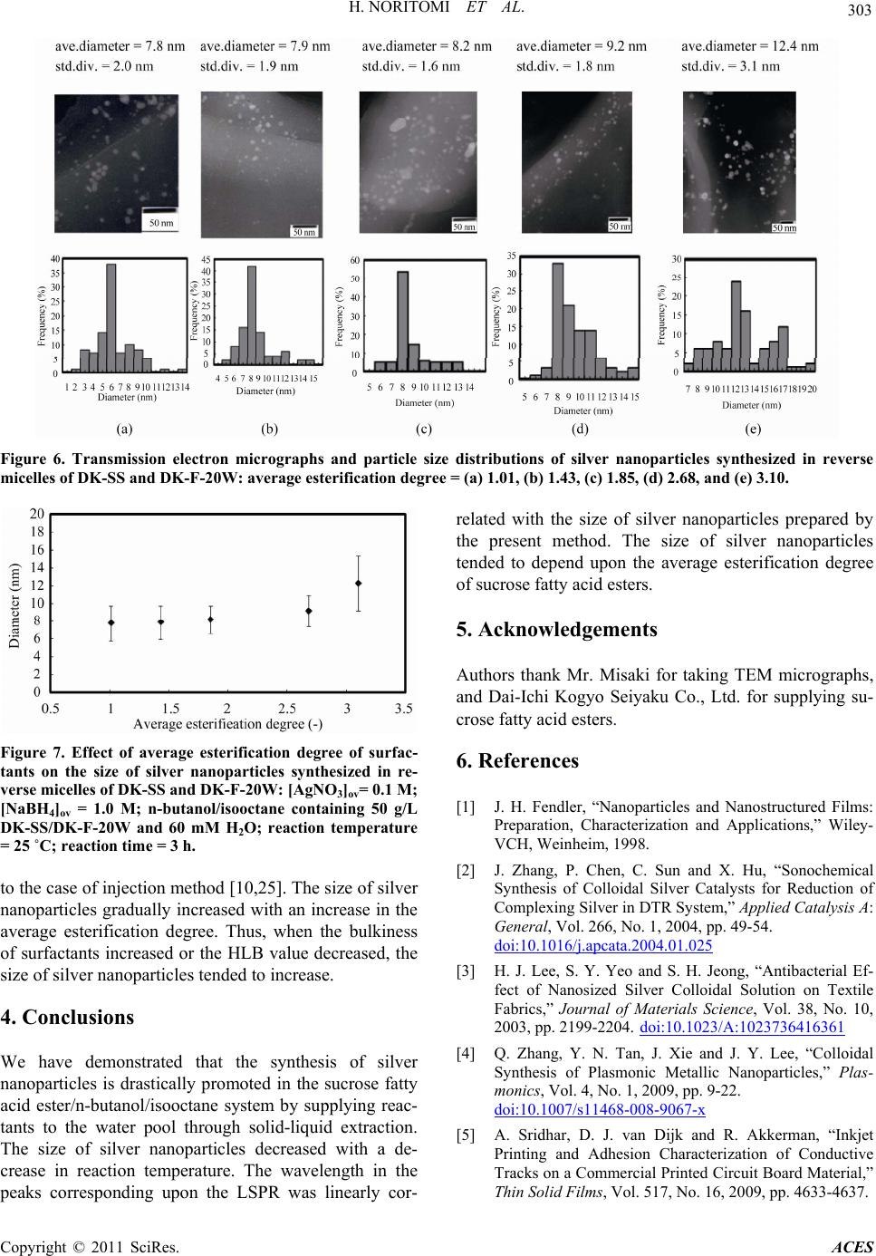 H. NORITOMI ET AL. 303 Figure 6. Transmission electron micrographs and particle size distributions of silver nanoparticles synthesized in reverse micelles of DK-SS and DK-F-20W: average esterification degree = (a) 1.01, (b) 1.43, (c) 1.85, (d) 2.68, and (e) 3.10. Figure 7. Effect of average esterification degree of surfac- tants on the size of silver nanoparticles synthesized in re- verse micelles of DK-SS and DK-F-20W: [AgNO3]ov= 0.1 M; [NaBH4]ov = 1.0 M; n-butanol/isooctane containing 50 g/L DK-SS/DK-F-20W and 60 mM H2O; reaction temperature = 25 ˚C; reaction time = 3 h. to the case of injection method [10,25]. The size of silver nanoparticles gradually increased with an increase in the average esterification degree. Thus, when the bulkiness of surfactants increased or the HLB value decreased, the size of silver nanoparticles tended to increase. 4. Conclusions We have demonstrated that the synthesis of silver nanoparticles is drastically promoted in the sucrose fatty acid ester/n-butanol/isooctane system by supplying reac- tants to the water pool through solid-liquid extraction. The size of silver nanoparticles decreased with a de- crease in reaction temperature. The wavelength in the peaks corresponding upon the LSPR was linearly cor- related with the size of silver nanoparticles prepared by the present method. The size of silver nanoparticles tended to depend upon the average esterification degree of sucrose fatty acid esters. 5. Acknowledgements Authors thank Mr. Misaki for taking TEM micrographs, and Dai-Ichi Kogyo Seiyaku Co., Ltd. for supplying su- crose fatty acid esters. 6. References [1] J. H. Fendler, “Nanoparticles and Nanostructured Films: Preparation, Characterization and Applications,” Wiley- VCH, Weinheim, 1998. [2] J. Zhang, P. Chen, C. Sun and X. Hu, “Sonochemical Synthesis of Colloidal Silver Catalysts for Reduction of Complexing Silver in DTR System,” Applied Catalysis A: General, Vol. 266, No. 1, 2004, pp. 49-54. doi:10.1016/j.apcata.2004.01.025 [3] H. J. Lee, S. Y. Yeo and S. H. Jeong, “Antibacterial Ef- fect of Nanosized Silver Colloidal Solution on Textile Fabrics,” Journal of Materials Science, Vol. 38, No. 10, 2003, pp. 2199-2204. doi:10.1023/A:1023736416361 [4] Q. Zhang, Y. N. Tan, J. Xie and J. Y. Lee, “Colloidal Synthesis of Plasmonic Metallic Nanoparticles,” Plas- monics, Vol. 4, No. 1, 2009, pp. 9-22. doi:10.1007/s11468-008-9067-x [5] A. Sridhar, D. J. van Dijk and R. Akkerman, “Inkjet Printing and Adhesion Characterization of Conductive Tracks on a Commercial Printed Circuit Board Material,” Thin Solid Films, Vol. 517, No. 16, 2009, pp. 4633-4637. Copyright © 2011 SciRes. ACES  304 H. NORITOMI ET AL. doi:10.1016/j.tsf.2009.03.133 [6] J. Chen, Y. Luo, Y. Liang, J. Jiang, G. Shen and R. Yu, “Surface-Enhanced Raman Scattering for Immunoassay Based on the Biocatalytic Production of Silver Nanopar- ticles,” Analytical Sciences, Vol. 25, No. 3, 2009, pp. 347-352. doi:10.2116/analsci.25.347 [7] V. Uskokovic and M. Drofenik, “Synthesis of Materials within Reverse Micelles,” Surface Review and Letters, Vol. 12, No. 2, 2005, pp. 239-277. doi:10.1142/S0218625X05007001 [8] P. L. Luisi, “Enzymes Hosted in Reverse Micelles in Hydrocarbon Solution,” Angewandte Chemie, Vol. 24, No. 6, 1985, pp. 439-528. doi:10.1002/anie.198504393 [9] A. Kitahara, T. Tamura and S. Matsumura, “Effect of Water on Stability of Carbon Black Dispersed in Nona- queous Aerosol OT Solutions,” Chemistry Letters, Vol. 8, No. 9, 1979, pp. 1127-1128. [10] H. Noritomi, N. Igari, K. Kagitani, Y. Umezawa, Y. Mu- ratsubaki and S. Kato, “Synthesis and Size Control of Silver Nanoparticles Using Reverse Micelles of Sucrose Fatty Acid Esters,” Colloid and Polymer Science, Vol. 288, No. 8, 2010, pp. 887-891. doi:10.1007/s00396-010-2214-x [11] N. Otomo, “Self-Organized Structures of Food Emulsifi- ers and Their Applications,” Japan Journal of Food En- gineering, Vol. 4, No. 1, 2003, pp. 1-9. [12] A. Henglein, “Physicochemical Properties of Small Metal Particles in Solution: ‘Microelectrode’ Reactions, Chemi- sorption, Composite Metal Particles, and the Astom-to- Metal Transition,” Journal of Physical Chemistry, Vol. 97, No. 21, 1993, pp. 5457-5471. doi:10.1021/j100123a004 [13] H. Noritomi, S. Ito, N. Kojima, S. Kato and K. Nagahama, “Forward and Backward Extractions of Cytochrome C Using Reverse Micellar System of Sucrose Fatty Acid Ester,” Colloid and Polymer Science, Vol. 284, No. 6, 2006, pp. 604-610. doi:10.1007/s00396-005-1398-y [14] A. Taleb, C. Petit and M. P. Pileni, “Synthesis of Highly Monodisperse Silver Nanoparticles from AOT Reverse Micelles: A Way to 2D and 3D Self-Organization,” Che- misty of Materials, Vol. 9, No. 4, 1997, pp. 950-959. doi:10.1021/cm960513y [15] W. Zhang, X. Qiao, J. Chen and H. Wang, “Preparation of Silver Nanoparticles in Water-in-Oil AOT Reverse Micelles,” Journal of Colloid and Interface Science, Vol. 302, No. 1, 2006, pp. 370-373. doi:10.1016/j.jcis.2006.06.035 [16] R. P. Bagwe and K. C. Khilar, “Effects of Intermicellar Exchange Rate on the Formation of Silver Nanoparticles in Reverse Microemulsions of AOT,” Langmuir, Vol. 16, No. 3, 2000, pp. 905-910. doi:10.1021/la980248q [17] W. L. Hinze, “Organized Assemblies in Chemical Analy- sis Vol. 1: Reverse Micelles,” JAI Press, London, 1994. [18] C. Tojo, M. C. Blanco and M. A. Lopez-Quintela, “The Influence of Reactant Excess and Film Flexibility on the Mechanism of Nanoparticle Formation in Microemul- sions: A Monte Carlo Simulation,” Langmuir, Vol. 14, No. 24, 1998, pp. 6835-6839. doi:10.1021/la980693l [19] J. P. Cason, M. E. Miller, J. B. Thompson and C. B. Ro- berts, “Solvent Effects on Copper Nanoparticle Growth Behavior in AOT Reverse Micelle Systems,” Journal of Physical Chemistry B, Vol. 105, No. 12, 2001, pp. 2297- 2302. doi:10.1021/jp002127g [20] C. Petit, P. Lixon and M. P. Pileni, “In-Situ Synthesis of Silver Nanocluster in AOT Reverse Micelles,” Journal of Physical Chemistry, Vol. 97, No. 49, 1993, pp. 12974- 12983. doi:10.1021/j100151a054 [21] K. P. Charle, F. Frank and W. Schulze, “The Optical Properties of Silver Microcrystallites in Dependence on Size and the Influence of the Matrix Environment,” Berichte der Bunsengesellschaft für physikalische Chemie, Vol. 88, No. 4, 1984, pp. 354-350. [22] D. J. Shaw, “Introduction to Colloid and Surface Chem- istry,” Butterworth-Heinemann, Oxford, 1992. [23] J. N. Israelachvili, “Intermolecular and Surface Forces,” Academic Press, London, 1985. [24] D. J. Mitchell and B. W. Ninham, “Micelles, Vesicles, and Microemulsions,” Journal of the Chemical Society, Faraday Transactions 2: Molecular and Chemical Phy- sics, Vol. 77, No. 4, 1981, pp. 601-629. [25] H. Noritomi, K. Kagitani, Y. Muratsubaki and S. Kato, “Effect of Composition of Sucrose Fatty Acid Esters on Formation of Palladium Nanoparticles in Reverse Mi- celles,” Colloid and Polymer Science, Vol. 287, No. , 2009, pp. 795-799. Copyright © 2011 SciRes. ACES |