

Paper Menu >>
Journal Menu >>
 Open Journal of Pathology, 2011, 1, 1-3 doi:10.4236/ojpathology.2011.11001 Published Online October 2011 (http://www.SciRP.org/journal/ojpathology) Copyright © 2011 SciRes. OJPathology Corticosteroid-Induced Multifocal and Extensive Lipomatosis: A Case Report Ilham Bouaddi1, Hanan Rkain1, Fadoua Allali1, Nazik Allali2, Najia Hajjaj-Hassouni1 1Department of Rheumatology B, El Ayachi Hospital, Rabat-Salé, Morocco; 2Department of Radiology, Avicenne Hospital, Rabat-Salé, Morocco. Email: [email protected] Received September 10th, 2011; revised October 12th, 2011; accepted October 23rd, 2011. ABSTRACT Introduction: Corticostero id-induced lipoma tosis are uncommon situation. We report a case of an extensive lipomato- sis in a rheumatoid arth ritis patient, who received a long term steroid therapy. Case report: A 49-year-old rheumatoid arthritis woman, who received for two years a mean dose of 15 mg/ day of prednisone equivalent, was admitted to the hospital for a six months history of progressive abdominal distension and dyspnea. Physical exam revealed typical symptoms of Cushing’s syndrome and an important increase of abdominal volume. Lipomatosis diagnosis with mesen- teric, mediastinal, retroperitoneal and epidural localizations, was rapidly confirmed by sonography and computed to- mography (CT) exams. Management was based on corticosteroid reduction as well as use of hygiene and dietary meas- ures. Therapeutic efficacy was noted on a clinical basis within one year. Discussion: lipomatosis occurs as a less known complication of long-term steroid therapy. Its localizations are numerous. Epidura l and mediastinal lip omatosis are more frequent than mesenteric or retroperitoneal ones. Most frequently asymptomatic, lipomatosis could sometimes be revealed by false symptoms. Medical treatment including corticosteroid with drawal or reduction and calorie re- striction, can lead to clinical improvement. Surgery is reseved in advanced forms with serious complications. Keywords: Lipomatosis , Mesenteric, Retroperitoneal, Mediastinal, Epidural, Corticosteroids 1. Introduction In rheumatoid arthritis (RA), oral corticosteroids are ef- fective for the short term relief of symptoms. Their toxic- ity requires that they are used only when necessary, at the lowest dose possible and for the shortest duration of time. Long-term use of corticosteroids can lead to several com- plications. The Cushing’s syndrome is the best model illustrating the adverse effects of corticosteroids. The lipomatosis steroid-induced is less known and was recen- tly described [1]. We report a case of extensive lipoma- tosis in a rheumatoid arthritis patien t and discuss charac- teristics of this pathology. 2. Case Report A 49-year-old rheumatoid arthritis woman, who received for two years a mean dose of 15 mg/day of prednisone equivalent, was admitted to the hospital for a six months history of progressive abdominal distension and dyspnea. Physical exam revealed and overweight (Body mass in- dex at 29), typical symptoms of Cushing’s syndrome and an important increase of abdominal volume (Figure 1). Biological exams showed no abnormalities. An ab- dominopelvic and thoracic computed tomography (CT) scan showed collections of fat with an attenuation of –125 to –1 00 Hounsfield units. This d eposition of adipo - se tissue was in mesenteric, mediastinal, retroperitoneal and epidural sites (Figures 2 and 3). Extensive and mul- tifocal lipomatosis diagnosis was retained. Since patient was not under Disease-modifying antirheumatic drugs (DMAR- Ds), RA treatment was adjusted permitting to reduce progressively the daily dose of corticosteroids. Hygiene and dietary measures were also given to patient who was overweight. Within one year, therapeutic effi- cacy was noted on a clinical basis with 10 cm decrease of the umbilical perimeter. 3. Discussion Lipomatosis represents a diffuse overgrowth of mature adipose tissue. It can be associated with obesity, steroid ingestion, or Cushing’s syndr ome or ma y be idiopat hic [1]. Lipomatosis usually occurs for glucocorticoids dosages above 0.5 mg/kg/day of prednisone equivalent [1-3]. The duration of treatment and cumulative dose seem to be the 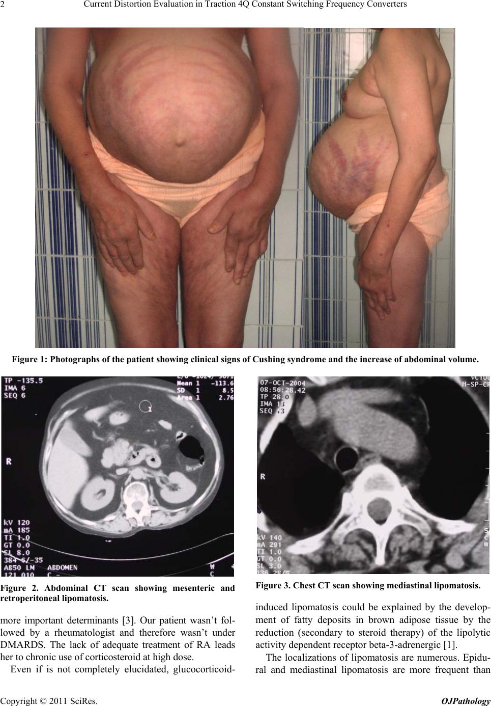 Current Distortion Evaluation in Traction 4Q Constant Switching Frequency Converters 2 Figure 1: Photographs of the patient showing clinical signs of Cushing syndrome and the increase of abdominal volume. Figure 2. Abdominal CT scan showing mesenteric and retroperitoneal lipomatosis. more important determinants [3]. Our patient wasn’t fol- lowed by a rheumatologist and therefore wasn’t under DMARDS. The lack of adequate treatment of RA leads her to chronic use of corti cost e roi d at hi g h dose. Even if is not completely elucidated, glucocorticoid- Figure 3. Chest CT scan showing mediastinal lipomatosis. induced lipomatosis could be explained by the develop- ment of fatty deposits in brown adipose tissue by the reduction (secondary to steroid therapy) of the lipolytic activity dependent receptor beta-3-adrenergic [1]. The localizations of lipomatosis are numerous. Epidu- ral and mediastinal lipomatosis are more frequent than Copyright © 2011 SciRes. OJPathology  Corticosteroid-Induced Multifocal and Extensive Lipomatosis: A Case Report 3 mesenteric or retroperitoneal ones. Lipomatosis is usu- ally asymptomatic nevertheless; many nonspecific symptoms could reveal this pathology [4,8-10]. In our patient persistent dyspnea was the principal symptom. Diagnosis of “Corticosteroid-induced multifocal and extensive lipomatosis” in our case was retained by Com- puterized tomography. This imaging and magnetic reso- nance imaging are the principals tools to identify the dif- ferents localizations of lipomatosis [1,5]. Treatment of lipomatosis is essentially medical. It is based on the reduction or discontinuation of corticoste- roid therapy if possible and the prescription of a hypo- caloric diet, with restriction for four weeks of fast-acting carbohydrate [6,7]. This treatment helps to reduce the hyperinsulinemia and promotes lipolysis in adipose tis- sue [6,7]. A more rational approach and concerted effort should be applied in situations of corticosteroids use to minimize their adverse effects and to evict a lipomatosis complication. Our patient had a good evolution after diet and progressive reducing of glucocorticoids thanks to DMARDS onset. Surgery is required for complicated or rapidly evolving forms [1]. The risks of surgery are im- portant in these patients; it should be discussed taking into account the clinical symptoms, the location and ex- tent [2]. 4. Conclusions The lipomatosis steroid-induced is probably an underesti- mated entity because of the prevalence of asymptomatic forms. This diagnosis should be discussed in the context of long-term corticosteroid. The scanner and MRI are the key diagnostic tests, esp ecially in the early stages. Medi- cal treatment is the first line treatment. Surgery is reser- ved for complicated shapes. REFERENCES [1] S. Humblot, J. C. Weber, A. S. Korganow, B. Hammann, J. L. Pasquali and T. Martin, “Les Lipomatoses Induites par la Corticothérapie,” La Revue de Médecine Interne, Vol. 18, No. 5, 1997, pp. 396-401. [2] L. Dujardin, C. Marcelli, C. Hérisson and L. Simon, “La Lipo-Matose Epidurale: Une Complication de la Cortico- thérapie au Long Court, Apropos de Deux Cas,” La Revue de Médecine Interne, Vol. 17, No. 7, 1996, pp. 563-567. [3] G. R. Fogel, “Cunningham PY 3rd, Esses SI. Spinal Epi- dural Lipomatosis: Case Reports, Literature Review and Metaanalysis,” Spine Journal, Vol. 5, No. 2, 2005, pp. 202-211. [4] K. Q. Nguyen, C. Hoeffel, L. H. Le and H. T. Phan, “Me- diastinal Lipomatosis,” South Medecine Journal, Vol. 91, 1998, pp. 1169-1172. [5] J. Hierholzer, G. Bendorf, T. Lehmann, G. H. Schneider, P. Podrabski, B. Sander, et al., “Epidural Lipomatosis: Case Report and Literature Review,” Neuroradiology, Vol. 38, No. 4, 1996, pp. 343-348. [6] F. Laroche, P. Chemouilli, R. Carlier, D. Doyon and G. “Saïd Efficacité du Traitement Médical dans une Com- pression Médullaire par Lipomatose Epidurale Compli- quant une Corticothérapie Prolongée,” Rev Rhum Ed Fr, Vol. 60, No. 11, 1993, pp. 846-849. [7] W. E. George Jr, M. Wilmot, A. Greenhouse and M. Ham- meke, “Medical Management of Steroid-Induced Epi- dural Lipomatosis,” The New England Journal of Medi- cine, Vol. 308, No. 6, 1983, pp. 316-319. doi:10.1056/NEJM198302103080605 [8] V. Gilsanz, P. W. Brill, B. S. Wolf, “Increased Retroperi- toneal Fat: A Sign of Corticosteroid Therapy,” Radiology, Vol. 123, 1977, pp. 147-148. [9] M. Domínguez Domínguez, J. E. Camacho Gonzalez, A. Palacios Martinez, J. M. Gonzalez Montano, R. Quintero Rodriguez and R. Ibanez Suarez, “Pelvic Lipomatosis: Cause of Bladder Rupture,” Actas Urologicsa Espanolas, Vol. 25, No. 8, 2001, pp. 592-595. [10] A. H. Zargar, B. A. Laway, S. R. Masoodi, M. H. Bhat, M. I. Bashir, A. I. Wani and N. A. Wani, “Diffuse Ab- dominal Lipomatosis,” The Journal of The Association of Physicians India, Vol. 51, 2003, pp. 621-622. Copyright © 2011 SciRes. OJPathology |