

Paper Menu >>
Journal Menu >>
 Pharmacology & Pharmacy, 2011, 2, 347-353 doi:10.4236/pp.2011.24045 Published Online October 2011 (http://www.SciRP.org/journal/pp) Copyright © 2011 SciRes. PP 347 Vazoactive Effects of Oxidative Stress Elicited by Hydrogen Peroxide in the Human Umbilical Artery: An in Vitro Study Ipek Duman*, Necdet Dogan Department of Pharmacology, Meram Faculty of Medicine, Selcuk University, Konya, Turkey. Email: *[email protected] Received June 5th, 2011; revised July 20th, 2011; accepted August 30th, 2011. ABSTRACT The vasoactive effects of oxidative stress induced by hydrogen peroxide (H2O2) on human umbilical artery strips as well as the possible mechanisms involved are studied. Contraction responses to cumulative H2O2 (10–7 M - 3 × 10⁻² M) in endothelium intact and denuded umbilical arteries and responses to cumulative H2O2 after incubation with L-NAME (10–4 M) (n = 8), indomethacin (10–5 M) (n = 8) and verapamil (10–6) (n = 8) were recorded. Responses elicited with cumulative H2O2 in Ca2+ free extracellular medium and the responses to cumulative Ca2+ (10–4 M - 2 × 10⁻³ M) after H2O2 (10–3 M) induced contraction were also studied. The Emax for each experiment was calculated. p < 0.05 was con- sidered as significant. H2O2 elicited contraction was greater in endothelium denuded artery strips compared to endo- thelium intact strips (p < 0.05). Compared to control, incubation with L-NAME significantly augmented (p < 0.05), while verapamil and indomethacin inhibited the contractions elicited by cumulative H2O2 (p < 0.05). Ca2+ free ex- tracellular medium caused decreases in cumulative H2O2 elicited contractions and cumulative Ca2+ caused concentra- tion dependent increases in the contraction caused by a single bolus of H2O2 (p < 0.05). Exposure to H2O2 causes con- centration-dependent constriction in human umbilical arteries. The presence of the endothelium and NOS enzyme acti- vation influences the H2O2 responses. Removal of the endothelium increases the H2O2 elicited contractions more than incubation with L-NAME suggesting beside NO, other endothelial vasodilators are also involved in vascular tonus of the umbilical arteries. Both intracellular and extracellular Ca2+ ions and constrictor cyclooxygenase metabolites play a role in the contractile responses elicited by H2O2 in human umbilical arteries. Keywords: Umbilical Arteries, Hydrogen Peroxide, Indomethacin, L-NAME, Oxidative Stress, Pre-Eclampsia, Reactive Oxygen Species, Verapamil 1. Introduction Pre-eclampsia (PE) is a major cause of fetal growth re- striction and perinatal complications. In PE, there is in- creased resistance to placental circulation which leads to reduced uteroplacental blood flow followed by placental dysfunction and intrauterine fetal growth restriction [1,2]. Umbilical blood vessels are not innervated therefore the control of umbilical blood flow depends entirely on vasoreactive substances either released locally or pre- sented in the circulation [3]. Pregnancy is a state of oxi- dative stress, characterized by the placental production of reactive oxygen species (ROS) including superoxide and hydrogen peroxide (H2O2) [4,5]. It is considered that during normal pregnancy, the rate of production of ROS is offset by their elimination by abundant antioxidant defenses. However in PE and preterm labor, due to ex- cessive oxidative stress and lipid peroxidation (LPO) ROS overpowers antioxidant defenses, leading to reduc- tion of uteroplacental blood flow [5,6]. H2O2 is a power- ful by product of LPO and is used as a model of oxida- tive stress. H2O2 easily crosses cell membranes and lead to cellular oxidative damage. LPO has been studied in- tensively over decades and remains to be a hot topic in biological research but still there is little information on the effects of LPO on human umbilical arteries [7-9]. This in vitro study was designed to assess the effects of H2O2 used as a model of oxidative stress on human um- bilical arteries as well as the possible mechanisms in- volved. 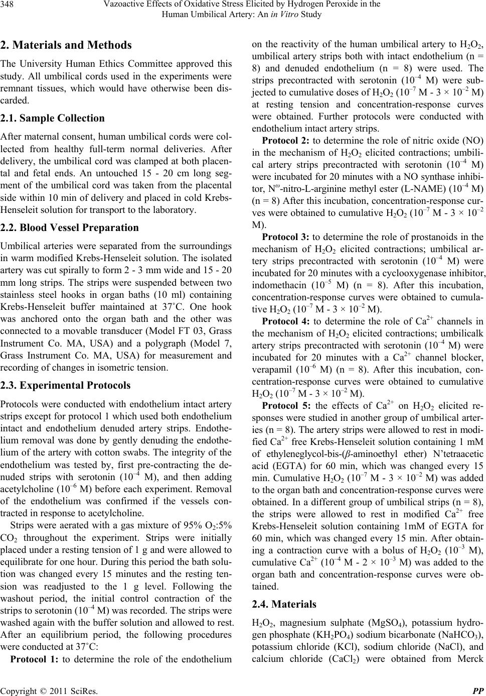 Vazoactive Effects of Oxidative Stress Elicited by Hydrogen Peroxide in the 348 Human Umbilical Artery: An in Vitro Study 2. Materials and Methods The University Human Ethics Committee approved this study. All umbilical cords used in the experiments were remnant tissues, which would have otherwise been dis- carded. 2.1. Sample Collection After maternal consent, human umbilical cords were col- lected from healthy full-term normal deliveries. After delivery, the umbilical cord was clamped at both placen- tal and fetal ends. An untouched 15 - 20 cm long seg- ment of the umbilical cord was taken from the placental side within 10 min of delivery and placed in cold Krebs- Henseleit solution for transport to the laboratory. 2.2. Blood Vessel Preparation Umbilical arteries were separated from the surroundings in warm modified Krebs-Henseleit solution. The isolated artery was cut spirally to form 2 - 3 mm wide and 15 - 20 mm long strips. The strips were suspended between two stainless steel hooks in organ baths (10 ml) containing Krebs-Henseleit buffer maintained at 37˚C. One hook was anchored onto the organ bath and the other was connected to a movable transducer (Model FT 03, Grass Instrument Co. MA, USA) and a polygraph (Model 7, Grass Instrument Co. MA, USA) for measurement and recording of changes in isometric tension. 2.3. Experimental Protocols Protocols were conducted with endothelium intact artery strips except for protocol 1 which used both endothelium intact and endothelium denuded artery strips. Endothe- lium removal was done by gently denuding the endothe- lium of the artery with cotton swabs. The integrity of the endothelium was tested by, first pre-contracting the de- nuded strips with serotonin (10–4 M), and then adding acetylcholine (10–6 M) before each experiment. Removal of the endothelium was confirmed if the vessels con- tracted in response to acetylcholine. Strips were aerated with a gas mixture of 95% O2:5% CO2 throughout the experiment. Strips were initially placed under a resting tension of 1 g and were allowed to equilibrate for one hour. During this period the bath solu- tion was changed every 15 minutes and the resting ten- sion was readjusted to the 1 g level. Following the washout period, the initial control contraction of the strips to serotonin (10–4 M) was recorded. The strips were washed again with the buffer solution and allowed to rest. After an equilibrium period, the following procedures were conducted at 37˚C: Protocol 1: to determine the role of the endothelium on the reactivity of the human umbilical artery to H2O2, umbilical artery strips both with intact endothelium (n = 8) and denuded endothelium (n = 8) were used. The strips precontracted with serotonin (10–4 M) were sub- jected to cumulative doses of H2O2 (10–7 M - 3 × 10⁻² M) at resting tension and concentration-response curves were obtained. Further protocols were conducted with endothelium intact artery strips. Protocol 2: to determine the role of nitric oxide (NO) in the mechanism of H2O2 elicited contractions; umbili- cal artery strips precontracted with serotonin (10–4 M) were incubated for 20 minutes with a NO synthase inhibi- tor, Nω-nitro-L-arginine methyl ester (L-NAME) (10–4 M) (n = 8) After this incubation, concentration-response cur- ves were obtained to cumulative H2O2 (10–7 M - 3 × 10⁻² M). Protocol 3: to determine the role of prostanoids in the mechanism of H2O2 elicited contractions; umbilical ar- tery strips precontracted with serotonin (10–4 M) were incubated for 20 minutes with a cyclooxygenase inhibitor, indomethacin (10–5 M) (n = 8). After this incubation, concentration-response curves were obtained to cumula- tive H2O2 (10–7 M - 3 × 10⁻² M). Protocol 4: to determine the role of Ca2+ channels in the mechanism of H2O2 elicited contractions; umbilicalk artery strips precontracted with serotonin (10–4 M) were incubated for 20 minutes with a Ca2+ channel blocker, verapamil (10–6 M) (n = 8). After this incubation, con- centration-response curves were obtained to cumulative H2O2 (10–7 M - 3 × 10⁻² M). Protocol 5: the effects of Ca2+ on H2O2 elicited re- sponses were studied in another group of umbilical arter- ies (n = 8). The artery strips were allowed to rest in modi- fied Ca2+ free Krebs-Henseleit solution containing 1 mM of ethyleneglycol-bis-(β-aminoethyl ether) N’tetraacetic acid (EGTA) for 60 min, which was changed every 15 min. Cumulative H2O2 (10–7 M - 3 × 10⁻² M) was added to the organ bath and concentration-response curves were obtained. In a different group of umbilical strips (n = 8), the strips were allowed to rest in modified Ca2+ free Krebs-Henseleit solution containing 1mM of EGTA for 60 min, which was changed every 15 min. After obtain- ing a contraction curve with a bolus of H2O2 (10–3 M), cumulative Ca2+ (10–4 M - 2 × 10⁻³ M) was added to the organ bath and concentration-response curves were ob- tained. 2.4. Materials H2O2, magnesium sulphate (MgSO4), potassium hydro- gen phosphate (KH2PO4) sodium bicarbonate (NaHCO3), potassium chloride (KCl), sodium chloride (NaCl), and calcium chloride (CaCl2) were obtained from Merck Copyright © 2011 SciRes. PP 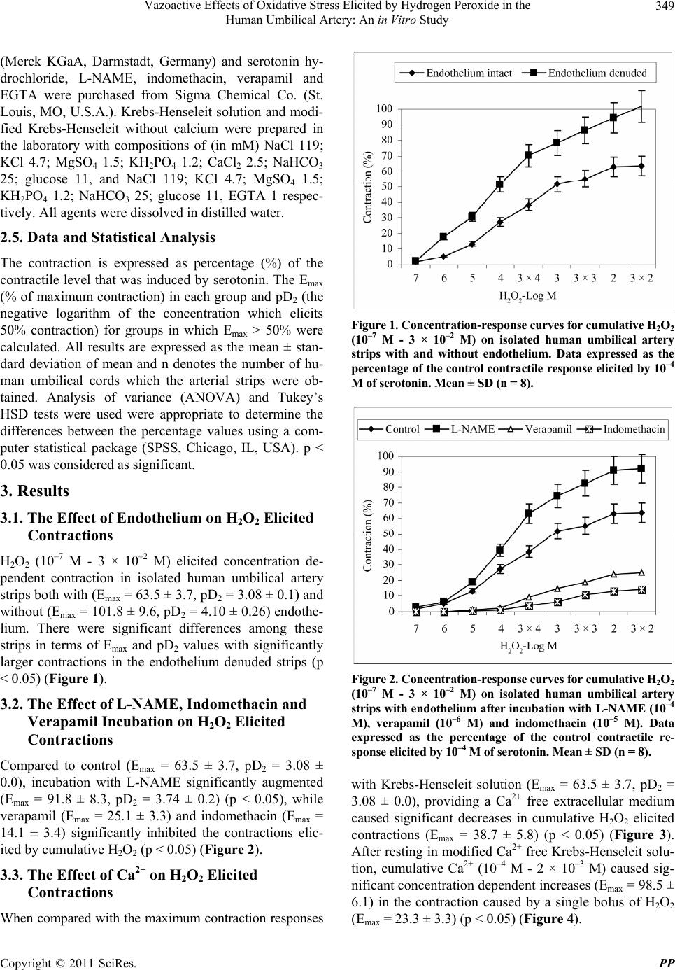 Vazoactive Effects of Oxidative Stress Elicited by Hydrogen Peroxide in the 349 Human Umbilical Artery: An in Vitro Study (Merck KGaA, Darmstadt, Germany) and serotonin hy- drochloride, L-NAME, indomethacin, verapamil and EGTA were purchased from Sigma Chemical Co. (St. Louis, MO, U.S.A.). Krebs-Henseleit solution and modi- fied Krebs-Henseleit without calcium were prepared in the laboratory with compositions of (in mM) NaCl 119; KCl 4.7; MgSO4 1.5; KH2PO4 1.2; CaCl2 2.5; NaHCO3 25; glucose 11, and NaCl 119; KCl 4.7; MgSO4 1.5; KH2PO4 1.2; NaHCO3 25; glucose 11, EGTA 1 respec- tively. All agents were dissolved in distilled water. 2.5. Data and Statistical Analysis The contraction is expressed as percentage (%) of the contractile level that was induced by serotonin. The Emax (% of maximum contraction) in each group and pD2 (the negative logarithm of the concentration which elicits 50% contraction) for groups in which Emax > 50% were calculated. All results are expressed as the mean ± stan- dard deviation of mean and n denotes the number of hu- man umbilical cords which the arterial strips were ob- tained. Analysis of variance (ANOVA) and Tukey’s HSD tests were used were appropriate to determine the differences between the percentage values using a com- puter statistical package (SPSS, Chicago, IL, USA). p < 0.05 was considered as significant. 3. Results 3.1. The Effect of Endothelium on H2O2 Elicited Contractions H2O2 (10–7 M - 3 × 10⁻² M) elicited concentration de- pendent contraction in isolated human umbilical artery strips both with (Emax = 63.5 ± 3.7, pD2 = 3.08 ± 0.1) and without (Emax = 101.8 ± 9.6, pD2 = 4.10 ± 0.26) endothe- lium. There were significant differences among these strips in terms of Emax and pD2 values with significantly larger contractions in the endothelium denuded strips (p < 0.05) (Figure 1). 3.2. The Effect of L-NAME, Indomethacin and Verapamil Incubation on H2O2 Elicited Contractions Compared to control (Emax = 63.5 ± 3.7, pD2 = 3.08 ± 0.0), incubation with L-NAME significantly augmented (Emax = 91.8 ± 8.3, pD2 = 3.74 ± 0.2) (p < 0.05), while verapamil (Emax = 25.1 ± 3.3) and indomethacin (Emax = 14.1 ± 3.4) significantly inhibited the contractions elic- ited by cumulative H2O2 (p < 0.05) (Figure 2). 3.3. The Effect of Ca2+ on H2O2 Elicited Contractions When compared with the maximum contraction responses Figure 1. Concentration-response curves for cumulative H2O2 (10–7 M - 3 × 10⁻² M) on isolated human umbilical artery strips with and without endothelium. Data expressed as the percentage of the control contractile response elicited by 10–4 M of serotonin. Mean ± SD (n = 8). Figure 2. Concentration-response curves for cumulative H2O2 (10–7 M - 3 × 10⁻² M) on isolated human umbilical artery strips with endotheli um after incubation with L-NAME (10 –4 M), verapamil (10–6 M) and indomethacin (10–5 M). Data expressed as the percentage of the control contractile re- sponse elicited by 10–4 M of serotonin. Mean ± SD (n = 8). with Krebs-Henseleit solution (Emax = 63.5 ± 3.7, pD2 = 3.08 ± 0.0), providing a Ca2+ free extracellular medium caused significant decreases in cumulative H2O2 elicited contractions (Emax = 38.7 ± 5.8) (p < 0.05) (Figure 3). After resting in modified Ca2+ free Krebs-Henseleit solu- tion, cumulative Ca2+ (10–4 M - 2 × 10⁻³ M) caused sig- nificant concentration dependent increases (Emax = 98.5 ± 6.1) in the contraction caused by a single bolus of H2O2 (Emax = 23.3 ± 3.3) (p < 0.05) (Figure 4). Copyright © 2011 SciRes. PP 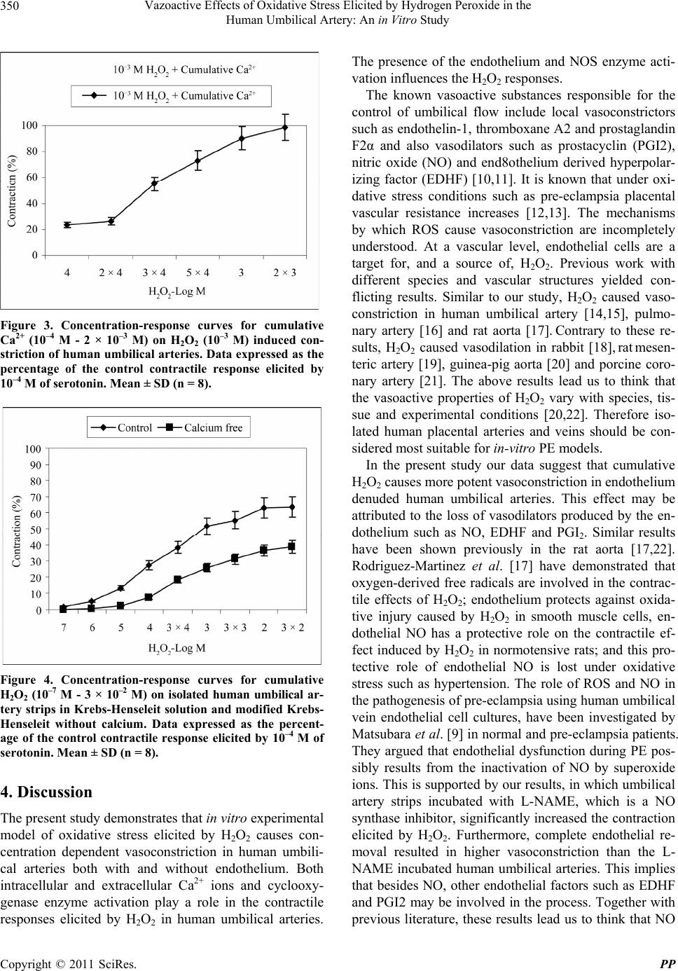 Vazoactive Effects of Oxidative Stress Elicited by Hydrogen Peroxide in the 350 Human Umbilical Artery: An in Vitro Study Figure 3. Concentration-response curves for cumulative Ca2+ (10–4 M - 2 × 10⁻³ M) on H2O2 (10–3 M) induced con- striction of human umbilical arteries. Data expressed as the percentage of the control contractile response elicited by 10–4 M of serotonin. Mean ± SD (n = 8). Figure 4. Concentration-response curves for cumulative H2O2 (10–7 M - 3 × 10⁻² M) on isolated human umbilical ar- tery strips in Krebs-Henseleit solution and modified Krebs- Henseleit without calcium. Data expressed as the percent- age of the control contractile response elicited by 10–4 M of serotonin. Mean ± SD (n = 8). 4. Discussion The present study demonstrates that in vitro experimental model of oxidative stress elicited by H2O2 causes con- centration dependent vasoconstriction in human umbili- cal arteries both with and without endothelium. Both intracellular and extracellular Ca2+ ions and cyclooxy- genase enzyme activation play a role in the contractile responses elicited by H2O2 in human umbilical arteries. The presence of the endothelium and NOS enzyme acti- vation influences the H2O2 responses. The known vasoactive substances responsible for the control of umbilical flow include local vasoconstrictors such as endothelin-1, thromboxane A2 and prostaglandin F2α and also vasodilators such as prostacyclin (PGI2), nitric oxide (NO) and end8othelium derived hyperpolar- izing factor (EDHF) [10,11]. It is known that under oxi- dative stress conditions such as pre-eclampsia placental vascular resistance increases [12,13]. The mechanisms by which ROS cause vasoconstriction are incompletely understood. At a vascular level, endothelial cells are a target for, and a source of, H2O2. Previous work with different species and vascular structures yielded con- flicting results. Similar to our study, H2O2 caused vaso- constriction in human umbilical artery [14,15], pulmo- nary artery [16] and rat aorta [17]. Contrary to these re- sults, H2O2 caused vasodilation in rabbit [18], rat mese n - teric artery [19], guinea-pig aorta [20] and porcine coro- nary artery [21]. The above results lead us to think that the vasoactive properties of H2O2 vary with species, tis- sue and experimental conditions [20,22]. Therefore iso- lated human placental arteries and veins should be con- sidered most suitable for in-vitro PE models. In the present study our data suggest that cumulative H2O2 causes more potent vasoconstriction in endothelium denuded human umbilical arteries. This effect may be attributed to the loss of vasodilators produced by the en- dothelium such as NO, EDHF and PGI2. Similar results have been shown previously in the rat aorta [17,22]. Rodriguez-Martinez et al. [17] have demonstrated that oxygen-derived free radicals are involved in the contrac- tile effects of H2O2; endothelium protects against oxida- tive injury caused by H2O2 in smooth muscle cells, en- dothelial NO has a protective role on the contractile ef- fect induced by H2O2 in normotensive rats; and this pro- tective role of endothelial NO is lost under oxidative stress such as hypertension. The role of ROS and NO in the pathogenesis of pre-eclampsia using human umbilical vein endothelial cell cultures, have been investigated by Matsubara et al. [9] in normal and pre-eclampsia patients. They argued that endothelial dysfunction during PE pos- sibly results from the inactivation of NO by superoxide ions. This is supported by our results, in which umbilical artery strips incubated with L-NAME, which is a NO synthase inhibitor, significantly increased the contraction elicited by H2O2. Furthermore, complete endothelial re- moval resulted in higher vasoconstriction than the L- NAME incubated human umbilical arteries. This implies that besides NO, other endothelial factors such as EDHF and PGI2 may be involved in the process. Together with previous literature, these results lead us to think that NO Copyright © 2011 SciRes. PP  Vazoactive Effects of Oxidative Stress Elicited by Hydrogen Peroxide in the 351 Human Umbilical Artery: An in Vitro Study acts as a negative modulator against H2O2 elicited con- tractions in placental arteries and also may be protective against oxidative insult [17,19,22]. Lelung et al. [3] have demonstrated that sodium nitro- prusside, an exogenous source of NO, only elicits small significant relaxations in isolated human umbilical arter- ies. They are in the opinion that NO might not be the major mediator responsible for vasodilation in umbilical arteries. Previous work also suggests that, instead of NO prostacyclin was the main factor mediating the endothe- lium dependent relaxation in human umbilical arteries [10,14,23]. Klockenbush et al. [24] have suggested that rather than NO, prostacyclin plays a major role in the vasoreactivity of umbilical and fetal circulation. H2O2 elicited vasoreactivity can be relaxation or constriction depending on the basal tonus of isolated pulmonary ar- teries, and these effects are mediated by phospholipase A2 activation leading to prostacyclin or thromboxane A2 release [25]. In the present study the H2O2 elicited con- traction in umbilical arteries were significantly inhibited in the presence of indomethacin, a cyclooxygenase en- zyme inhibitor. These data suggest that increased con- strictor cyclooxygenase metabolites as a result of in- creased arachidonic acid metabolism partially mediate the constrictive effects of H2O2 in the vascular smooth muscle of human umbilical arteries. Previous research shows that prostaglandin H2 and more possibly throm- boxane A2 are involved in the contractile response [26, 27]. These findings are in accordance with studies which, showed that H2O2 increases prostaglandin F2α and thromboxane A2 in rat aorta smooth muscle [28,29]. Ca2+ is essential for the contraction of smooth muscles and it is believed that intracellular Ca2+ homeostasis plays a major role in antioxidant activity [30-33]. The harmful effects of ROS can influence the ion channels or ion pumps, which maintain low Ca2+ levels under normal conditions [33,34]. It is known from previous work that in different tissues H2O2 can increase intracellular Ca2+ by promoting mobilization of Ca2+ and Ca2+ influx [31,33, 34]. In isolated rat cardiomyocytes Gen et al. [33] have shown that H2O2 increases intracellular Ca2+ in a dose dependent manner. In our study we have found that H2O2 induced contractions increase in a dose dependent pattern when cumulative Ca2+ was added after a single bolus of H2O2 in human umbilical arteries. It has been reported that NO modulates Ca2+-channel activity in vascular smooth muscle and induces relaxa- tion [35]. Incubation with verapamil, a Ca2+-channel an- tagonist significantly decreased but did not totally block H2O2 induced contractions. Sotnikova [22] and Yang et al. [36] have reported similar results in rat aorta with calcium antagonists. In the present study, both incubation with verapamil and resting the arteries in Ca2+-free me- dium attenuated but did not prevent H2O2 induced con- tractions. These findings suggest that both intracellular and extracellular Ca2+ mediate these contractions. It has been suggested previously that the decrease caused by ROS on membrane resistance may depolarize the cells and thus activate voltage sensitive Ca2+ channels and lead to an increase in intracellular Ca2+ [31,32]. Also, Ca2+ channel blockers are thought to be ineffective in blocking mobilization of Ca2+ from intracellular stores but effec- tively block the influx of extracellular Ca2+ via the L type Ca2+ channels [33]. 5. Conclusions In conclusion, in this in-vitro model for oxidative stress we have demonstrated that exposure to H2O2 causes concentration-dependent constriction in human umbilical arteries. Removal of the endothelium increased the H2O2 elicited contractions more than incubation with L-NAME suggesting beside NO, other endothelial vasodilators are also involved in vascular tonus of the umbilical arteries. Inhibition of H2O2 elicited contractions with indometha- cin suggests that increased constrictor cyclooxygenase metabolites partially mediate the constrictive effects of H2O2 in the vascular smooth muscle of human umbilical arteries. Incubation with verapamil, and Ca2+ free me- dium significantly decreased but did not totally block H2O2 induced contractions suggesting both intracellular and extracellular Ca2+ involvement in H2O2 elicited con- tractions. REFERENCES [1] T. Todros, A. Sciarrone, E. Piccoli, C. Guiot, P. Kauf- mann and J. Kingdom, “Umbilical Doppler Waveforms and Placental Villous Angiogenesis in Pregnancies Com- plicated by Fetal Growth Restriction,” Obstetrics & Gy- necology, Vol. 93, No. 4, 1999, pp. 499-503. doi:10.1016/S0029-7844(98)00440-2 [2] A. Fleischer, H. Schulman, G. Farmakides, L. Bracero, L. Grunfeld, B. Rochelson and M. Koenigsberg, “Uterine artery Doppler Velocimetry in Pregnant Women with Hypertension,” American Journal of Obstetrics & Gyne- cology, Vol. 154, No. 4, 1986, pp. 806-813. [3] S. W. Leung, A. Quan, T. T. Lao and R. Y. Man, “Effi- cacy of Different Vasodilators on Human Umbilical Arte- rial Smooth Muscle under Normal and Reduced Oxygen Conditions,” Early Human Development, Vol. 82, No. 7, 2006, pp. 457-462. doi:10.1016/j.earlhumdev.2005.11.009 [4] T. A. Mills, M. Wareing, A. A. H. Shennan, L. Poston, P. N. Baker and S. L. Greenwood, “Acute and Chronic Modulation of Placental Chorionic Plate Artery Reactiv- ity by Reactive Oxygen Species,” Free Radical Biology & Medicine, Vol. 47, No. 2, 2009, pp. 159-166. Copyright © 2011 SciRes. PP  Vazoactive Effects of Oxidative Stress Elicited by Hydrogen Peroxide in the 352 Human Umbilical Artery: An in Vitro Study doi:10.1016/j.freeradbiomed.2009.04.019 [5] N. Rani, R. Dhingra, D. S. Arya, M. Kalaivani, N. Bhatla and R. Kumar, “Role of Oxidative Stress Markers and Antioxidants in the Placenta of Preeclamptic Patients,” Journal of Obstetrics and Gynaecology Research, Vol. 36, No. 6, 2010, pp. 1189-1194. doi:10.1111/j.1447-0756.2010.01303.x [6] A. Cinkaya, H. L. Keskin, U. Buyukkagnici, T. Gungor, E. A. Keskin, A. F. Avsar and U. Bilge, “Maternal Plas- ma Total Antioxidant Status in Preterm Labor,” Journal of Obstetrics and Gynaecology Research, Vol. 36, No. 6, 2010, pp. 1185-1188. doi:10.1111/j.1447-0756.2010.01300.x [7] A. Negre-Salvayre, N. Auge, V. Ayala, H. Basaga, J. Boada, R. Brenke, S. Chapple, G. Cohen, J. Feher, T. Grune, G. Lengyel, G. E. Mann, R. Pamplona, G. Poli, M. Portero-Otin, Y. Riahi, R. Salvayre, S. Sasson, J. Serrano, O. Shamni, W. Siems, R. C. Siow, I. Wiswedel, K. Zarkovic and N. Zarkovic, “Pathological Aspects of Lipid Peroxidation,” Free Radical Research, Vol. 44, No. 10, 2010, pp. 1125-1171. doi:10.3109/10715762.2010.498478 [8] T. Grune, N. Zarkovic and K. Kalliopi, “Lipid Peroxida- tion Sesearch in Europe and the COST B35 Action ‘Lipid Peroxidation Associated Disorders’,” Free Radical Re- search, Vol. 44, No. 10, 2010, pp. 1095-1097. doi:10.3109/10715762.2010.504395 [9] K. Matsubara, Y. Matsubara, S. Hyodo, T. Katayama and M. Ito, “Role of Nitric Oxide and Reactive Oxygen Spe- cies in the Pathogenesis of Preeclampsia,” Journal of Ob- stetrics and Gynaecology Research, Vol. 36, No. 2, 2010, pp. 239-247. doi:10.1111/j.1447-0756.2009.01128.x [10] L. Carbillon, M. Uzan and S.Uzan, “Pregnancy, Vascular Tone, and Maternal Hemodynamics: A Crucial Adapta- tion,” Obstetrical & Gynecological Survey, Vol. 55, No. 9, 2000, pp. 574-581. doi:10.1097/00006254-200009000-00023 [11] T. Matoba and H. Shimokawa, “Hydrogen Peroxide Is an Endothelium-Derived Hyperpolarizing Factor in Animals and Humans,” Journal of Pharmacological Sciences, 2003, Vol. 92, No. 1, pp. 1-6. doi:10.1254/jphs.92.1 [12] K. Rytlewski, H. Huras, K. Kusmierska, A. Jaworowski, T. Gornisiewicz, P. Ossowski and A. Reron, “Doppler Velocimetry of the Materno-Fetal Circulation in Preterm Delivered Pregnancies Complicated with Hypertension,” Neuroendocrinology Letters, Vol. 30, No. 3, 2009, pp. 403-408. [13] R. Cruz-Martinez and F. Figueras, “The Role of Doppler and Placental Screening,” Best Practice & Research Cli- nical Obstetrics & Gynaecology, Vol. 23, No. 6, 2009, pp. 845-855. doi:10.1016/j.bpobgyn.2009.08.007 [14] Y. Okatani, K. Watanabe and Y. Sagara, “Effect of Nitric Oxide, Prostacyclin and Tromboxane on the Vasospastic Action of Hydrogen Peroxide on Human Umbilical Ar- tery,” Acta Obstetricia et Gynecologica Scandinavica, Vol. 76, No. 6, 1997, pp. 515-520. [15] Y. Okatani, K. Watanabe, A. Wakatsuki, S. Tamura and Y. Sagara, “Effects of Superoxide and Peroxynitrite on Vascular Tension in the Human Umbilical Artery,” Acta Obstetricia et Gynecologica Scandinavica, Vol. 77, No. 9, 1998, pp. 883-888. doi:10.1080/j.1600-0412.1998.770902.x [16] N. Jin, C. S. Packer and R. A. Rhoades, “Reactive Oxy- gen-Mediated Contraction in Pulmonary Arterial Smooth Muscle: Cellular Mechanisms,” Canadian Journal of Physiology and Pharmacology, Vol. 69, No. 3, 1991, pp. 383-388. doi:10.1139/y91-058 [17] M. A. Rodríguez-Martínez, E. C. García-Cohen, A. B. Baena, R. González, M. Salaíces and J. Marín, “Contractile Responses Elicited by Hydrogen Peroxide in Aorta From Normotensive and Hypertensive Rats. Endothelial Modu- lation and Mechanism Involved,” British Journal of Phar- macology, Vol. 125, No. 6, 1998, pp. 1329-1335. doi:10.1038/sj.bjp.0702200 [18] T. Hattori, J. Kajikuri, H. Katsuya and T. Itoh, “Effects of H2O2 on Membrane Potential of Smooth Muscle Cells in Rabbit Mesenteric Resistance Artery,” European Journal of Pharmacology, Vol. 464, No. 2-3, 2003, pp. 101-109. doi:10.1016/S0014-2999(03)01427-4 [19] Y. J. Gao, S. Hirota, D. W. Zhang, L. J. Janssen and R. M. Lee, “Mechanisms of Hydrogen-Peroxide-Induced Bipha- sic Response in Rat Mesenteric Artery,” British Journal of Pharmacology, Vol. 138, No. 6, 2003, pp. 1085-1092. [20] S. Fujimoto, M. Mori and H. Tsushima, “Mechanisms Underlying the Hydrogen Peroxide-Induced, Endothelium- Independent Relaxation of the Norepinephrine-Contrac- tion in Guinea-Pig Aorta,” European Journal of Pharma- cology, Vol. 459, No. 1, 2003, pp. 65-73. doi:10.1016/S0014-2999(02)02825-X [21] R. S. Barlow and R. E. White, “Hydrogen Peroxide Relax- es Porcine Coronary Arteries by Stimulating BKCa Chan- nel Activity,” American Journal of Physiology: Heart and Circulatory Physiology, Vol. 275, No. 4, 1998, pp. H1283- H1289. [22] R. Sotnikova, “Investigation of the Mechanisms Under- lying H2O2-Evoked Contraction in the Isolated Rat Aorta,” General Pharmacology, Vol. 31, No. 1, 1998, pp. 115-119. doi:10.1016/S0306-3623(97)00392-3 [23] G. Bodelsson and M. Stjernquist, “Endothelium-Depen- dent Relaxation to Substance P in Human Umbilical Ar- tery Is Mediated via Prostanoid Synthesis,” Human Re- production, Vol. 9, No. 4, 1994, pp. 733-737. [24] W. Klockenbusch, H. Strobach and K. Schrör, “Any Phy- siological Role for Prostacyclin in Regulation of Fetal Vessel Tone?” Agents & Actions, Vol. 37, No. 1, 1992, pp. 361-368. [25] D. W. Sheehan, E. C. Giese, S. F. Gugino, J. A. Russell, “Characterization and Mechanisms of H2O2-Induced Con- tractions of Pulmonary Arteries,” American Journal of Physiology: Heart and Circulatory Physiology, Vol. 264, No. 5, 1993, pp. H1542-H1547. [26] K. Watanabe, Y. Okatani and Y. Sagara, “Potentiating Effect of Hydrogen Peroxide on the Serotonin-Induced Vasocontraction in Human Umbilical Artery,” Acta Ob- Copyright © 2011 SciRes. PP  Vazoactive Effects of Oxidative Stress Elicited by Hydrogen Peroxide in the Human Umbilical Artery: An in Vitro Study Copyright © 2011 SciRes. PP 353 stetricia et Gynecologica Scandinavica, Vol. 75, No. 9, 1996, pp. 783-789. doi:10.3109/00016349609054704 [27] M. S. Wolin, C. A. Davidson, P. M. Kaminski, R. P. Fayngersh and K. M. Mohazzab-H., “Oxidant-Nitric Ox- ide Signalling Mechanisms in Vascular Tissue,” Bio- chemistry (Moscow), Vol. 63, No. 7, 1998, pp. 810-816. [28] S. López-Ongil, G. Torrecillas, D. Pérez-Sala, L. González- Santiago, M. Rodríguez-Puyol and D. Rodríguez-Puyol, “Mechanisms Involved in the Contraction of Endothelial Cells by Hydrogen Peroxide,” Free Radical Biology & Medicine, Vol. 26, No. 5-6, 1999, pp. 501-510. doi:10.1016/S0891-5849(98)00223-8 [29] P. Srivastava, M. Rajanikanth, S. A. V. Raghavan and M. Dikshit, “Role of Endogenous Reactive Oxygen Derived Species and Cyclooxygenages Mediators in 5-Hydroxy- tryptamine-Induced Contractions in Rat Aorta: Relation- ship to Nitric Oxide,” Pharmacological Research, Vol. 45, No. 5, 2002, pp. 375-382. doi:10.1006/phrs.2001.0859 [30] J. Daut, N. B. Standen and M. T. Nelson, “The Role of Membrane Potential of Endothelial and Smooth Muscle Cells in the Regulation of Coronary Blood Flow,” Jour- nal of Cardiovascular Electrophysiology, Vol. 5, No. 2, 1994, pp. 154-181. doi:10.1111/j.1540-8167.1994.tb01156.x [31] K. Bielefeldt, C. A. Whiteis, R. V. Sharma, F. M. Abboud and J. L. Conklin, “Reactive Oxygen Species and Cal- cium Homeostasis in Cultured Human Intestinal Smooth Muscle Cells,” American Journal of Physiology: Heart and Circulatory Physiology, Vol. 272, No. 6, 1997, pp. G1439-G1450. [32] A. Roveri, M. Coassin, M. Maiorino, A. Zamburlini, F. T. van Amsterdam, E. Ratti and F. Ursini, “Effect of Hy- drogen Peroxide on Calcium Homeostasis in Smooth Muscle Cells,” Archives of Biochemistry and Biophysics, Vol. 297, No. 2, 1992, pp. 265-270. doi:10.1016/0003-9861(92)90671-I [33] W. Gen, M. Tani, J. Takeshita J, Y. Ebihara and K. Tamaki, “Mechanisms of Ca2+ Overload Induced by Ex- tracellular H2O2 in Quiescent Isolated Rat Myocytes,” Basic Research in Cardiology, Vol. 96, No. 6, 2001, pp. 623-629. doi:10.1007/s003950170014 [34] J. Tang and J. H. Zhang, “Mechanisms of (Ca2+)i Eleva- tion by H2O2 in Islets of Rats,” Life Sciences, Vol. 68, No. 4, 2000, pp. 475-481. doi:10.1016/S0024-3205(00)00944-9 [35] L. A. Blatter and W. G. Wier, “Nitric Oxide Decreased [Ca2+]i in Vascular Smooth Muscle by Inhibition of the Calcium Current,” Cell Calcium, Vol. 15, No. 2, 1994, pp. 122-131. doi:10.1016/0143-4160(94)90051-5 [36] D. Yang, M. Feletou, C. M. Boulanger, H. F. Wu, N. Levens, J. N. Zhang and P. M. Vanhoutte, “Oxygen-De- rived Free Radicals Mediate Endothelium-Dependent Contractions to Acetylcholine in Aortas from Spontane- ously Hypertensive Rats,” British Journal of Pharmacol- ogy, Vol. 136, No. 1, 2002, pp. 104-110. doi:10.1038/sj.bjp.0704669 |