

Paper Menu >>
Journal Menu >>
 Vol.3, No.10, 620-622 (2011) Health doi:10.4236/health.2011.310104 C opyright © 2011 SciRes. Openly accessible at http://www.scirp.org/journal/HEALTH/ Primary breast leiomyosarcoma: case report and literature review Fatima Zahra EL M’ra bet1,2*, Hanane El kac emi1, Omar El Mesbahi2, Khal id El Hasso uni1, Brahim khalil El Gu eddari1 1Radiotherapy unit, National Institute of Oncology, Rabat, Morocco; *Corresponding Author: [email protected] 2Departement of medical oncology, university hospital Hassan II, Fez, Morocco. Received 4 April 2011; revised 16 May 2011; accepted 29 May 2011. ABSTRACT PURPOSE: Leiomyosarcoma of the breast is extremely rare. To date, the factors that are predictive of patient prognosis have not been identified. To clarify the nature of leiomyosa- rcoma of the breast, and also to establish the proper treatment strategy, we report this case We report this case while discussing The Cli- nical presentation, diagnosis, therapy and path- ologic feature. INTRODUCION: Sarcomas com- prise less than 1% of all primary breast neo- plasms and only a minority of these are leio- myosarcomas. There were only 35 cases being reported in English literature. The mainstay of treatment is surgical excision with clear mar- gins and longterm followup is essential. Its pro- gnosis is better compared to other breast sar- coma. CASE PRESENTATION: We report a case of primary leiomyosarcoma of the breast occ- urring in young female patient which was succ- essfully treated by surgery and radiotherapy. CONCLUSION: According to literature data, primary leiomyosarcoma is characterized by a better prognosis compared with other breast sarcomas, To date, the factors that are predic- tive of patient prognosis have not been iden- tified. The rarity and diagnostic difficulty impo- sed a multidisciplinary approach. Keywords: leiomyosarcoma; Breast; Immunohistochemistry; Mastectomy 1. INTRODUCTION Sarcomas of the breast account for less than 1% of all malignant tumors of the breast. Leiomyosarcomas bel- ong to a less common subgroup (5% to 10%) of sar- comas of the breast [1]. There were only 35 cases being reported in English literature. Confirming the diagnosis on either immunohistochemistry is important as it is sometimes difficult to differentiate these pathologically from poorly differentiated sarcomatoid carcinomas. To date, the factors that are predictive of patient prognosis have not been identified. [2] In this report, we describe case with this uncommon Tumor, which was treated successfully in The Institute of National Oncology Rabat, Morocco. 2. CASE REPORT A 40-year-old female patient, without any significant history, who presented a left breast lump, it has been pre- sent for 17 months. There was no other breast complaint and no family history of breast cancer. Physical exam- ination found a tumor of 6 cm in diameter in the lower inner quadrant of her right breast with ipsilateral axillary lymph node. The left breast was unremarkable. Mam- mography showed a dense, well circumscribed mass with little evidence of local invasion and the lump was easily detectable by ultrasound. An incisional biopsy was performed, the histological exam showed a sarcomatous proliferation, Immunohistochemically, the tumor cells were diffusely and strongly positive for α-smooth mus- cle actin and vimentin (Figure 1), while negative for cytokeratin, CD34 and S-100 protein ,which concludes with primary breast leiomyosarcoma . Investigations including Liver echography, chest scan and bone scan were showed no evidence of another primary tumor or metastatic disease. The patient under- went a radical mastectomy with lymph node dissection. Additional immunohistochemical examen routinely perf- ormed in breast carcinomas (oestrogen, progesteron rec- eptors, Her 2) also proved to be negative. Tumor cells showed strong positivity for vimentin, desmin and smoo- th muscle actin (SMA). Axillary lymph nodes were free of metastases Axillary. Subsequently the patient had rec- eived adjuvant radiotherapy to 50 Gray, the patient is in good control until now. A follow-up examination revea- led no evidence of distant metastasis or local recurrences 8 years after the treatment. 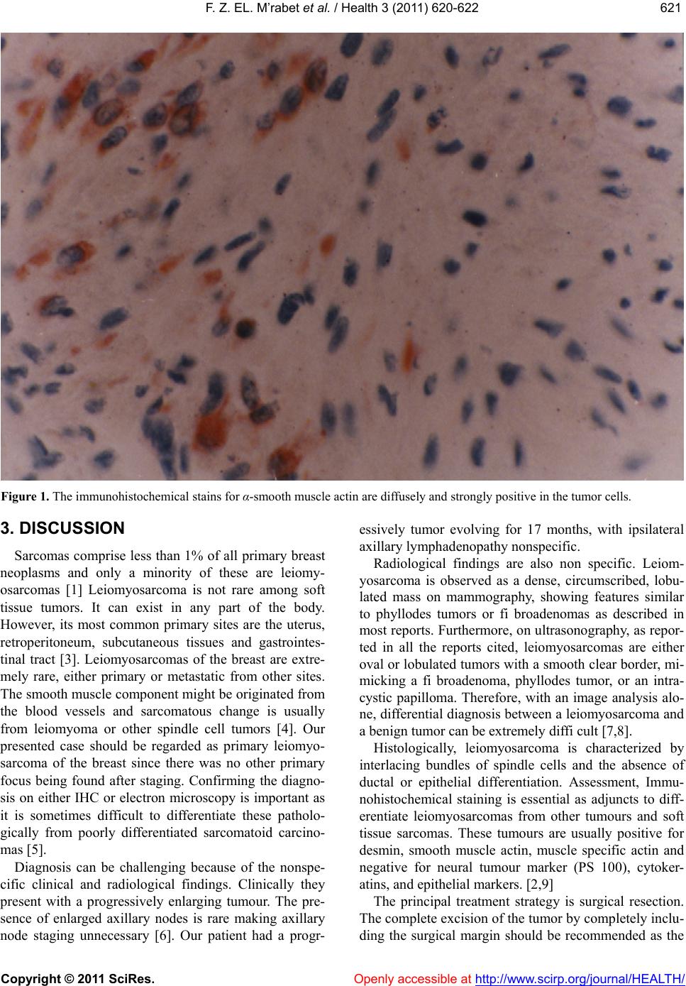 F. Z. EL. M’rabet et al. / Health 3 (2011) 620-622 Copyright © 2011 SciRes. Openly accessible at http://www.scirp.org/journal/HEALTH/ 621621 Figure 1. The immunohistochemical stains for α-smooth muscle actin are diffusely and strongly positive in the tumor cells. 3. DISCUSSION Sarcomas comprise less than 1% of all primary breast neoplasms and only a minority of these are leiomy- osarcomas [1] Leiomyosarcoma is not rare among soft tissue tumors. It can exist in any part of the body. However, its most common primary sites are the uterus, retroperitoneum, subcutaneous tissues and gastrointes- tinal tract [3]. Leiomyosarcomas of the breast are extre- mely rare, either primary or metastatic from other sites. The smooth muscle component might be originated from the blood vessels and sarcomatous change is usually from leiomyoma or other spindle cell tumors [4]. Our presented case should be regarded as primary leiomyo- sarcoma of the breast since there was no other primary focus being found after staging. Confirming the diagno- sis on either IHC or electron microscopy is important as it is sometimes difficult to differentiate these patholo- gically from poorly differentiated sarcomatoid carcino- mas [5]. Diagnosis can be challenging because of the nonspe- cific clinical and radiological findings. Clinically they present with a progressively enlarging tumour. The pre- sence of enlarged axillary nodes is rare making axillary node staging unnecessary [6]. Our patient had a progr- essively tumor evolving for 17 months, with ipsilateral axillary lymphadenopathy nonspecific. Radiological findings are also non specific. Leiom- yosarcoma is observed as a dense, circumscribed, lobu- lated mass on mammography, showing features similar to phyllodes tumors or fi broadenomas as described in most reports. Furthermore, on ultrasonography, as repor- ted in all the reports cited, leiomyosarcomas are either oval or lobulated tumors with a smooth clear border, mi- micking a fi broadenoma, phyllodes tumor, or an intra- cystic papilloma. Therefore, with an image analysis alo- ne, differential diagnosis between a leiomyosarcoma and a benign tumor can be extremely diffi cult [7,8]. Histologically, leiomyosarcoma is characterized by interlacing bundles of spindle cells and the absence of ductal or epithelial differentiation. Assessment, Immu- nohistochemical staining is essential as adjuncts to diff- erentiate leiomyosarcomas from other tumours and soft tissue sarcomas. These tumours are usually positive for desmin, smooth muscle actin, muscle specific actin and negative for neural tumour marker (PS 100), cytoker- atins, and epithelial markers. [2,9] The principal treatment strategy is surgical resection. The complete excision of the tumor by completely inclu- ding the surgical margin should be recommended as the  F. Z. EL. M’rabet et al. / Health 3 (2011) 620-622 Copyright © 2011 SciRes. Openly accessible at http://www.scirp.org/journal/HEALTH/ 622 best treatment. Most authors advice either radical or sim- ple mastectomy arguing that wide excision is associated with higher rate of recurrence [2,5]. Though axillary node dissection is not necessary, it has been performed in many of the reported cases because of uncertain pre- operative diagnosis or if there were clinically palpable nodes [6]. In our case, the patient underwent a radical mastectomy with lymph node dissection for the presence of clinically palpable axillary lymph nodes, lymph nodes were free of metastases Axillary. There is no evidence describing whether radiation therapy and/or chemothe- rapy improve the disease-free rate or overall survival [10]. In general, the prognosis of patients with leiomy- osarcoma of the breast is considered to be better than that for patients with other sarcomas of the breast [11] 4. CONCLUSIONS Leiomyosarcoma of the breast is extremely rare. To date, the factors that are predictive of patient prognosis have not been identified. Since local recurrences and distant metastases could occur after a relatively long period, a long-term follow-up is necessary. 5. CONSENT Written informed consent was obtained from the pa- tient for publication of this case report. 6. COMPETING INTERESTS The authors declare that they have no competing in- terests. REFERENCES [1] Abd El-All, H.S. (2006) Breast spindle cell tumours: About eight cases. Diagnostic Pathology, 1, 13. doi:10.1186/1746-1596-1-13 [2] De la Pena, J, Wapnir, I. (2008) Leiomyosarcoma of the breast in a patient with a 10-year-history of cyclo- phosphamide exposure: A case report. Cases Journal, 1, 301. doi:10.1186/1757-1626-1-301 [3] Cohen, D.T, Oliva, E., Hahn, P.F., Fuller, A.F. Jr., Lee, S.I. (2007) Uterine smooth-muscle tumors with unusual growth patterns: Imaging with pathologic correlation. American Roentgen Ray Society, 188, 246-255. doi:10.2214/AJR.05.1070 [4] Szekely, E., Madaras, L., Kulka, J., Jaraym B., Nagy, L. (2001) Leiomyosarcoma of the female breast. Pathology & Oncology Research, 7, 151-153. doi:10.1007/BF03032583 [5] Munitiz, V., Rios, A., Canovas, J., et al. (2004) Primitive leiomyosarcoma of the breast: case report and review of the literature. Breast, 13, 72-76. doi:10.1016/j.breast.2003.09.004 [6] Wong, L.-C., Huang, P.-C., Luh, S.-P., Huang, C.-S. (2008) Primary leiomyosarcoma of the nipple-areola complex: Report of a case and review of literature. Journal of Zhejiang University: Science B, 9, 109-113. doi:10.1631/jzus.B0720246 [7] Gupta, R.K. (2007) Needle aspiration cytology and imm- unohistologic findings in a case of leiomyosarcoma of the breast. Diagnostic Cytopathology, 35, 254-256. doi:10.1002/dc.20618 [8] Jayaram, G., Jayalakshmi, P., Yip, C.H. (2005) Leiomy- osarcoma of the breast: Report of a case with fine needle aspiration cytologic, histologic and immunohistochem- ical features. Acta Cytologica, 49, 656-660. doi:10.1159/000326256 [9] Lee, J., Li, S., Torbenson, M., et al. (2004) Leio- myosarcoma of the breast: A pathologic and comparative genomic hybridization study of two cases. Cancer Genetics and Cytogenetic, 149, 53-57. doi:10.1016/S0165-4608(03)00286-3 [10] Stafyla, V.K., Gauvin, J.M., Farley, D.R. (2004) A 53-year-old woman with a leiomyosarcoma of the breast. Current Surgery, 61, 572-575. doi:10.1016/j.cursur.2004.05.008 [11] Liang, W.C., Sickle-Santanello, B.J., Nims, T.A., et al. (2003) Primary leiomyosarcoma of the breast: A case report with review of the literature, The Breast Journal, 9, 494-496. doi:10.1046/j.1524-4741.2003.09613.x |