

Paper Menu >>
Journal Menu >>
 Journal of Biomaterials and Nanobiotechnology, 2011, 2, 472-475 doi:10.4236/jbnb.2011.24057 Published Online October 2011 (http://www.SciRP.org/journal/jbnb) Copyright © 2011 SciRes. JBNB Preparation and Antibacterial Activity of Silver Nanoparticles Ratan Das1, Sneha Gang2, Siddhartha Sankar Nath3* 1Department of Physics, Assam University , Silchar, India; 2Department of Biotechnology, Assam University, Silchar, India; 3Central Instrumentation Center, Assam University, Silchar, India. Email: *[email protected] Received January 1st, 2011; revised June 27th, 2011; accepted July 28th, 2011. ABSTRACT Uniform silver nanoparticles have been prepared through the chemical reduction of silver ions by ethanol in presence of sodium linoleate. TEM micrograph shows a uniform distribution of the particles with an average size of 12 nm. Further, the antimicrobial activity of silver nanoparticles shows that these nanoparticles can be used as effective growth inhibitors against Staphylococcus Basillus, Staphylloccoccus Aureus, and Pseudimonas Aureginosa. Keywords: Linoleic Acid, Absorption Band, Colloid, Antimicrobial, Microorganisms 1. Introduction Noble metal nanoparticles show unique electronic, opti- cal, magnetic and chemical properties, which differ con- siderably from those of the corresponding bulk materials [1-3] and hence preparations of noble metal nanoparti- cles are of technological importance. Recently inorganic nanoparticles protected by organic ligands have attracted much interest due to their diverse technological applica- tions [4-6]. In the present investigation, silver nanoparti- cles have been synthesized through the chemical reduc- tion of silver ions by ethanol using linoleic acid as a cap- ping agent, which is then dispersed in chloroform to form homogeneous colloidal solution [7] to study the antimicrobial activity of fatty acid (linoleic acid) capped silver nanoparticles. The important advantage is that the silver nanoparticles prepared by this simple reduction process remain stable for one month without any ag- glomeration. The prepared silver nanoparticles have been examined using Transmission Electron Microscope (TEM) and Fourier Transform Infrared Spectroscopy (FTIR). These studies reveal that average size of freshly prepared silv er nanoparticles is 12 nm with a narrow size distribution. 2. Materials and Methods Uniform silver nanoparticles can be obtained through the reduction of silver ions by ethanol at a temperature of 90˚C under atmospheric conditions in presence of li- noleic acid and sodium linoleate [7]. In this reduction method, 20 ml of aqueous solution containing silver ni- trate (0.6 g of AgNO 3), 2 g sodium linoleate (C18 H32ONa), 12 ml ethanol (C2H5OH) and 2.5 ml linoleic acid (C18 H32 O2) are added in a capped tube under continuous agi- tation. The system is kept at the temperatures 90˚C for 2 hours. In the aqueous solution of silver nitrate, sodium linoleate and the mixture of linoleic acid and ethanol are added in order. Ethanol in the solution phases reduced silver ions into silver nanoparticles. The linoleic acid caps the silver nanoparticles along with the reduction process thereby stabilizes the nanoparticles. In this sim- ple reduction process, the role of linoleic acid is to pro- tect the silver nanoparticles from aggolomeration, by making a layer over them with its alkyl chains on the outside giving a hydrophilic surroundings to the nanopar- ticles and hence the produced nanoparticles gain hydro- phobic surfaces [7]. In this way, capping these particle linoleic acid stabilise them for one month. The product, collected at the bottom of vessel after cooling to room temperature, is dispersed in chloroform to form a homo- genous colloidal solution of silver nanoparticles, which is reddish brown in colour as shown in Figure 1(a) with the structure of linoleic acid as shown in Figure 1(b). 3. Results 3.1. TEM Image Analysis Size and shape of the silver nanoparticles have been ob- tained from TEM micrograph, which was performed on a JEM 1000C X II model instrument. TEM micrograph of the prepared colloidal solution of silver nanoparticles is shown in the Figure 2(a), which indicates that the size 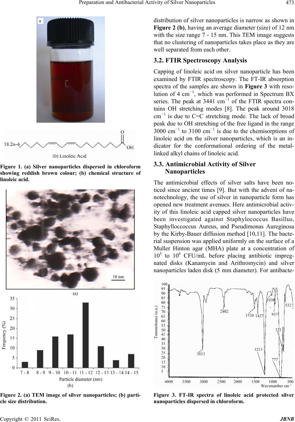 Preparation and Antibacterial Activity of Silver Nanoparticles473 Figure 1. (a) Silver nanoparticles dispersed in chloroform showing reddish brown colour; (b) chemical structure of linoleic acid. (b) Figure 2. (a) TEM image of silver nanoparticles; (b) parti- cle size distribution. distribution of silver nanoparticles is narrow as shown in Figure 2 (b), having an average diameter (size) of 12 nm with the size range 7 - 15 nm. This TEM image suggests that no clustering of nanoparticles takes place as they are well separated from each other. 3.2. FTIR Spectroscopy Analysis Capping of linoleic acid on silver nanoparticle has been examined by FTIR spectroscopy. The FT-IR absorption spectra of the samples are shown in Figure 3 with reso- lution of 4 cm−1, which was performed in Spectrum BX series. The peak at 3441 cm−1 of the FTIR spectra con- tains OH stretching modes [8]. The peak around 3018 cm−1 is due to C=C stretching mode. The lack of broad peak due to OH stretching of the free ligand in the range 3000 cm−1 to 3100 cm−1 is due to the chemisorptions of linoleic acid on the silver nanoparticles, which is an in- dicator for the conformational ordering of the metal- linked alkyl chains of linoleic acid. 3.3. Antimicrobial Activity of Silver Nanoparticles The antimicrobial effects of silver salts have been no- ticed since ancient times [9]. But with the advent of na- notechnology, the use of silver in nanoparticle form has opened new treatment avenues. Here antimicrobial activ- ity of this linoleic acid capped silver nanoparticles have been investigated against Staphylococcus Basillus, Staphylloccoccus Aureus, and Pseudimonas Aureginosa by the Kirby-Bauer diffusion method [10,11]. The bacte- rial suspension was applied uniformly on the surface of a Muller Hinton agar (MHA) plate at a concentration of 105 to 106 CFU/mL before placing antibiotic impreg- nated disks (Kanamycin and Arithromycin) and silver nanoparticles laden disk (5 mm diameter). For antibacte- Figure 3. FT-IR spectra of linoleic acid protected silver nanoparticles dispersed in chloroform. Copyright © 2011 SciRes. JBNB  Preparation and Antibacterial Activity of Silver Nanoparticles 474 rial study silver nanoparticles laden disk have been pre- pared by keeping 10 disks in 5 ml colloidal solution of silver nanoparticles for two days. These disks absorb the silver nanoparticles and become dry and hence there is no presence of chloroform. So there is no impact of sol- vent to the bacteria. The plates with the discs were incu- bated at 35˚C for 24 h, after which the average diameter of the inhibitio n zon e surround ing th e disk was measured with a ruler. Figure 4 shows plates to which Staphylo- coccus Basillus and Staphylloccoccus Aureus bacterial suspension were applied with nanoparticles laden disk and antibiotic impregnated disks. The diameter of inhibi- tion zones around the disk containing silver nanoparticles in S. Basillus, S. Aureus, and Pseudimonas Aureginosa bacterial suspension are 9 mm, 11 mm, 10 mm respec- tively. This test shows that silver nanoparticles are nearly 70%, 85% and 60% effective compare to Kanamycin and Arithromycin respectively. It is observed that the pres- ence of silver nanoparticles inhibited bacterial growth by more than 97%. The mechanism of the bactericidal effect of silver na- noparticles is not very well-known. It is believed that cellular proteins become inactive after treatment with silver nanoparticles [12]. It is also believed that silver nanoparticles after penetration into the bacteria have in- activated their enzymes, generating hydrogen peroxide and caused bacterial cell death [11]. Heavy metals are toxic and react with proteins, therefore they bind protein molecules; as a result cellular metabolism is inhibited causing death of microorganism [12]. It is known that silver sources such as silver nitrate and silver sulfadiaz- ine release Ag+ only [12] but high activity of silver nanoparticles is attributed to the release of Ag0 and Ag+ clusters when they dissolve [12]. Our experimental result shows that linoleic acid capped silver nanoparticles can be used as effective growth inhibitors in various micro- Figure 4. Shows silver nanoparticles laden disk (a) and an- tibiotic impregnated disk (b, c) placed on the surface of the Staphylococcus Basillus(1) and Staphylloccoccus Aureus(2) bacterial suspension on Muller Hinton agar (MHA) plate after incubation at 35˚C for 24 h. organisms, making them applicable to diverse medical medicines and antimicrobial control systems. 4. Discussion Uniform linoleic acid capped silver nanoparticles have been prepared through the reduction of silver ions by ethanol. TEM micrograph reveals that the prepared nano- particles are spherical in shape with average size of 12 nm having nearly uniform distribution and FTIR spectra confirms the capping of linoleic acid on nanoparticles surfaces. These linoleic acid capped silver nanoparticles are tested for its antimicrobial activity and the result shows that silver nanoparticles can be used as effective growth inhibitors in various microorganisms thereby applicable to diverse medical devices. 5. Acknowledgements Authors thanks to Dr B. Dkhar (S.O.), NEHU, Shillong, India, Dr. A. K. Paul, NEHU, Shillong, India, and Prof. Arun Chottapadhay, IIT, Guwahati, Assam, India for their suggestions and assistance during the work. REFERENCES [1] D. L. Feldheim and C. A. Foss, “Metal nanoparticles: Synthesis, Characterization and Applications,” Marcel Dekker Inc., New York, 2002. [2] G. Cao, “Nanostructures and Nanomaterials,” Edited by Imperial College Press, London, 2004. [3] C. P. Poole and F. J. Owens, “Introduction to Nanotech- nology,” Edited by Wiley Interscience Publication, New Jersey, 2005. [4] M. Brust, M. Walker, D. Bethell, D. J. Schiffrin and R. J. Whyman, “Synthesis of Thiol Derivatised Gold Nano- particles in a Two Phase Liquid/Liquid System,” Journal of the Chemical Society, Chemical Communications, Vol. 7, No. 7, 1994, pp. 801-802. doi:10.1039/c39940000801 [5] A. S. Nair and T. Y. Pradeep, “Halocarbon Mineraliza- tion and Catalytic Destruction by Metal Nanoparticles,” Current Science, Vol. 84, No. 12, 2003, pp. 1560-1564. [6] Y. Fang, “Optical Absorption of Nanoscale Colloidal Sil- ver: Aggregate Band and Adsorbate-Silver Surface Ban d,” Journal of Physical Chemistry, Vol. 108, No. 10, 1998, pp. 4315-4318. doi:10.1063/1.475831 [7] X. Wang, J. Zhuang, Q. Peng and Y. Li, “A General Stra- tegy for Nanocrystal Synthesis,” Nature, Vol. 437, No. 7055, 2005, pp. 121-124. doi:10.1038/nature03968 [8] M. D. Porter, T. B. Bright, D. L. Allara, and C. E. D. Chi- dsey, “Chemical Functionality in Self-Assembled Mono- layers and Electrochemistry,” Journal of the American Chemical Society, Vol. 109, No. 12, 1987, pp. 3559- 3568. doi:10.1021/ja00246a011 [9] M. Bahadory, “Synthesis of Noble Metal Nanoparticles,” Dissertation, Drexel University, Philadelphia, 2008. [10] S. Pal, Y. Kyung and J. M. Song, “Does the Antibacterial C opyright © 2011 SciRes. JBNB  Preparation and Antibacterial Activity of Silver Nanoparticles Copyright © 2011 SciRes. JBNB 475 Activity of Silver Nanoparticles Depend on the Shape of the Nanoparticle? A Study of the Gram-Negative Bacte- rium Escherichia coli,” Applied and Environmental Mi- crobiology, Vol. 73, No. 6, 2007, pp. 1712-1720. doi:10.1128/AEM.02218-06 [11] M. Raffi, F. Hussain, T. M. Bhatti, J. I. Akhter, A. Hameed and M. M. Hasan, “Antibacterial Characterization of Silver Nanoparticles against E. coli ATCC-15224,” Journal of Materials Science and Technology, Vol. 24, No. 2, 2008, pp. 192-196. [12] M. Kokkoris, C. C. Trapalis, S. Kossionides, R. Vlastou, B. Nsouli, R. Grotzschel, S. Spartalis, G. Kordas and T. Paradellis, “RBS and HIRBS studies of nanostructured AgSiO2 Sol-Gel Thin Coatings,” Nuclear Instruments and Methods B, Vol. 188, No. 1-4, 2002, pp. 67-72. doi:10.1016/S0168-583X(01)01020-5 |