

Paper Menu >>
Journal Menu >>
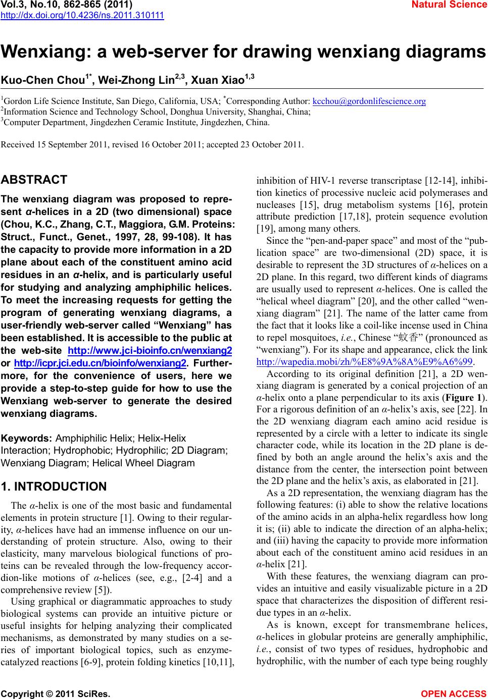 Vol.3, No.10, 862-865 (2011) Natural Science http://dx.doi.org/10.4236/ns.2011.310111 Copyright © 2011 SciRes. OPEN ACCESS Wenxiang: a web-server for drawing wenxiang diagrams Kuo-Chen Chou1*, Wei-Zhong Lin2,3, Xuan Xiao1,3 1Gordon Life Science Institute, San Diego, California, USA; *Corresponding Author: [email protected] 2Information Science and Technology School, Donghua University, Shanghai, China; 3Computer Department, Jingdezhen Ceramic Institute, Jingdezhen, China. Received 15 September 2011, revised 16 October 2011; accepted 23 October 2011. ABSTRACT The wenxiang diagram was proposed to repre- sent α-helices in a 2D (two dimensional) space (Chou, K.C., Zhang, C.T., Maggiora, G.M. Proteins: Struct., Funct., Genet., 1997, 28, 99-108). It has the capacity to provide more information in a 2D plane about each of the constituent amino acid residues in an α-helix, and is particularly useful for studying and analyzing amphiphilic helices. To meet the increasing requests for getting the program of generating wenxiang diagrams, a user-friendly web-server called “Wenxiang” has been established. It is accessible to the public at the web-site http ://www. jci- bioinfo.cn/w enxiang2 or http://icpr.j ci.edu.cn/bioinfo/w enxiang2. Further- more, for the convenience of users, here we provide a step-to-step guide for how to use the Wenxiang web-server to generate the desired wenxiang diagrams. Keywords: Amphiphilic Helix; Helix-Helix Interaction; Hydrophobic; Hydrophilic; 2D Diagram; Wenxiang Diagram; Helical Wheel Diagram 1. INTRODUCTION The α-helix is one of the most basic and fundamental elements in protein structure [1]. Owing to their regular- ity, α-helices have had an immense influence on our un- derstanding of protein structure. Also, owing to their elasticity, many marvelous biological functions of pro- teins can be revealed through the low-frequency accor- dion-like motions of α-helices (see, e.g., [2-4] and a comprehensive review [5]). Using graphical or diagrammatic approaches to study biological systems can provide an intuitive picture or useful insights for helping analyzing their complicated mechanisms, as demonstrated by many studies on a se- ries of important biological topics, such as enzyme- catalyzed reactions [6-9], protein folding kinetics [10,11], inhibition of HIV-1 reverse transcriptase [12-14], inhibi- tion kinetics of processive nucleic acid polymerases and nucleases [15], drug metabolism systems [16], protein attribute prediction [17,18], protein sequence evolution [19], among many others. Since the “pen-and-paper space” and most of the “pub- lication space” are two-dimensional (2D) space, it is desirable to represent the 3D structures of α-helices on a 2D plane. In this regard, two different kinds of diagrams are usually used to represent α-helices. One is called the “helical wheel diagram” [20], and the other called “wen- xiang diagram” [21]. The name of the latter came from the fact that it looks like a coil-like incense used in China to repel mosquitoes, i.e., Chinese “蚊香” (pronounced as “wenxiang”). For its shape and appearance, click the link http://wapedia.mobi/zh /%E8%9A%8A%E9%A6%99. According to its original definition [21], a 2D wen- xiang diagram is generated by a conical projection of an α-helix onto a plane perpendicular to its axis (Figure 1). For a rigorous definition of an α-helix’s axis, see [22]. In the 2D wenxiang diagram each amino acid residue is represented by a circle with a letter to indicate its single character code, while its location in the 2D plane is de- fined by both an angle around the helix’s axis and the distance from the center, the intersection point between the 2D plane and the helix’s axis, as elaborated in [21]. As a 2D representation, the wenxiang diagram has the following features: (i) able to show the relative locations of the amino acids in an alpha-helix regardless how long it is; (ii) able to indicate the direction of an alpha-helix; and (iii) having the capacity to provide more information about each of the constituent amino acid residues in an α-helix [21]. With these features, the wenxiang diagram can pro- vides an intuitive and easily visualizable picture in a 2D space that characterizes the disposition of different resi- due types in an α-helix. As is known, except for transmembrane helices, α-helices in globular proteins are generally amphiphilic, i.e., consist of two types of residues, hydrophobic and hydrophilic, with the number of each type being roughly 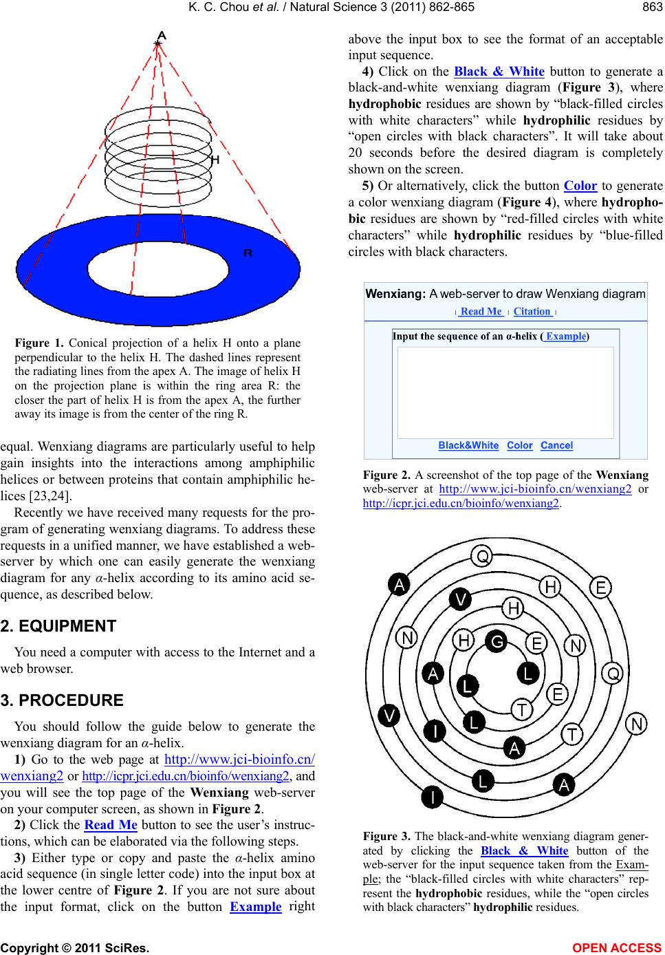 K. C. Chou et al. / Natural Science 3 (2011) 862-865 Copyright © 2011 SciRes. OPEN ACCESS 863 863 Figure 1. Conical projection of a helix H onto a plane perpendicular to the helix H. The dashed lines represent the radiating lines from the apex A. The image of helix H on the projection plane is within the ring area R: the closer the part of helix H is from the apex A, the further away its image is from the center of the ring R. equal. Wenxiang diagrams are particularly useful to help gain insights into the interactions among amphiphilic helices or between proteins that contain amphiphilic he- lices [23,24]. Recently we have received many requests for the pro- gram of generating wenxiang diagrams. To address these requests in a unified manner, we have established a web- server by which one can easily generate the wenxiang diagram for any α-helix according to its amino acid se- quence, as described below. 2. EQUIPMENT You need a computer with access to the Internet and a web browser. 3. PROCEDURE You should follow the guide below to generate the wenxiang diagram for an α-helix. 1) Go to the web page at http://www.jci-bioinfo.cn/ wenxiang2 or http://icpr.jci.edu.cn/bioinfo/wenxiang2, and you will see the top page of the Wenxiang web-server on your computer screen, as shown in Figure 2. 2) Click the Read Me button to see the user’s instruc- tions, which can be elaborated via the following steps. 3) Either type or copy and paste the α-helix amino acid sequence (in single letter code) into the input box at the lower centre of Figure 2. If you are not sure about the input format, click on the button Example right above the input box to see the format of an acceptable input sequence. 4) Click on the Black & White button to generate a black-and-white wenxiang diagram (Figure 3), where hydrophobic residues are shown by “black-filled circles with white characters” while hydrophilic residues by “open circles with black characters”. It will take about 20 seconds before the desired diagram is completely shown on the screen. 5) Or alternatively, click the button Color to generate a color wenxiang diagram (Figure 4), where hydropho- bic residues are shown by “red-filled circles with white characters” while hydrophilic residues by “blue-filled circles with black characters. Figure 2. A screenshot of the top page of the Wenxiang web-server at http://www.jci-bioinfo.cn/wenxiang2 or http://icpr.jci.edu.cn/bioinfo/wenxiang2. Figure 3. The black-and-white wenxiang diagram gener- ated by clicking the Black & White button of the web-server for the input sequence taken from the Exam- ple; the “black-filled circles with white characters” rep- resent the hydrophobic residues, while the “open circles with black characters” hydrophilic residues. 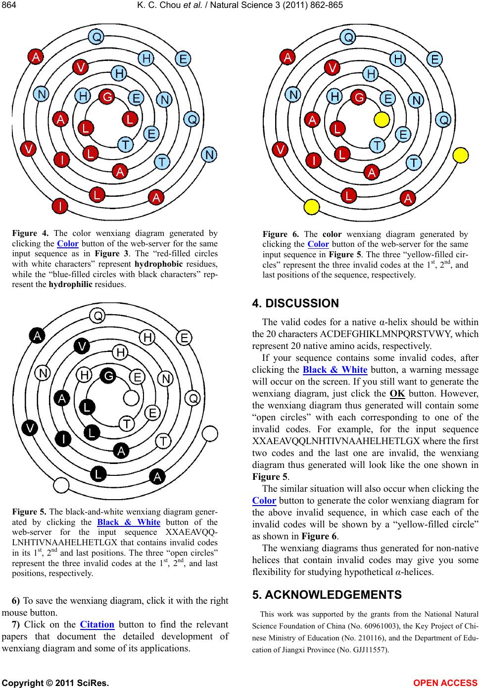 K. C. Chou et al. / Natural Science 3 (2011) 862-865 Copyright © 2011 SciRes. OPEN ACCESS 864 Figure 4. The color wenxiang diagram generated by clicking the Color button of the web-server for the same input sequence as in Figure 3. The “red-filled circles with white characters” represent hydrophobic residues, while the “blue-filled circles with black characters” rep- resent the hydrophilic residues. Figure 5. The black-and-white wenxiang diagram gener- ated by clicking the Black & White button of the web-server for the input sequence XXAEAVQQ- LNHTIVNAAHELHETLGX that contains invalid codes in its 1st, 2nd and last positions. The three “open circles” represent the three invalid codes at the 1st, 2nd, and last positions, respectively. 6) To save the wenxiang diagram, click it with the right mouse button. 7) Click on the Citation button to find the relevant papers that document the detailed development of wenxiang diagram and some of its applications. Figure 6. The color wenxiang diagram generated by clicking the Color button of the web-server for the same input sequence in Figure 5. The three “yellow-filled cir- cles” represent the three invalid codes at the 1st, 2nd, and last positions of the sequence, respectively. 4. DISCUSSION The valid codes for a native α-helix should be within the 20 characters ACDEFGHIKLMNPQRSTVWY, which represent 20 native amino acids, respectively. If your sequence contains some invalid codes, after clicking the Black & White button, a warning message will occur on the screen. If you still want to generate the wenxiang diagram, just click the OK button. However, the wenxiang diagram thus generated will contain some “open circles” with each corresponding to one of the invalid codes. For example, for the input sequence XXAEAVQQLNHTIVNAAHELHETLGX where the first two codes and the last one are invalid, the wenxiang diagram thus generated will look like the one shown in Figure 5. The similar situation will also occur when clicking the Color button to generate the color wenxiang diagram for the above invalid sequence, in which case each of the invalid codes will be shown by a “yellow-filled circle” as shown in Figure 6. The wenxiang diagrams thus generated for non-native helices that contain invalid codes may give you some flexibility for studying hypothetical α-helices. 5. ACKNOWLEDGEMENTS This work was supported by the grants from the National Natural Science Foundation of China (No. 60961003), the Key Project of Chi- nese Ministry of Education (No. 210116), and the Department of Edu- cation of Jiangxi Province (No. GJJ11557).  K. C. Chou et al. / Natural Science 3 (2011) 862-865 Copyright © 2011 SciRes. OPEN ACCESS 865 865 REFERENCES [1] Richardson, J.S. (1981) The anatomy and taxonomy of protein structure. Advances in Protein Chemistry, 34, 167-339. doi:10.1016/S0065-3233(08)60520-3 [2] Chou, K.C. (1983) Identification of low-frequency modes in protein molecules. Biochemical Journal, 215, 465-469. PMID 6362659 [3] Chou, K.C. (1989) Low-frequency resonance and coop- erativity of hemoglobin. Trends in Biochemical Sciences, 14, 212. doi:10.1016/0968-0004(89)90026-1 [4] Martel, P. (1992) Biophysical aspects of neutron scatter- ing from vibrational modes of proteins. Progress in Bio- physics and Molecular Biology, 57, 129-179. doi:10.1016/0079-6107(92)90023-Y [5] Chou, K.C. (1988) Review: Low-frequency collective motion in biomacromolecules and its biological functions. Biophysical Chemistry, 30, 3-48. doi:10.1016/0301-4622(88)85002-6 [6] Chou, K.C. and Forsen, S. (1980) Graphical rules for enzyme-catalyzed rate laws. Biochemical Journal, 187, 829-835. PMID 7188428 [7] Zhou, G.P. and Deng, M.H. (1984) An extension of Chou’s graphical rules for deriving enzyme kinetic equa- tions to system involving parallel reaction pathways. Biochemical Journal, 222, 169-176. PMID 6477507 [8] Chou, K.C. (1989) Graphic rules in steady and non- steady enzyme kinetics. Journal of Biological Chemistry, 264, 12074-12079. PMID 2745429 [9] Andraos, J. (2008) Kinetic plasticity and the determina- tion of product ratios for kinetic schemes leading to mul- tiple products without rate laws: New methods based on directed graphs. Canadian Journal of Chemistry, 86, 342-357. doi:10.1139/v08-020 [10] Chou, K.C. (1990) Review: Applications of graph theory to enzyme kinetics and protein folding kinetics. Steady and non-steady state systems. Biophysical Chemistry, 35, 1-24. doi:10.1016/0301-4622(90)80056-D [11] Shen, H.B., Song, J.N. and Chou, K.C. (2009) Prediction of protein folding rates from primary sequence by fusing multiple sequential features. Journal of Biomedical Sci- ence and Engineering (JBiSE), 2, 136-143 (openly ac- cessible at http://www.scirp.org/journal/jbise). doi:10.4236/jbise.2009.23024 [12] Althaus, I.W., Chou, J.J., Gonzales, A.J., Diebel, M.R., Chou, K.C., Kezdy, F.J., Romero, D.L., Aristoff, P.A., Tarpley, W.G. and Reusser, F. (1993) Steady-state kinetic studies with the non-nucleoside HIV-1 reverse transcrip- tase inhibitor U-87201E. Journal of Biological Chemistry, 268, 6119-6124. PMID 7681060 [13] Althaus, I.W., Gonzales, A.J., Chou, J.J., Diebel, M.R., Chou, K.C., Kezdy, F.J., Romero, D.L., Aristoff, P.A., Tarpley, W.G. and Reusser, F. (1993) The quinoline U-78036 is a potent inhibitor of HIV-1 reverse transcrip- tase. Journal of Biological Chemistry, 268, 14875-14880. PMID 7686907 [14] Althaus, I.W., Chou, J.J., Gonzales, A.J., Diebel, M.R., Chou, K.C., Kezdy, F.J., Romero, D.L., Aristoff, P.A., Tarpley, W.G. and Reusser, F. (1993) Kinetic studies with the nonnucleoside HIV-1 reverse transcriptase inhibitor U-88204E. Biochemistry, 32, 6548-6554. doi:10.1021/bi00077a008 [15] Chou, K.C., Kezdy, F.J. and Reusser, F. (1994) Review: Steady-state inhibition kinetics of processive nucleic acid polymerases and nucleases. Analytical Biochemistry, 221, 217-230. doi:10.1006/abio.1994.1405 [16] Chou, K.C. (2010) Graphic rule for drug metabolism systems. Current Drug Metabolism, 11, 369-378. doi:10.2174/138920010791514261 [17] Xiao, X., Wang, P. and Chou, K.C. (2008) Predicting protein structural classes with pseudo amino acid com- position: An approach using geometric moments of cel- lular automaton image. Journal of Theoretical Biology, 254, 691-696. doi:10.1016/j.jtbi.2008.06.016 [18] Xiao, X., Wang, P. and Chou, K.C. (2009) GPCR-CA: A cellular automaton image approach for predicting G- protein-coupled receptor functional classes. Journal of Computational Chemistry, 30, 1414-1423. doi:10.1002/jcc.21163 [19] Wu, Z.C., Xiao, X. and Chou, K.C. (2010) 2D-MH: A web-server for generating graphic representation of pro- tein sequences based on the physicochemical properties of their constituent amino acids. Journal of Theoretical Biology, 267, 29-34. doi:10.1016/j.jtbi.2010.08.007 [20] Schiffer, M. and Edmundson, A.B. (1967) Use of helical wheels to represent the structures of proteins and to iden- tify segments with helical potential. Biophysical Journal, 7, 121-135. doi:10.1016/S0006-3495(67)86579-2 [21] Chou, K.C., Zhang, C.T. and Maggiora, G.M. (1997) Disposition of amphiphilic helices in heteropolar envi- ronments. Proteins: Structure, Function, and Genetics, 28, 99-108. doi:10.1002/(SICI)1097-0134(199705)28:1<99::AID-PR OT10>3.0.CO;2-C [22] Chou, K.C., Nemethy, G. and Scheraga, H.A. (1984) Energetic approach to packing of a-helices: 2. General treatment of nonequivalent and nonregular helices. Jour- nal of American Chemical Society, 106, 3161-3170. doi:10.1021/ja00323a017 [23] Zhou, G.P. (2011) The disposition of the LZCC protein residues in wenxiang diagram provides new insights into the protein-protein interaction mechanism. Journal of Theoretical Biology, 284, 142-148. doi:10.1016/j.jtbi.2011.06.006 [24] Zhou, G.P. (2011) The structural determinations of the leucine zipper coiled-coil domains of the cGMP-De- pendent protein kinase I alpha and its interaction with the myosin binding subunit of the myosin light chains phos- phase. Proteins & Peptide Letters, 18, 966-978. PMID 21592084 |