

Paper Menu >>
Journal Menu >>
 Chinese Medicine, 2011, 2, 103-108 doi:10.4236/cm.2011.23017 Published Online September 2011 (http://www.SciRP.org/journal/cm) Copyright © 2011 SciRes. CM Immunopotentiating Activity of Dendrobium Species in Mouse Splenocytes David T. W. Lau, Michel K. T. Poon, Hoi Yan Leung, Kam Ming Ko Division of Life Science, Hong Kong University of Science & Technology, Hong Kong, China E-mail: [email protected] Received April 4, 2011; revised April 25, 2011; accepted May 13, 2011 Abstract This study aimed to explore a pharmacological activity marker for quality assurance of Dendrobium species. The immunopotentiating activity in aqueous extracts prepared from four Dendrobium species, including D. officinalis, was assessed by an in vitro assay of concanavalin A (Con A)-stimulated proliferation of mouse splenocytes. Four samples of commercially available Dendrobii Caulis were also analyzed for comparison. The results indicated that the aqueous extract of D. officinalis produced immunopotentiating action, as evi- denced by the increase in Con A-stimulated proliferation of mouse splenocytes, with the extent of stimula- tion being more prominent than those of other tested Dendrobium species and Dendrobii Caulis samples. In conclusion, an in vitro immunopotentiation assay may be used for assessing the pharmacological activity of Dendrobium species. The finding that D. officinalis produced a more potent immunopotentiating action is consistent with its “yin-nourishing” action in Chinese medicine, which is more effective than other Den- drobium species in clinical use. Keywords: Dendrobium offincalis, Dendrobii Caulis, Splenocyte Proliferation, Concanavalin A 1. Introduction Dendrobium species was regarded as a first-rate herb in Shen-nong Bencao Jing (Cano n on Medicinal Herbs, 200 B.C.) and has a long history of usage in China. Over the past few decades, the dried stem of Dendrobium species (Dendrobii Caulis, also called Shi Hu in Chinese) has been used for clearing “heat” and nourishing “yin” in the practice of Chinese medicine [1]. A recent study showed that the administration of an aqueous extract of Den- drobium candidum ameliorated the dry-mouth symptom in patients suffering from Sjögren syndrome [2]. Phar- macological studies have demonstrated that the aqueous extract and/or polysaccharides of Dendrobii Caulis pos- sess antioxidant [3,4], immuno-stimulating [5-7], anti- tumor [8,9], anti-microbial [9], anti-hyperlipidemia [10] and anti-hyperglycemic [11] activities. The widespread application of Dendrobii Caulis has raised a concern re- garding the identity of Dendrobium species as herbal source. In this regard, the record of Dendrobii Caulis in Chinese Pharmacopoeia has been constantly amended over the past 20 years. While five Dendrobium species, including D. loddigesii Rolfe, D. fimbriatum Hook and D. nobile Lindl., were listed in the 1995 and 2000 editions, a more extended list of Dendrobium species was gener- ated in the 2005 edition. As such, up to 74 Dendrobium species can be used as Dendrobii Caulis in China. In the 2010 edition of Chinese Pharmacopoeia [12], D. Offici- nalis has been singled out as a distinct herb originated from D. officinale Kimura et Migo. Not surprisingly, D. Officinalis is relatively scarce in abundance and deemed to possess a stronger therapeutic potential than other Dendrobium species [13,14]. As more than 45 Dendrobium species have been iden- tified to possess therapeutic potential [15-18], the devel- opment of chemical and DNA fingerprinting methods for species authentication, particularly for those recorded in Chinese Pharmacopoeia, has been an area of intensive research [19-26]. In addition to chemical markers, the measurement of pharmacological activity may offer a complementary and functionally relevant quality assess- ment for herbal materials [27]. In this connection, “yin- nourishing” tonic herbs (including Herba Dendrobii) have been found to produce immunopotentiating effect on mouse splenocytes both in vivo and in vitro [28]. In the present study, using an in vitro assay of conc anavalin A (Con A)-stimulated proliferation of mouse splenocytes, we assessed the immunopotentiating activity of aqueous  D. T. W. LAU ET AL. 104 extracts prepared from four Dendrobium species includ- ing D. Officinalis, with an objective of exploring a pha- rmacological activity marker for quality assurance. Four samples of commercially available Dendrobii Caulis were also analyzed for comparison. 2. Materials and Methods 2.1. Chemicals and Plant Materials RPMI-1640 medium, heat inactivated fetal bovine serum (FBS) and Con A were purchased from Sigma Chemical Co. (St. Louis, MO, USA). MTT (3-[4,5-dimethylthiazol- 2-yl]-2,5-diphenyl tetrazolium bromide)-based cell pro- liferation kit was purchased from Boehringer Mann- heim Gmbh (Mannheim, Germany). All other chemicals were of analytical grade. Four Dendrolium species, namely, D. loddigesii Rolfe, D. fimbriatum Hook, D. nobile Lindl. and D. officinalis Kimura et Migo, were obtained from an ornamental flora shop in Hong Kong, and they were authenticated by floral structure with reference to China Flora. Four samples of Dendrobium Caulis were randomly bought from local herbal stores. Voucher specimens of Dendrobium plants and Dendrobii Caulis have been deposited in the Division of Life Science, The Hong Kong University o f Science & Tech- nology (HK UST), Hong Kon g SAR, Chi na. 2.2. Plant/Herbal Extraction Fresh stems of Dendrobium plants or Dendrobii Caulis (i.e., dried stems of Dendrobium species) were cut into small pieces and extracted by 10 volumes (w/v) of dou- ble distilled water at 60˚C for 2 h. The extraction proce- dure was repeated once, and the pooled aqueous extracts were dried by lyophilization. The lyophilized powders (i.e. Dendrobium extracts) were stored at 20˚C prior to use for experiment. 2.3. Measurement of in Vitro Immunopotentiating Activity in Con A-Stimulated Mouse Splenocytes Procedures involved in isolation of mouse splenocytes were conducted under aseptic conditions. Splenic tissue obtained from adult female ICR mice (25 - 28 g) was teased with a plastic syringe in a culture dish (60 mm) containing 10 mL of RPMI-1640 medium, and it was gently strained through a 200 mesh stainless steel sieve to remove clumps to produce a cell suspension. The product was then left to stand on ice for 5 min to remove tissue fragments. The cell suspension was centrifuged at 600 × g for 10 min an d washed 3 times with RMPI-1 640 medium, and it was finally resuspended in RPMI-1640 medium supplemented with 10% FBS at a concentration of 1 × 10⁷ viable cells/mL. The viability of isolated splenocytes, as determined by Trypan blue exclusion test, was found to be higher than 95%. Mouse splenocytes were cultured in medium with Con A in the presence or absence of Dendrobium extract in 96-well microtiter plates (flat bottom) in a final volume of 100 L. Con A (prepared in phosphate buffered saline) was added at final concentrations of 0.5, 1 and 2 g/mL, respectively. Aliquots of respective Dendrobium extract (10 L in aqueous solution) were added at increasing final concen- trations ranging from 1.6 to 100 g/mL. Control wells were added with 10 L of sterilized double-distilled wa- ter only. Splenocytes were then cultured for 72 h at 37˚C in a humidified atmosphere of 5% CO2 in air. At th e end of the culture period, the extent of splenocyte prolifera- tion was determined colorimetrically by MTT-based cell proliferation assay. An aliquot (10 L) of MTT labeling reagent was added to each well under dark conditions. After 4 h of incubation, 100 L of solubilization buffer (10% SDS in 0.01 M HCl) was added, and the mixtures were incubated in 5% CO2 at 37˚C overnight for dis- solving the colored crystals. The extent of splenocyte proliferation was determined by measuring the absorb- ance at 570 nm using Victor V2 Multi-label Counter (Perkin Elmer, Turku, Finland). Stimulation index (SI) was calculated using the equation: SI = mean absorbance of cells stimulated with Con A/ mean absorbance of cells not stimulated with Con A. The extent of Con A-stimu- lated proliferation of isolated splenocytes was estimated by computing the area under the curve (AUC1) of the graph plotting stimulatory indices against Con A con- centrations. Data of AUC1 were expressed as the per- centage of non-Dendrobium extract-treated control, and AUC2 was computed from a graph plotting percentages of control against tested concentrations of Dendrobium extract. The immunopotentiating activity was expressed as the difference in AUC2 values between the Dendro- bium extract-untreated control and Dendrobium-treated group. 2.4. Measurements of Interleukin-2 (IL-2), IL-6 and Interferon- (INF- ) Levels Isolated mouse splenocytes were cultured with Den- drobium extract (25, 50, 100 g/mL, final concentration) in the presence or absence of Con A (2 g/mL) for 24 h. IL-2, IL-6 and INF- levels in the culture medium was then measured using ELISA kits (InvitrogenTM Mouse IL-2 ELISA kit, Invitrogen, Frederick, MD, USA; Invi- trogenTM Mouse IL-6 ELISA kit, Invitrogen, Camarillo, CA, USA; ELISAPRO kit for mouse IFN- , MABTECH, Copyright © 2011 SciRes. CM 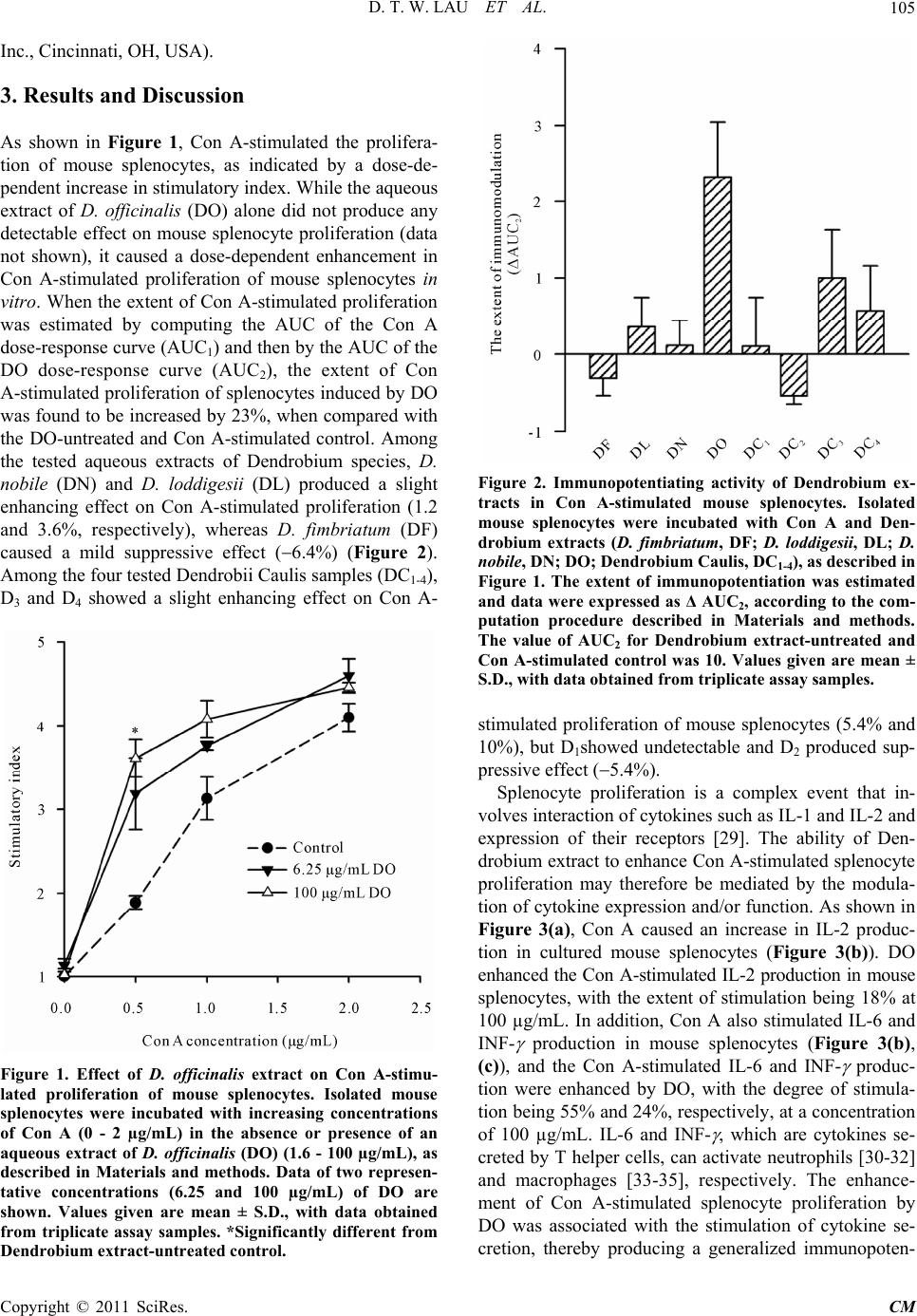 D. T. W. LAU ET AL.105 Inc., Cincinnati, OH, USA). 3. Results and Discussion As shown in Figure 1, Con A-stimulated the prolifera- tion of mouse splenocytes, as indicated by a dose-de- pendent increase in stimulatory index. While the aqueous extract of D. officinalis (DO) alone did not produce any detectable effect on mouse splenocyte proliferation (data not shown), it caused a dose-dependent enhancement in Con A-stimulated proliferation of mouse splenocytes in vitro. When the extent of Con A-stimulated proliferation was estimated by computing the AUC of the Con A dose-response curve (AUC1) and then by the AUC of the DO dose-response curve (AUC2), the extent of Con A-stimulated proliferation of splenocytes induced by DO was found to be increased by 23%, when compared with the DO-untreated and Con A-stimulated control. Among the tested aqueous extracts of Dendrobium species, D. nobile (DN) and D. loddigesii (DL) produced a slight enhancing effect on Con A-stimulated proliferation (1.2 and 3.6%, respectively), whereas D. fimbriatum (DF) caused a mild suppressive effect (6.4%) (Figure 2). Among the four tested Dendrobii Caulis samples (DC1-4), D3 and D4 showed a slight enhancing effect on Con A- Figure 1. Effect of D. officinalis extract on Con A-stimu- lated proliferation of mouse splenocytes. Isolated mouse splenocytes were incubated with increasing concentrations of Con A (0 - 2 µg/mL) in the absence or presence of an aqueous extract of D. officinalis (DO) (1.6 - 100 µg/mL), as described in Materials and methods. Data of two represen- tative concentrations (6.25 and 100 µg/mL) of DO are shown. Values given are mean ± S.D., with data obtained from triplicate assay samples. *Significantly different from Dendrobium extract-untreated control. Figure 2. Immunopotentiating activity of Dendrobium ex- tracts in Con A-stimulated mouse splenocytes. Isolated mouse splenocytes were incubated with Con A and Den- drobium extracts (D. fimbriatum, DF; D. loddigesii, DL; D. nobile, DN; DO; Dendrobium Caulis, DC1-4), as described in Figure 1. The extent of immunopotentiation was estimated and data were expressed as Δ AUC2, according to the com- putation procedure described in Materials and methods. The value of AUC2 for Dendrobium extract-untreated and Con A-stimulated control was 10. Values given are mean ± S.D., with data obtained from triplicate assay samples. stimulated proliferation of mouse splenocytes (5.4% and 10%), but D1showed undetectable and D2 produced sup- pressive effect (5.4%). Splenocyte proliferation is a complex event that in- volves interaction of cytokines such as IL-1 and IL-2 and expression of their receptors [29]. The ability of Den- drobium extract to enhan ce Con A-stimulated splenocyte proliferation may therefore be mediated by the modula- tion of cytokine expression and/or function. As shown in Figure 3(a), Con A caused an increase in IL-2 produc- tion in cultured mouse splenocytes (Figure 3(b)). DO enhanced the Con A-stimulated IL-2 production in mouse splenocytes, with the extent of stimulation being 18% at 100 µg/mL. In addition, Con A also stimulated IL-6 and INF- production in mouse splenocytes (Figure 3(b), (c)), and the Con A-stimulated IL-6 and INF- produc- tion were enhanced by DO, with the degree of stimula- tion being 55% and 24%, respectively, at a concentration of 100 µg/mL. IL-6 and INF- , which are cytokines se- creted by T helper cells, can activate neutrophils [30-32] and macrophages [33-35], respectively. The enhance- ment of Con A-stimulated splenocyte proliferation by DO was associated with the stimulation of cytokine se- cretion, thereby producing a generalized immunopoten- Copyright © 2011 SciRes. CM 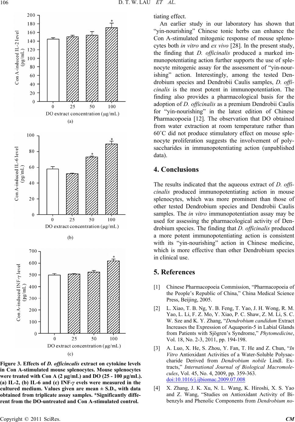 D. T. W. LAU ET AL. 106 (a) (b) (c) Figure 3. Effects of D. officincalis extract on cytokine levels in Con A-stimulated mouse splenocytes. Mouse splenocytes were treated with Con A (2 µg/mL) and DO (25 - 100 µg/mL). (a) IL-2, (b) IL-6 and (c) INF-γ evels were measured in the cultured medium. Values given are mean ± S.D., with data obtained from triplicate assay samples. *Significantly diffe- rent from the DO-untreated and Con A-stimulated control. tiating effect. An earlier study in our laboratory has shown that “yin-nourishing” Chinese tonic herbs can enhance the Con A-stimulated mitogenic response of mouse spleno- cytes both in vitro and ex vivo [28]. In the present study, the finding that D. officinalis produced a marked im- munopotentiating action further supports the use of sple- nocyte mitogenic assay for the assessment of “yin-nour- ishing” action. Interestingly, among the tested Den- drobium species and Dendrobii Caulis samples, D. offi- cinalis is the most potent in immunopotentiation. The finding also provides a pharmacological basis for the adoption of D. officinalis as a premium Dendrobii Caulis for “yin-nourishing” in the latest edition of Chinese Pharmacopoeia [12]. The observation that DO obtained from water extraction at room temperature rather than 60˚C did not produce stimulatory effect on mouse sple- nocyte proliferation suggests the involvement of poly- saccharides in immunopotentiating action (unpublished data). 4. Conclusions The results indicated that the aqueous extract of D. offi- cinalis produced immunopotentiating action in mouse splenocytes, which was more prominent than those of other tested Dendrobium species and Dendrobii Caulis samples. The in vitro immunopotentiation assay may be used for assessing the pharmacological activity of Den- drobium species. The finding that D. officinalis produ ced a more potent immunopotentiating action is consistent with its “yin-nourishing” action in Chinese medicine, which is more effective than other Dendrobium species in clinical use. 5. References [1] Chinese Pharmacopoeia Commission, “Pharmacopoeia of the People’s Republic of China,” China Medical Science Press, Beijing, 2005. [2] L. Xiao, T. B. Ng, Y. B. Feng, T. Yao, J. H. Wong, R. M. Yao, L. Li, F. Z. Mo, Y. Xiao, P. C. Shaw, Z. M. Li, S. C. W. Sze and K. Y. Z hang, “Dendrobium candidum Extract Increases the Expression of Aquaporin-5 in Labial Glands from Patients with Sjögren’s Syndrome,” Phytomedicine, Vol. 18, No. 2-3, 2011, pp. 194-198. [3] A. Luo, X. He, S. Zhou, Y. Fan, T. He and Z. Chun, “In Vitro Antioxidant Activities of a Water-Soluble Polysac- charide Derived from Dendrobium nobile Lindl. Ex- tracts,” International Journal of Biological Macromole- cules, Vol. 45, No. 4, 2009, pp. 359-363. doi:10.1016/j.ijbiomac.2009.07.008 [4] X. Zhang, J. K. Xu, N. L. Wang, K. Hiroshi, X. S. Yao and Z. Wang, “Studies on Antioxidant Activity of Bi- benzyls and Phenolic Components from Dendrobium no- Copyright © 2011 SciRes. CM  D. T. W. LAU ET AL.107 bile,” Chinese Pharmaceutical Journal, Vol. 43, 2008, pp. 829-832. [5] J. H. Wang, J. P. Luo, X. Q. Zha and B. J. Feng, “Com- parison of Antitumor Activities of Different Polysaccha- ride Fractions from the Stems of Dendrobium nobile Lindl,” Carbohydrate Polymers, Vol. 79, No. 1, 2010, pp. 114-118. doi:10.1016/j.carbpol.2009.07.032 [6] X. Q. Zha, J. P. Luo, S. Z. Luo and S. T. Jiang, “Structure Identification of a New Immunostimulating Polysaccha- ride from the Stems of Dendrobium huoshanense,” Car- bohydrate Polym ers, Vol. 69, No. 1, 2007, pp. 86-93. [7] X. Q. Zha, J. P. Luo and S. T. Jiang, “Induction of Im- munomodulating Cytokines by Polysaccharides from Dendrobium huoshanense,” Pharmaceutical Biology, Vol. 45, No. 1, 2007, pp. 71-76. doi:10.1080/13880200601028420 [8] J. H. Wang, X. Q. Zha, J. P. Luo and X. F. Yang, “An Acetylated Galactomannoglucan from the Stems of Den- drobium nobile Lindl,” Carbohydrate Research, Vol. 345, No. 8, 2010, pp. 1023-1027. doi:10.1016/j.carres.2010.03.005 [9] D. Uma, S. Selvi, D. Devipriy a, S. Murugan and S. Suja, “Antitumor and Antimicrobial Activities and Inhibition of in-Vitro Lipid Peroxidation by Dendrobium nobile,” African Journal of Biotechnology, Vol. 8, No. 10, 2009, pp. 2289-2293. [10] X. Y. Li, Q. H. Gong , Q. Wu, Y. F. Lu, F. Jin, Y. F. Li and J. S. Shi, “Effects of Dendrobium nobile Polyose on Hy- perlipemia and Liver Fatty Degeneration in Rats,” Chinese Pharmaceutical Journal, Vol. 45, 2010, pp. 1142-1144. [11] Y. Zhao, Y. O. Son, S. S. Kim, Y. S. Jang and J. C. Lee, “Antioxidant and Anti-Hyperglycemic Activity of Poly- saccharide Isolated from Dendrobium chrysotox um Lindl,” Journal of Biochemistry and Molecular Biology, Vol. 40, No. 5, 2007, pp. 670-677. doi:10.5483/BMBRep.2007.40.5.670 [12] Chinese Pharmacopoeia Commission, “Pharmacopoeia of the People’s Republic of China,” China Medical Science Press, Beijing, 2010. [13] X. Lin, C. W. Sze, Y. Tong, Z. Zhang, Y. Feng, J. P. Chen, T. B. Ng, L. Xiao, P. Shaw and K. Y. Zhang, “Protective Effect of Dendrobium officinale Polysaccha- rides on Experimental Sjögren’s Syndrome,” Journal of Complementary and Integrative Medicine, Vol. 7, No. 1, 2010, Article 14. doi:10.2202/1553-3840.1342 [14] Y. Wu and J. Si, “Present Status and Sustainable Devel- opment of Dendrobium officinal Industry,” China Jour- nal of Chinese Materia Medica, Vol. 35, No. 15, 2010, pp. 2033-2037. [15] W. L. Sha and J. Y. Luo, “Study of the Chinese drug Shi- Hu (Dendrobium). I. Inve stigation of Botanical Origin and the Drug,” Acta Pharmaceutica Sinica, Vol. 15, No. 6, 1980, pp. 351-357. [16] G. X. Ma, G. J. Xu, L. S. Xu and M. F. Li, “Survey and Identification of Commercial Samples of Shihu (Dendro- bium Sw.) (III),” Chinese Traditional and Herbal Drugs, Vol. 20, 1995, pp. 370-373. [17] J. Liu, T. He and Z. Chun, “DNA Molecular Identifica- tion of Herba Dendrobii and Its Adulterant Species Based on ITS Sequence Analysis,” China Journal of Chinese Materia Medica, Vol. 34, No. 22, 2009, pp. 2853-2856. [18] Z. Q. Yuan, J. Y. Zhang and T. Liu, “Phy logenetic Rela- tionship of China Dendrobium Species Based on the Se- quence of the Internal Transcribed Spacer of Ribosomal DNA,” Biologia Plantarum, Vol. 53, No. 1, 2009, pp. 155-158. doi:10.1007/s10535-009-0024-0 [19] J. Liu, T. He and Z. Chun, “Advance in Application of DNA Molecular Markers on Dendrobium,” Chinese Journal of Applied and Environmental Biology, Vol. 14, No. 6, 2008, pp. 855-862. [20] Y. P. Li, C. Qing, T. T. Fang, Y. Liu and Y. G. Chen, “Chemical Constituents of Dendrobium chrysotoxum,” Chemistry of Natural Compounds, Vol. 45, No. 3, 2009, pp. 414-416. doi:10.1007/s10600-009-9329-7 [21] H. J. Ou, J. L. Cheng, X. X. Li, R. T. Zhan, Y. Z. Liang, H. Xu and P. Yan, “HPLC Fingerprint of Flavonoids and Phenols of Dendrobium nobile,” Journal of Chinese Me- dicinal Materials, Vol. 32, No. 6, 2009, pp. 871-874. [22] S. G. Sha o, L. Han, Y. H. Ma, J. S hen, W. C. Zhang and X. Y. Ding, “Analysis and Authentication of cpDNA psbA- trnH Regions of Dendrobiu m Species of Fengdous,” Acta Pharmaceutica Sinica, Vol. 44, 2009, pp. 1173-1178. [23] S. J. Wu, Y. S. Liu, T. W. Chen, C. C. Ng, W. S. Tzeng and Y. T. Shyu, “Differentiation of Medicinal Den- drobium Species (Orchidaceae) Using Molecular Markers and Scanning Electron Microscopy,” Journal of Food and Drug Analysis, Vol. 17, No. 6, 2009, pp. 474-488. [24] H. H. Fan, T. C. Li, J. Qiu, Y. Lin and Y. P. Cai, “Com- parison of SRAP and RAPD Markers for Genetic Analy- sis of Plants in Dendrobium sw.,” Chinese Traditional and Herbal Drugs, Vol. 41, No. 4, 2010, pp. 627-632. [25] S. G. Feng, X. Hu, H. Y. Zhao and H. Z. Wang, “Appli- cation of DNA Molecular Marker in Study on Den- drobium officinale,” Chinese Traditional and Herbal Drugs, Vol. 41, 2010, pp.499-502. [26] D. Xue, S. Feng, H. Zhao, H. Jiang, B. Shen, N. Shi, J. Lu, J. Liu and H. Wang, “ The Linkage Maps of Den- drobium Species Based on RAPD and SRAP Markers,” Journal of Genetics and Genomics, Vol. 37, No. 3, 2010, pp. 197-204. doi:10.1016/S1673-8527(09)60038-2 [27] K. M. Ko and K. M. Ng, “Processing Control of Chinese Herbal Products: An Ultimate Approach for Quality As- surance,” Asia-Pacific Biotechnology News, Vol. 8, No. 23, 2004, pp. 1338-1341. [28] T. K. Yim and K. M. Ko, “Antioxidant and Immuno- Modulatory Activities of Chinese Tonifying Herbs, in Vi- tro and ex Vivo,” Pharmaceutical Biology, Vol. 40, No. 5, 2002, pp. 329-335. doi:10.1076/phbi.40.5.329.8457 [29] K. A. Smith, “Interleukin-2: Inception, Impact, and Im- plications,” Science, Vol. 240, No. 4856, 1988, pp. 1169- 1176. doi:10.1126/science.3131876 [30] J. Parkin and B. Cohen, “An Overview of the Immune System,” The Lancet, Vol. 357, No. 4856, 2001, pp. 1777 - 1789. Copyright © 2011 SciRes. CM  D. T. W. LAU ET AL. Copyright © 2011 SciRes. CM 108 [31] S. D. Kobayashi, J. M. Voyich, C. Burlak and F. R. DeLeo, “Neutrophils in the Innate Immune Response,” Archivum Immunologiae et Therapia Experimentalis (Warsz), Vol. 53, No. 6, 2005, pp. 505-517. [32] C. Guzmán, C. Hallal-Calleros, L. López-Griego and J. Morales-Montor, “Interleulin-6: A Cytokine with a Plei- otropic Role in the Neuroimmunoendocrine Network,” The Open Neuroendocrinology Journal, Vol. 3, 2010, pp. 152-160. [33] S. Gordon, “Alternative Activation of Macrophages,” Nature Review Immunology, Vol. 3, 2003, pp. 23-35. doi:10.1038/nri978 [34] D. M. Mosser, “The Many Faces of Macrophage Activa- tion,” Journal of Leukocyte Biology, Vol. 73, No. 2, 2003, pp. 209-212. doi:10.1189/jlb.0602325 [35] C. Pujol, J. P. Grabenstein, R. D. Perry and J. B. Bliska, “Replication of Yersinia pestis in Interferon Gamma-Ac- tivated Macrophages Requires ripA, a Gene Encoded in the Pigmentation Locus,” Proceedings of the National Academy of Sciences, Vol. 102, No. 36, 2005, pp. 12909- 12914. |