

Paper Menu >>
Journal Menu >>
 International Journal of Clinical Medicine, 2011, 2, 463-468 doi:10.4236/ijcm.2011.24078 Published Online September 2011 (http://www.SciRP.org/journal/ijcm) Copyright © 2011 SciRes. IJCM 463 Ongoing Blood Pressure Change in Both Upper Extremities: An Unusual Presentation of Aortic Dissection Hung Yi Chen Department of Cardiology, Taipei City Hospital-Heping Branch, Chinese Taipei. Email: [email protected].net, [email protected] Received May11st, 2011; revised June 29th, 2011; accepted July 30th, 2011. ABSTRACT Aortic dissection is a critical condition requiring immediate assessment and management. Patients with this condition usually present with severe chest pain and high blood pressure. However, because of the variety of presenting symp- toms and features, it is a challenge to identify th is condition, and patients are frequently misdiagnosed. The potentially critical course of aortic dissectio n can resu lt in tragedy. We p resent the case of a 46-year-old woman who initia lly pre- sented with a light headache and sensory loss in her right upper limb. She had a medical history of hypertension with- out regular medication, and her blood pressure (BP) was 110/67 mmHg on arrival. Four days later, she was sent to the emergency department again because she experienced transient loss of consciousness lasting for a few minutes. Her BP was 94/57 mmHg in the right arm and 89/54 in the left arm. She was admitted to the hospital, and the pulses in both upper limbs were impalpable on the following day. Chest magnetic resonance imaging (MRI) was arranged, and sub- sequently, aortic dissection was diagnosed. The case presented with unusual characteristics, which increased th e diffi- culty in immediate correct diagnosis. Keywords: Aortic Dissection, Blood Press ur e , Pulseless Upper Extremities 1. Introduction Aortic dissection is a potential fatal condition and early diagnosis is critical to the prognosis. The management demands either surgical repair the dissected aorta or medical reduce arterial shear forces on the torn aortic site. Early diagnosis with minimal loss of time is the princip le for prompt management. However, misdiagnosis remains an unresolved problem because of myriad and unpre- dictable clinical presentation. Widened mediastinum in a chest X-ray is a common finding. The initial symptoms usually present with severe and abrupt chest pain. Physi- cal examination helpful in diagnosis is unstable blood pressure. Arterial hypertension is the most frequent pres- entation. Asymmetric pulses and varying blood pressure between different upper limbs are another indication of possible aortic dissection. Hemodynamic instability, shock, and syncope could be the less common manifesta- tions in acute aortic dissection. We described a case of acute aortic dissection presenting with progressive de- creased blood pressure detected in bilateral upper ex- tremities. The unusual presentations without typ ical clini- cal findings obscured the diagnosis. The possibility of diagnosis should be kept in mind to avoid catastrophe. 2. Case Report A 46-year-old woman who experienced headache and right upper limb numbness for 4 hours was admitted to our emergency department. She denied having any chest, back, abdominal, arm, or leg pain. She had neither syn- cope nor dyspnea. No other symptoms were noted at this time. She had a history of hypertension that was not treated with regular medication. She was a non-smoker and had no prior surgeries. No other aspect of her medi- cal history was significant. Physical examination revealed that the patient had a comfortable appearance, and was mild obesity. The pa- tient’s vital signs were as follows: blood pressure (BP), 110/67 mmHg; pulse rate, 74 beats/min; respiration rate, 18 breaths/min; body temperature, 37˚C. She was alert and conscious of time, people, and position. Cardiac auscultation revealed no murmur, gallops, or rubs. The lung sounds were clear during auscultation. The abdomen  Ongoing Blood Pressure Change in Both Upper Extremities: An Unusual Presentation of Aortic Dissection 464 was soft and non-tender with no masses during palpation. The results of other physical and neurological examina- tions were unremark able. Chest radiography showed car- diomegaly but no other significant findings. The results of electrocardiography were unremarkable. A brain computer tomography (CT) scan was performed, but the result showed no abnormalities. Th e results of laboratory tests, including a complete blood count and cardiac en- zyme, glucose, electrolyte, and renal function tests were normal. After consultation with a neuro logist, a d iagno sis of cerebrovascular disease was ruled out. The pa tient was initially recommended treatment for a tension migraine. After that, she was prescribed a medication and under- went follow-up at the outpatient department after obser- vation. Four days later, she was admitted to our emergency department again because of loss of consciousness. The episode lasted for 5 minutes in the early morning and culminated with spontaneous and full recovery. After recovering clear consciousness, the patient complained of dizziness and felt faint. At the second examination, her BP was 94/57 mmHg in the right arm and 89/54 in the left arm. Her pulse rate was 106 beats/min, respiratory rate was 20/min, and body temperature was 37˚C. Car- diac, respiratory, abdominal, and neurological examina- tions did not reveal further abnormalities, except for sensory loss in the right upper limb. There were no sig- nificant changes in her chest radiographs and electrocar- diographs in comparison with the previous findings. She was again recommended for admission to the inpatient unit. On the following day, her BP was difficult to detect in the upper arms and both radial arteries were impalpable. When measured in the lower limbs, her blood pressure was 190/95 mmHg in the right leg and 198/98 mmHg in the left leg. The patient did not report any further com- plaints, and the sensation of numbness in her right upper limb improved. There was neither further syncope nor development of other neurological defects. The patient stated that she was now feeling fine. Subsequently, sonography and Doppler scan of her upper extremities were performed under the impression of peripheral vascular obstruction. The results showed bilateral patent radial arteries with normal compressibil- ity. Carotid Doppler was performed, and it revealed in- creased intimal thickness without marked plaque forma- tion. In addition, both subclavian arteries were invisible, the left common carotid artery was co mpletely occluded, and inverted flow was noted in the left external carotid artery. Echocardiography showed good left ventricle contractility. Neither pericardial effusion nor significant valvular regurgitation was found. Under the impression of aortoarteritis, chest magnetic reson ance imaging (MRI) was performed. The results showed aortic dissection with an intimal flap from the orifice of the left common ca- rotid artery. The left common caro tid artery and left sub- clavian artery were almost occluded. A possible intimal flap in the right innominate artery with high-grade steno- sis of the right subclavian artery was also demonstrated (Figure 1). A CT scan was arranged to evaluate the ab- dominal aorta for surgical management, and the results showed similar findings. It showed aortic dissectio n fro m the transverse site, and the dissection extended to the level of the celiac trunk. There was an intimal flap in the right innominate artery and a small amount of dense fluid collected in the pericardial space (Figure 2). The disease was in progression, and the patient received surgical in- tervention on the following day. After confirmed the di- agnosis, surgical repair with aortic aneurysmectomy was performed and a synthetic graft was interposed between aortic root and aortic arch. Another two grafts between ascending aorta graft to right brachiocephalic artery and to left common carotid artery were placed. Then the other graft was interposed between ascending aorta and distal aortic arch. Valvuloplasty with aortic annulus pla- cation was performed finally. And the patient was dis- charged after ascending aorta total repair with stable condition. 3. Discussion Acute aortic dissection is a potentially fatal condition requiring immediate assessment and treatment. Immedi- ate surgical intervention is the treatment of choice for dissection originating in the ascending aorta, and early diagnosis is critical to the prognosis. A cute onset of chest pain is the most common initial presentation. Classical symptoms include an abrupt, severely painful tearing or ripping sensation in the chest or substernal area, which sometimes radiates to infrascapular, mid-back, or ab- dominal sites. However, pain may be absent or intermit- tent in aortic dissection, and, because of the myriad and unpredictable possible clinical presentations, misdiagno- sis remains an unresolved problem. The clinical presentation of th is condition may include symptoms and signs secondary to organ system involve- ment [1]. The presentations may be diverse and numer- ous, including neurological, cardiovascular, and gastro- intestinal manifestations. Neurologic symptoms are fre- quently associated with thoracic aortic dissection, which may be a clue to early diagnosis [2]. Although most of the neurological symptoms are associated with chest pain, painless aortic dissection may present with neurologic symptoms. The common neurologic presentations in dis- secting aortic aneurysm include acute stroke or peripheral ischemic neuropathy. Among these, stroke is the most common presentation. Acute stroke may develop when Copyright © 2011 SciRes. IJCM 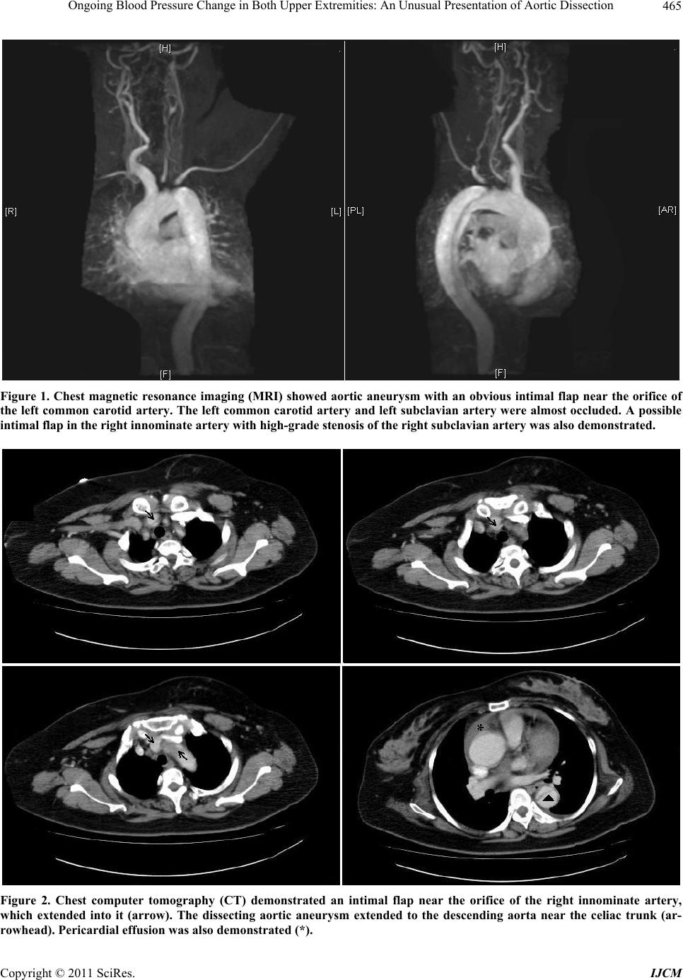 Ongoing Blood Pressure Change in Both Upper Extremities: An Unusual Presentation of Aortic Dissection Copyright © 2011 SciRes. IJCM 465 Figure 1. Chest magnetic resonance imaging (MRI) showed aortic aneurysm with an obvious intimal flap near the orifice of the left common carotid artery. The left common carotid artery and left subclavian artery were almost occluded. A possible intimal flap in the right innominate artery with high-grade stenosis of the right subclavian artery was also demonstrated. Figure 2. Chest computer tomography (CT) demonstrated an intimal flap near the orifice of the right innominate artery, which extended into it (arrow). The dissecting aortic aneurysm extended to the descending aorta near the celiac trunk (ar- owhead). Pericardial effusion was also demonstrated (*). r  Ongoing Blood Pressure Change in Both Upper Extremities: An Unusual Presentation of Aortic Dissection 466 the aortic dissection extends to the innominate artery or common carotid arteries. Other clinical manifestations, such as transient cerebral hypoperfusion caused by al- tered cerebrovascular flow, may present as syncope [3]. As for spinal cord ischemia and peripheral nerve in- volvement, paraplegia is the most common neurologic symptom caused by obstruction of spinal arteries; this symptom has been described in several case reports [4-6]. Spinal cord ischemia and ischemic peripheral neuropa- thies are more commonly associated with distal aortic dissection. The aortic dissections in most of these cases were located in the abdominal aorta and resulted in injury to the lower extremities. Possible manifestation s of these injuries include transverse myelitis, progressive myelo- pathy, anterior spinal cord syndrome, paraplegia, and qu ad r i p l e g i a [ 6 - 9 ] . Painless di ssecting aorta with isch emic peripheral neuropathy in upper limbs is very rare and is often ignored or misdiagnosed. Clinical presentations are varied and may result in neurologic symptoms, including paresthesia in the limbs, hoarseness of voice, and Horner syndrome [10,11]. The possible mechanisms underlying these symptoms include neuronal ischemia caused by obstruction of the branch artery, compression caused by the exp ansion of the false lume n, and leakag e of the aor- tic dissection aneurysm. Aortic dissection may also result in symptomatic ischemia, which most commonly occurs in a lower ex- tremity [12,13]. Clinicians sho uld co ns ider the possibility of aortic dissection in patients presenting with abrupt onset of chest pain and sudden loss of pulse in a lower extremity. However, the symptomatic ischemia caused by aortic dissection is not li mited to th e lower ex tremities. One previous report described a case of acute aortic dis- section in which the patient experienced numbness and paleness in the right arm, which was caused by acute occlusion of the right subclavian artery [14]. The patho- logic mechanism in that case was similar to that in our case, but the 2 cases had different clinical manifestations. In our case, right arm numbness without blood pressure differentials and without the c linical features of ischemia obscured the initial diagnosis. The value of different blood pressure assessed between bilateral upper extremi- ties believed to be clinically s ignificant is greater than 10 mm Hg in systolic blood pressure [15]. Asymmetric pulses and varying blood pressures between different upper limbs are strong indicators of possible aortic dis- section. Other possible condition included coarctation aorta involving aortic arch, inflammatory aortitis as syphilitic aortitis, arotic arch syndrome, supravalvular aortic stenosis, and subclavian steal syndrome [16-20]. Comparing the blood pressure values in both upper ex- tremities is important determination in the diagnosis of pathology involving the aortic arch or upper-extremity arteries. In fact, asymmetric blood pressures are the most specific physical signs of aortic dissection and have been reported in 38% of patients with aortic dissections [21]. Aortic dissection involving both subclavian arteries is rare, and it is extremely rare when the clinical manifesta- tion includes absent pulses in both upper extremities and lacks neurol ogic defects. Most patients with aortic dissection present with hy- pertension; however, hemodynamic instability with the clinical features of circulatory shock may also be a sign of aortic dissection. Hypotension and shock in acute aor- tic dissection are secondary to acute severe aortic regur- gitation, low cardiac output because of cardiac tampo- nade, aortic rupture, or left ventricular systolic dysfunc- tion with coronary artery involvement [22,23]. Echocar- diography did not reveal any special condition in our case, and this obscured the diagnosis. Fortunately, the true diagnosis was confirmed by MRI, and a prompt sur- gical intervention was arranged without any problems. Emergent surgical repair is indicated to avoid life- threatening events when aortic dissection involving as- cending aorta. The standard operation for type A dissec- tion is to perform an ascending aortic replacement with open distal hemi-arch anastomosis. Residual aortic an- eurysm with gradually enlargement in patients who had previously undergone ascending aortic repair had been noted [24,25]. Using an open total arch/elephant-trunk approach or hybrid endovascular approaches provide a method to compress the false lumen in distal descending thoracic aorta [26]. Preliminary showed hybrid arch pro- cedures have benefit in elderly and high risk patients, but, this benefit is not as pronounced in younger [27-29]. Further outcome data will be necessary in the future. In our case, after the surgical procedure, she was discharged under stable condition and kept follow-up at out-patient department. 4. Conclusions The case demonstrated the uncommon clinical evolution of aortic dissection. The most distal intimal tear in a dis- secting aortic aneurysm where the blood is presumed to return to the circulation (reentry tears) or multiple intimal tears without interarms blood pressure differences may occur in the cases of aortic dissection. Knowledge of the clinical manifestations and the possible involvement of aortic arch vessels are the critical factors in diagnosis. Early blood pressure measurement of other extremities and image study are indicated to assist the diagnosis of this condition. REFERENCES [1] I. A. Khan and C. K. Nair, “Clinical, Diagnostic, and Copyright © 2011 SciRes. IJCM  Ongoing Blood Pressure Change in Both Upper Extremities: An Unusual Presentation of Aortic Dissection467 Management Perspectives of Aortic Dissection,” Chest, Vol. 122, No. 1, 2002, pp. 311-328. doi:10.1378/chest.122.1.311 [2] J. L. Prendes, “Neurovascular Syndromes in Aortic Dis- section,” American Family Physician, Vol. 23, No. 6, 1981, pp. 175-179. [3] T. P. Kuhlmun and R. P. Powers, “Painless Aortic Dis- section: An Unusual Cause of Syncope,” Annals of Emergency Medicine, Vol. 13, No. 7, 1984, pp. 549-551. doi:10.1016/S0196-0644(84)80528-4 [4] J. Inamasu, S. Hori, M. Yokoyama, T. Funabiki, K. Aoki and N. Aikawa, “Paraplegia Caused by Painless Acute Aortic Dissection,” Spinal Cord, Vol. 38, No. 11, 2000, pp. 702-704. [5] E. M. Donovan, G. K. Seidel and A. Cohen, “Painless Aortic Dissectoin Presenting as High Paraplegia: A Case Report,” Archives of Physical Medicine and Rehabilita- tion, Vol. 81, No. 10, 2000, pp. 1436-1438. doi:10.1053/apmr.2000.7158 [6] S. A. Rosen, “Painless Aortic Dissection Presenting as Spinal Cord Ischemia,” Annals of Emergency Medicine, Vol. 17, No. 8, 1988, pp. 840-842. doi:10.1016/S0196-0644(88)80567-5 [7] S. F. Holloway, P. B. Fayad, R. G. Kalb, J. B. Guarnaccia and S. G. Waxman, “Painless Aortic Dissection Present- ing as a Myelopathy,” Journal of the Neurological Sci- ences, Vol. 120, 1993, pp. 141-144. [8] C. Beach and D. Manthey, “Painless Acute Aortic Dis- section Presenting as Left Lower Extremity Numbness,” The American Journal of Emergency Medicine, Vol. 16, No. 1, 1998, pp. 49-51. doi:10.1016/S0735-6757(98)90064-X [9] J.B. Joo and A. J. Cummings, “Acute Thoracoabdominal Aortic Dissection Presenting as Painless, Transient Pa- ralysis of the Lower Extremities: A Case Report,” The Journal of Emergency Medicine, Vol. 19, No. 4, 2000, pp. 333-337. doi:10.1016/S0736-4679(00)00264-X [10] W. R. Greenwood and M. D. Robinson, “Painless Dissec- tion of the Thoracic Aorta,” The American Journal of Emergency Medicine, Vol. 4, No. 4, 1986, pp. 330-333. doi:10.1016/0735-6757(86)90303-7 [11] I. A. Khan, N. Wattanasauwan and A. W. Ansari, “Painless Aortic Dissection Presenting as Hoarseness of Voice: Cardiovocal Syndrome; Ortner’s Syndrome,” The American Journal of Emergency Medicine, Vol. 17, No. 4, 1999, pp. 361-363. doi:10.1016/S0735-6757(99)90087-6 [12] I. M. Schoon, J. Holm and G. Sudow, “Lower-Extremity Ischemia in Aortic Dissection: Report of Three Cases,” Scandinavian Journal of Thoracic and Cardiovascular Surgery, Vol. 19, No. 1, 1985, pp. 93-95. [13] T. J. White 3rd, M. L. Pinstein, R. L. Scott and R. E. Gold, “Aortic Dissection Manifested as Leg Ischemi,” Ajr American Journal of Roentgenology, Vol. 135, No. 2, 1980, pp. 353-356. [14] R. Koushima, Y. Kikuchi, T. Sakurada and K. Kusajima, “A Case of Painless Standford Type A Acute Aortic Dis- section Complicating Acute Occlusion of the Right Sub- clavian Artery,” Kyobu Geka, Vol. 51, No. 3, 1998, pp. 226-230. [15] A. J. Singer and J. E. Hollander, “Blood Pressure: As- sessment of Interarm Differences,” Archives of Internal Medicine, Vol. 156, No. 17, 1996, pp. 2005-2008. [16] F. H. Ellis and O.T. Clagett, “Coarctation of the Aorta Proximal to the Left Subclavian Artery: Experience with Six Surgical Cases,” Annals of Surgery, Vol. 146, No. 2, 1957, pp. 145-151. [17] D. C. Levinson, D. T. Edmeades and G. C. Griffith, “Ab- dominal Pain in Dissecting Aneurysm of the Aorta,” The American Journal of Medicine, Vol. 8, No. 4, 1950, pp. 474-479. doi:10.1016/0002-9343(50)90229-4 [18] H. M. Korns and P. H. Guinand, “Inequality of Blood Pressure in the Brachial Arteries, with Especial Reference to Disease of the Arch of the Aorta,” Journal of Clinical Investigation, Vol. 12, No. 1, 1933, pp. 143-154. doi:10.1172/JCI100485 [19] R. D. Judge, R. D. Currier, W. A. Gracie and M. M. Fig- ley, “Takayasu’s Arteritis and the Aortic Arch Syn- drome,” The American Journal of Medicine, Vol. 32, 1962, pp. 379-392. doi:10.1016/0002-9343(62)90128-6 [20] A. G. Morrow, J. A. Waldhausen, R. L. Peters, R. D. Bloodwell and E. Braunwald, “Supravalvular Aortic Stenosis: Clinical, Hemodynamic and Pathologic Obser- vations,” Circulation, Vol. 20, 1959, pp. 1003-1010. [21] Y. von Kodolitsch, A. G. Schwartz and C. A. Nienaber, “Clinical Prediction of Acute Aortic Dissection,” Ar- chives of Internal Medicine, Vol. 160, No. 19, 2000, pp. 2977-2982. [22] M. J. Eisenberg, S. A. Rice, A. Paraschos, G. R. Caputo and N. B. Schiller, “The Clinical Spectrum of Patients with Aneurysms of the Ascending Aorta,” American Heart Journal, 125, No. 5, 1993, pp. 1380-1385. doi:10.1016/0002-8703(93)91011-3 [23] A. Carcia-Jimenez, A. Peraza-Torres and G. Martinez- Lopez, “Alvarez Dieguez I, Botana Alba CM. Cardiac Tamponade by Aortic Dissection in a Hospital without Cardiovascular Surgery,” Chest, Vol. 104, 1993, pp. 290- 291. [24] A. Geirsson, J. E. Babaria, D. Swarr, M. G. Keane, Y. J. Woo, W. Y. Szeto and A. Pochettino, “Fate of the Resi- dual Distal and Proximal Aorta after Acute Type A Dis- section Repair Using a Contemporary Surgical Re- construction Algorithm,” The Annals of Thoracic Surgery, Vol. 84, No. 6, 2007, pp. 1955-1964. doi:10.1016/j.athoracsur.2007.07.017 [25] J. C. Halstead, M. Meier, C. Etz, D. Spielvoqel, C. Bodian, M. Wurm, R. Shahani and R. B. Griepp, “The Fate of the Distal Aorta After Repair of Acute Type A Aortic Dissection,” The American Association for Tho- racic Surgery, Vol. 133, No. 1, 2007, pp. 127-135. doi:10.1016/j.jtcvs.2006.07.043. [26] F. S. Schoenhoff, J. Schmidli, F. S. Eckstein, P. A. Be rdat, F. F. Immer and T. P. Carrel, “The Frozen Elephant Trunk: An Interesting Hybrid Endovascular-Surgical Copyright © 2011 SciRes. IJCM  Ongoing Blood Pressure Change in Both Upper Extremities: An Unusual Presentation of Aortic Dissection Copyright © 2011 SciRes. IJCM 468 Technique to Treat Complex Pathologies of the Thoracic Aorta,” Journal of Vascular Surgery, Vol. 45, No. 3, 2007, pp. 597-599. doi:10.1016/j.jvs.2006.10.038 [27] T. Kim, T. D. Martin, W. A. Lee, P. Hess Jr., C. T. Klodell, C. G. Tribble, R. J. Feezor and T. M. Beaver, “Evoluation in the Management of the Total Thoracic Aorta,” The Journal of Thoracic and Cardiovascular Surgery, Vol. 137, No. 3, 2009,pp. 627-634. doi:10.1016/j.jtcvs.2008.11.012 [28] M. Czerny, R. Gottardi, D. Zimpfer, M. Schoder, M. Grabenwoger, J. Lammer, E. Wolner and M. Grimm, “Mid-Term Results of Supraaortic Transpositions for Ex- tended Endovascular Repair of Aortic Arch Pathologies,” European Journal of Cardiothoracic Surgery, Vol. 31, No. 4, 2007, pp. 623-627. doi:10.1016/j.ejcts.2006.12.030 [29] P. Bergeron, N. Mangialardi, P. Costa, P. Coulon, V. Douillez, E. Serreo, I. Tuccimei, C. Cavazzini, F. Mariotti, Y. Sun and J. Gay, “Great Vessel Management for En- dovascular Exclusion of Aortic Arch Aneurysms and Dissections,” European Journal of Cardiothoracic Sur- gery, Vol. 32, No. 1, 2006, pp. 38-45. doi:10.1016/j.ejvs.2005.12.023 |