

Paper Menu >>
Journal Menu >>
 Surgical Science, 2011, 2, 393-396 doi:10.4236/ss.2011.27086 Published Online September 2011 (http://www.SciRP.org/journal/ss) Copyright © 2011 SciRes. SS Robotic–Assisted Thymectomy in Patients with Nonthymomatous Myasthenia Gravis Iskander Al-Githmi Department of Surgery, Division of Cardiothoracic Surgery, King Abdulaziz University Ho spital, Jeddah, Saudi Arabia E-mail: [email protected] Received August 2, 2011; revised August 23, 2011; accepted September 1, 2011 Abstract Complete removal of the thymus and fatty tissue adherent to pericardium is crucial for treating myasthenia gravis. Our aim was to analyze our surgical and clinical results and early experience of robot-assisted thy- mectomy in patients with nonthymomatous myasthenia gravis. Between January 2008 and October 2010, 8 patients, all women, with nonthymomatous myasthenia gravis underwent robot-assisted thymectomy using a 3-port, left-sided approach. There were no operative mortality; 1 patient had left phrenic nerve injury. Com- plete remission was achieved in 25% of the patients at 18 months’ follow-up. There was no significant agreement between preoperative computed tomography and histopathology finding (kappa = 0.059; P = 0.85) and no significant correlation between age and duration of symptoms (P = 0.51). Robotic-assisted thymec- tomy is promising procedure. It can be performed safely and effectively. With follow-up greater than 12 months, 87.5% of the patients who underwent robot-assisted thymectomy demonstrated clinical improve- ment. Keywords: Myasthenia Gravis, Nonthymoma, Robot, Thymectomy 1. Introduction Myasthenia gravis is an autoimmune disease affecting postsynaptic nicotinic acetylcholine receptors at the neu- romuscular junction block producing neuromuscular transmission-producing muscular weakness of striated muscles. Thymectomy is an established therapy in treating my- asthenia gravis. Ferdinand Sauerbruch performed the first successful thymectomy in a patient with myasthenia gravis in 1911 in Zurich [1]. The efficacy of thymectomy depends on complete removal of all thymic tissue. In 1941, Blalock said, “Complete removal of all thymus tissue offers the best chance of altering the course of the disease” [2]. Multiple described surgical approaches to thymectomy include transcervical thymectomy [3], transsternal thy- mectomy [4], infrasternal subxiphoid mediastinoscopic thymectomy [5], and video-assisted thoracoscopic thy- mectomy [6-8]. Introduction of robotic surgical-assisted system contributed to the field of minimally invasive surgery [9]. Yoshino described the first robotic thymec- tomy in treating small thymoma [10]. However; ro- bot-assisted thymectomy has been reported with good short-term results. Only scant data are available on the long-term results of robot-assisted thymectomy for my- asthenia gravis. It is uncertain which patients may benefit from robot-assisted thymectomy. This study sought to analyze the surgical and clinical results of robot-assisted thymectomy in patients with nonthymomatous myasthe- nia gravis. 2. Materials and Methods Between January 2008 and October 2010, 8 patients all women (mean age, 28 y; range, 16 - 46 y) with nonthy- momatous myasthenia gravis referred by neurologists underwent robot-assisted thymectomy by 1 cardiotho- racic surgeon at King Abdulaziz University Hospital, Jeddah, Saudi Arabia. Preoperative patient clinical char- acteristics, duration of symptoms, preoperative acetyl- choline receptors antibody, preoperative Osserman clas- sification, and computed tomogram of chest finding are shown in (Table 1). The diagnosis of myasthenia gravis was based on clinical features, result of electromyography (decrement- 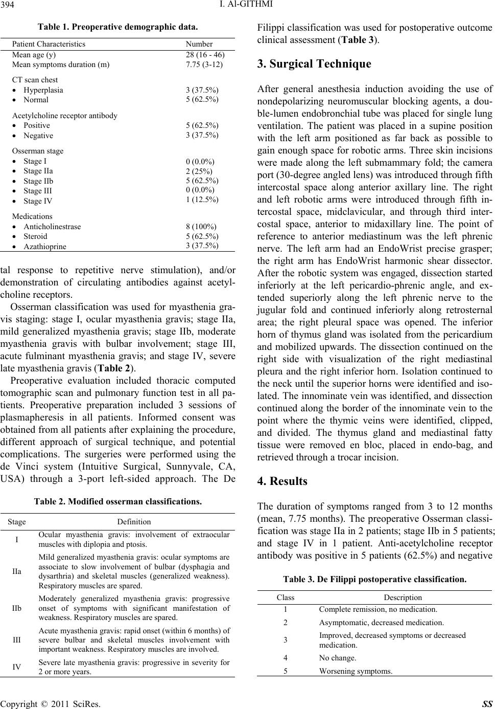 394 I. Al-GITHMI Table 1. Preoperative demographic data. Patient Characteristics Number Mean age (y) 28 (16 - 46) Mean symptoms duration (m) 7.75 (3-12) CT scan chest Hyperplasia Normal 3 (37.5%) 5 (62.5%) Acetylcholine receptor antibody Positive Negative 5 (62.5%) 3 (37.5%) Osserman stage Stage I Stage IIa Stage IIb Stage III Stage IV 0 (0.0%) 2 (25%) 5 (62.5%) 0 (0.0%) 1 (12.5%) Medications Anticholinestrase Steroid Azathioprine 8 (100%) 5 (62.5%) 3 (37.5%) tal response to repetitive nerve stimulation), and/or demonstration of circulating antibodies against acetyl- choline receptors. Osserman classification was used for myasthenia gra- vis staging: stage I, ocular myasthenia gravis; stage IIa, mild generalized myasthenia gravis; stage IIb, moderate myasthenia gravis with bulbar involvement; stage III, acute fulminant myasthenia gravis; and stage IV, severe late myasthenia gravis (Table 2). Preoperative evaluation included thoracic computed tomographic scan and pulmonary function test in all pa- tients. Preoperative preparation included 3 sessions of plasmapheresis in all patients. Informed consent was obtained from all patients after explaining the procedure, different approach of surgical technique, and potential complications. The surgeries were performed using the de Vinci system (Intuitive Surgical, Sunnyvale, CA, USA) through a 3-port left-sided approach. The De Table 2. Modified osserman classifications. Stage Definition I Ocular myasthenia gravis: involvement of extraocular muscles with diplopia and ptosis. IIa Mild generalized myasthenia gravis: ocular symptoms are associate to slow involvement of bulbar (dysphagia and dysarthria) and skeletal muscles (generalized weakness). Respiratory muscles are spared. IIb Moderately generalized myasthenia gravis: progressive onset of symptoms with significant manifestation of weakness. Respiratory muscles are spared. III Acute myasthenia gravis: rapid onset (within 6 months) of severe bulbar and skeletal muscles involvement with important weakness. Respiratory muscles are involved. IV Severe late myasthenia gravis: progressive in severity for 2 or more years. Filippi classification was used for postoperative outcome clinical assessment (Table 3). 3. Surgical Technique After general anesthesia induction avoiding the use of nondepolarizing neuromuscular blocking agents, a dou- ble-lumen endobronchial tube was placed for single lung ventilation. The patient was placed in a supine position with the left arm positioned as far back as possible to gain enough space for robotic arms. Three skin incisions were made along the left submammary fold; the camera port (30-degree angled lens) was introduced through fifth intercostal space along anterior axillary line. The right and left robotic arms were introduced through fifth in- tercostal space, midclavicular, and through third inter- costal space, anterior to midaxillary line. The point of reference to anterior mediastinum was the left phrenic nerve. The left arm had an EndoWrist precise grasper; the right arm has EndoWrist harmonic shear dissector. After the robotic system was engaged, dissection started inferiorly at the left pericardio-phrenic angle, and ex- tended superiorly along the left phrenic nerve to the jugular fold and continued inferiorly along retrosternal area; the right pleural space was opened. The inferior horn of thymus gland was isolated from the pericardium and mobilized upwards. The dissection continued on the right side with visualization of the right mediastinal pleura and the right inferior horn. Isolation continued to the neck until the superior horns were identified and iso- lated. The innominate vein was identified, and dissection continued along the border of the innominate vein to the point where the thymic veins were identified, clipped, and divided. The thymus gland and mediastinal fatty tissue were removed en bloc, placed in endo-bag, and retrieved through a trocar incision. 4. Results The duration of symptoms ranged from 3 to 12 months (mean, 7.75 months). The preoperative Osserman classi- fication was stage IIa in 2 patients; stage IIb in 5 patients; and stage IV in 1 patient. Anti-acetylcholine receptor antibody was positive in 5 patients (62.5%) and negative Table 3. De Filippi postoperative classification. Class Description 1 Complete remission, no medication. 2 Asymptomatic, decreased medication. 3 Improved, decreased symptoms or decreased medication. 4 No change. 5 Worsening symptoms. Copyright © 2011 SciRes. SS 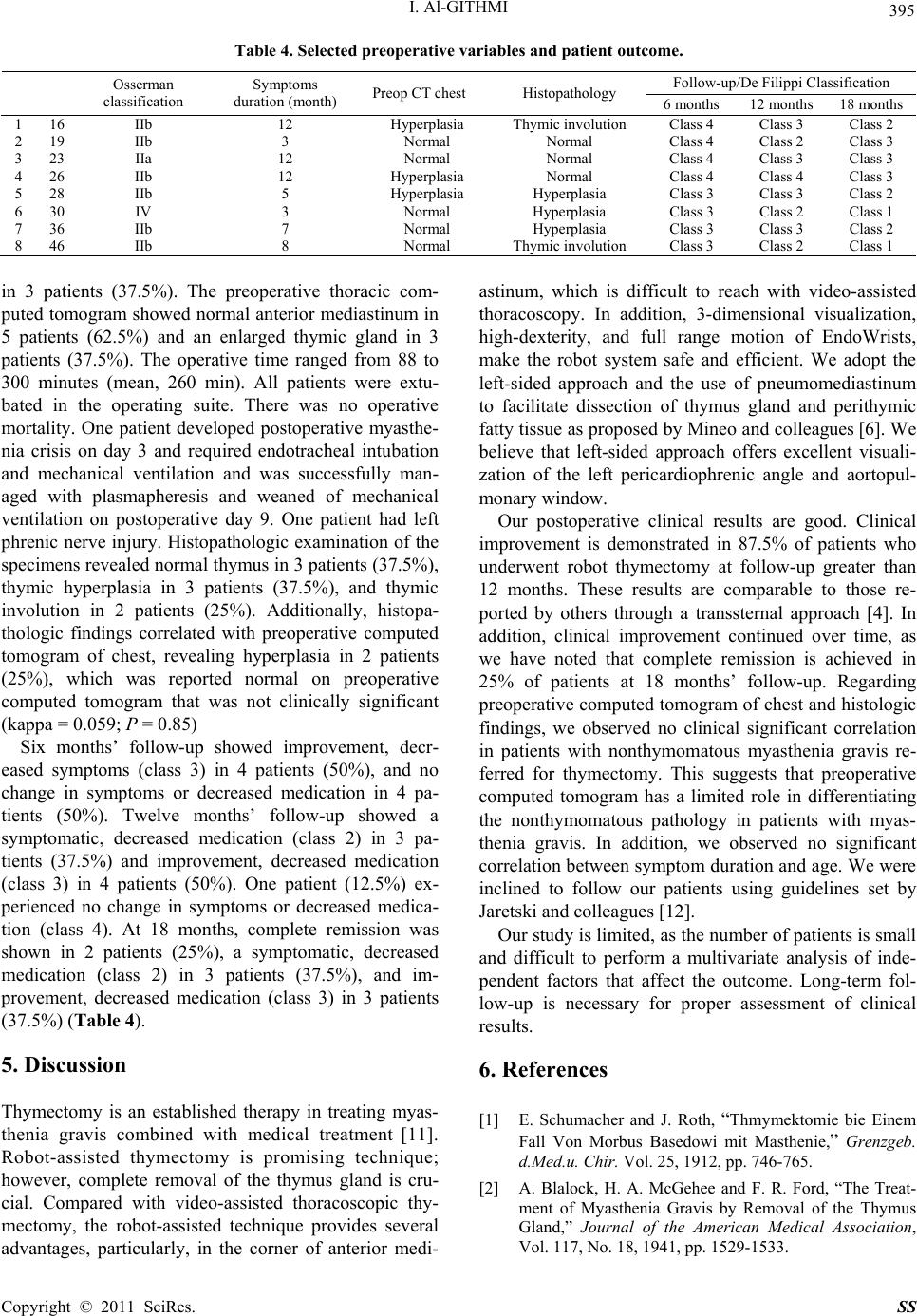 I. Al-GITHMI Copyright © 2011 SciRes. SS 395 Table 4. Selected preoperative variables and patient outcome. Follow-up/De Filippi Classification Osserman classification Symptoms duration (month) Preop CT chest Histopathology 6 months 12 months 18 months 1 16 IIb 12 Hyperplasia Thymic involution Class 4 Class 3 Class 2 2 19 IIb 3 Normal Normal Class 4 Class 2 Class 3 3 23 IIa 12 Normal Normal Class 4 Class 3 Class 3 4 26 IIb 12 Hyperplasia Normal Class 4 Class 4 Class 3 5 28 IIb 5 Hyperplasia Hyperplasia Class 3 Class 3 Class 2 6 30 IV 3 Normal Hyperplasia Class 3 Class 2 Class 1 7 36 IIb 7 Normal Hyperplasia Class 3 Class 3 Class 2 8 46 IIb 8 Normal Thymic involution Class 3 Class 2 Class 1 in 3 patients (37.5%). The preoperative thoracic com- puted tomogram showed normal anterior mediastinum in 5 patients (62.5%) and an enlarged thymic gland in 3 patients (37.5%). The operative time ranged from 88 to 300 minutes (mean, 260 min). All patients were extu- bated in the operating suite. There was no operative mortality. One patient developed postoperative myasthe- nia crisis on day 3 and required endotracheal intubation and mechanical ventilation and was successfully man- aged with plasmapheresis and weaned of mechanical ventilation on postoperative day 9. One patient had left phrenic nerve injury. Histopathologic examination of the specimens revealed normal thymus in 3 patients (37.5%), thymic hyperplasia in 3 patients (37.5%), and thymic involution in 2 patients (25%). Additionally, histopa- thologic findings correlated with preoperative computed tomogram of chest, revealing hyperplasia in 2 patients (25%), which was reported normal on preoperative computed tomogram that was not clinically significant (kappa = 0.059; P = 0.85) Six months’ follow-up showed improvement, decr- eased symptoms (class 3) in 4 patients (50%), and no change in symptoms or decreased medication in 4 pa- tients (50%). Twelve months’ follow-up showed a symptomatic, decreased medication (class 2) in 3 pa- tients (37.5%) and improvement, decreased medication (class 3) in 4 patients (50%). One patient (12.5%) ex- perienced no change in symptoms or decreased medica- tion (class 4). At 18 months, complete remission was shown in 2 patients (25%), a symptomatic, decreased medication (class 2) in 3 patients (37.5%), and im- provement, decreased medication (class 3) in 3 patients (37.5%) (Table 4). 5. Discussion Thymectomy is an established therapy in treating myas- thenia gravis combined with medical treatment [11]. Robot-assisted thymectomy is promising technique; however, complete removal of the thymus gland is cru- cial. Compared with video-assisted thoracoscopic thy- mectomy, the robot-assisted technique provides several advantages, particularly, in the corner of anterior medi- astinum, which is difficult to reach with video-assisted thoracoscopy. In addition, 3-dimensional visualization, high-dexterity, and full range motion of EndoWrists, make the robot system safe and efficient. We adopt the left-sided approach and the use of pneumomediastinum to facilitate dissection of thymus gland and perithymic fatty tissue as proposed by Mineo and colleagues [6]. We believe that left-sided approach offers excellent visuali- zation of the left pericardiophrenic angle and aortopul- monary window. Our postoperative clinical results are good. Clinical improvement is demonstrated in 87.5% of patients who underwent robot thymectomy at follow-up greater than 12 months. These results are comparable to those re- ported by others through a transsternal approach [4]. In addition, clinical improvement continued over time, as we have noted that complete remission is achieved in 25% of patients at 18 months’ follow-up. Regarding preoperative computed tomogram of chest and histologic findings, we observed no clinical significant correlation in patients with nonthymomatous myasthenia gravis re- ferred for thymectomy. This suggests that preoperative computed tomogram has a limited role in differentiating the nonthymomatous pathology in patients with myas- thenia gravis. In addition, we observed no significant correlation between symptom duration and age. We were inclined to follow our patients using guidelines set by Jaretski and colleagues [12]. Our study is limited, as the number of patients is small and difficult to perform a multivariate analysis of inde- pendent factors that affect the outcome. Long-term fol- low-up is necessary for proper assessment of clinical results. 6. References [1] E. Schumacher and J. Roth, “Thmymektomie bie Einem Fall Von Morbus Basedowi mit Masthenie,” Grenzgeb. d.Med.u. Chir. Vol. 25, 1912, pp. 746-765. [2] A. Blalock, H. A. McGehee and F. R. Ford, “The Treat- ment of Myasthenia Gravis by Removal of the Thymus Gland,” Journal of the American Medical Association, Vol. 117, No. 18, 1941, pp. 1529-1533.  396 I. Al-GITHMI [3] R. F. Calhoun, J. H. Ritter, T. J. Guthrie, A. Pestronk, B. F. Meyers, G. A. Patterson, M. S. Pohl and J. D. Cooper, “Results of Transcervical Thymectomy for Myasthenia Gravis in 100 Consecutive Patients,” Annals of Surgery, Vol. 230, No. 4, 1999, pp. 555-559. doi:10.1097/00000658-199910000-00011 [4] A. Masaoka, Y. Yamakawa, H. Niwa, I. Fukai, S. Kondo, M. Kobayashi, Y. Fujii and Y. Monden, “Extended Thy- mectomy for Myasthenia Gravis Patients: A 20-Year Re- view,” Annals of Thoracic Surgery , Vol. 62, No. 3, 1996, pp. 853-859. doi:10.1016/S0003-4975(96)00376-1 [5] A. Uchiyama, S. Shimizu, H. Murai, S. Kuroki, M. Okido and M. Tanaka, “Infrasternal Mediastinoscopic Thymec- tomy in Myasthenia Gravis: Surgical Results in 23 Pa- tients,” Annals of Thoracic Surgery, Vol. 72, No. 6, 2001, pp. 1902-1905. doi:10.1016/S0003-4975(01)03210-6 [6] T. C. Mineo, E. Pompeo, T. E. Lerut, G. Bernardi, W. Coosemans and I. Nofroni, “Thoracoscopic Thymectomy in Autoimmune Myasthesia: Results of Left-Sided Ap- proach,” Annals of Thoracic Surgery, Vol. 69, No. 5, 2000, pp. 1537-1541. doi:10.1016/S0003-4975(00)01237-6 [7] M. J. Mack, R. J. Landreneau, A. P. Yim, S. R. Hazelrigg and G. R. Scruggs, “Results of Video-Assisted Thymec- tomy in Patients with Myasthenia Gravis,” Journal of Thoracic and Cardiovascular Surgery, Vol. 112, No. 5, 1996, pp. 1352-1359. doi:10.1016/S0022-5223(96)70151-4 [8] R. Vizeteu, M. Damian, B. Smeu, A. Stoica, V. Culman, O. Băjenaru and A. Ene, “Video-Assisted Thoracoscopic Extended Thymectomy (VATET) with Cervical Ap- proach for Myasthenia Gravis—Initial Experience [in Romanian],” Chirurgia (Bucur). Vol. 105, No. 6, 2010, pp. 797-803. [9] M. Hashizume, K. Konishi, N. Tsutsumi, S. Yamaguchi and R. Shimabukuro, “A New Era of Robotic Surgery Assisted by a Computer-Enhanced Surgical System,” Surgery, Vol. 131, Supplement 1, 2002, pp. S330-S333. [10] I. Yoshino, M. Hashizume, M. Shimada, M. Tomikawa, M. Tomiyasu, R. Suemitsu and K. Sugimachi, “Thoraco- scopic Thymomectomy with the Da Vinci Com- puter-Enhanced Surgical System,” Journal of Thoracic and Cardiovascular Surgery, Vol. 122, No. 4, 2001, pp. 783-785. doi:10.1067/mtc.2001.115231 [11] A. E. Papatestas, L. I. Alpert and K. E. Osserman, “Stud- ies in Myasthenia Gravis: Effects of Thymectomy Results on 185 Patients with Nonthymomatous and Thymimatous Myasthenia Gravis,” American Journal of Medicine, Vol. 38, 1971, pp. 580-585. [12] A. Jaretzki, R. J. Barohn, R. M. Ernstoff, H. J. Kaminski, J. C. Keesey, A. S. Penn and D. B. Sanders, “Myasthenia Gravis: Recommendations for Clinical Research Stan- dards. Task Force of the Medical Scientific Advisory Board of the Myasthenia Gravis Foundation of America,” Annals of Thoracic Surgery, Vol. 70, No. 1, 2000, pp. 327-334. doi:10.1016/S0003-4975(00)01595-2 Copyright © 2011 SciRes. SS |