

Paper Menu >>
Journal Menu >>
 Surgical Science, 2011, 2, 388-392 doi:10.4236/ss.2011.27085 Published Online September 2011 (http://www.SciRP.org/journal/ss) Copyright © 2011 SciRes. SS A Technique of Bilateral Inguinal Hernia Repair Using 10 mm Single Port Access and Bioresorbable Composite Mesh Fixed with Endoclose Sutures: Three Cases Reported Wuttichai Thanapongsathorn HRH Princess Maha Chakri S i ri nd horn Medical Center , Department of Surgery, Faculty of Medicine, Srinakharinwirot University, Nakron-Nayok, Thailand E-mail: [email protected] Received April 22, 2011; revised May 3, 2011; accept ed May 24, 2011 Abstract Purpose: To report a novel technique of laparoscopic 10 mm Single Port Access IntraPeritoneal Onlay Mesh (SPA-IPOM) using bioresorbable composite mesh fixed with Endoclose suture (percutaneous subcutaneous suture) in 3 cases of bilateral inguinal hernia. Methods: Laparoscopic SPA-IPOM is done through a 10 mm trocar with one 10 mm instrument that has 5 mm optical lens and 5 mm channel for grasper. After inserting 10 mm trocar at umbilicus using semi-open technique, intraperitoneal anatomical landmark of inguinal her- nia is identified. A 10 × 15 cm pre-tied bioresorbable composite mesh is then placed to cover hernia defect and all three potential area of indirect, direct and femoral hernia. Using Endoclose™ needle, each pair of pre-tied sutures is retrieved percutaneously through a needle wound and extracorporeal tied with knot in subcutaneous space. After the upper half of mesh is sutured to the posterior surface of abdominal wall, the lower half of mesh is fixed by hernia tacker to Symphysis Pubis, Cooper Ligament and Iliopubic tract. Re- sults: Three men, average 48 year olds were operated by laparoscopic 10 mm SPA-IPOM for bilateral in- guinal hernia repair using bioresorbable composite mesh. Average operative time was 36 minutes. No imme- diate complication. All patients were discharge on the 2nd post-operative day and average 6 months follow up has no recurrence. Conclusions: Laparoscopic SPA-IPOM is an optional operation and is much easier to be performed. Benefits include operative time saving, cosmesis, early discharge and early return to work. Bio- resorbable composite mesh prevents bowel adhesion, however, is much more expensive. Long term follow up study for complications and recurrence is needed. Keywords: 10 mm Single Port Access, IntraPeritoneal Onlay Mesh, Percutaneous Subcutaneous Suture, Bioresorbable Composite Mesh, Bilateral Inguinal Hernia Repair 1. Introduction Hernias occur fairly frequently and are more common in adult males. Occasionally these can be life threatening or more frequently simply a painful nuisance. Nonetheless, the only true remedy is surgical repair. Over centuries surgical techniques have evolved until the present whereby laparoscopic approaches predominate. Several various methodologies have been introduced [1]. We now wish to add to this growing laparoscopic experience and offer three cases of bilateral inguinal hernia repair. To our knowledge, we are the first to report on utilizing a 10 mm single port access intraperitoneal onlay mesh [SPA-IPOM] incorporating bioresorbable composite mesh fixed with Endoclose suture (percutaneous subcutaneous suture) in bilateral inguinal hernia repair. 2. Cases Report A total of three Thai men average age 48 years old [range 32 - 58] presented with symptomatic bilateral in- guinal hernias. One was a bilateral indirect and the re- maining 2 bilateral direct inguin al hernias. All u nd erwent repair using the laparoscopic technique described in the next section after informed consent was obtained. The average operative time was 36 minutes [range 28 - 50] without any immediate complications. The patients have to be admitted due to health in suran ce system of the 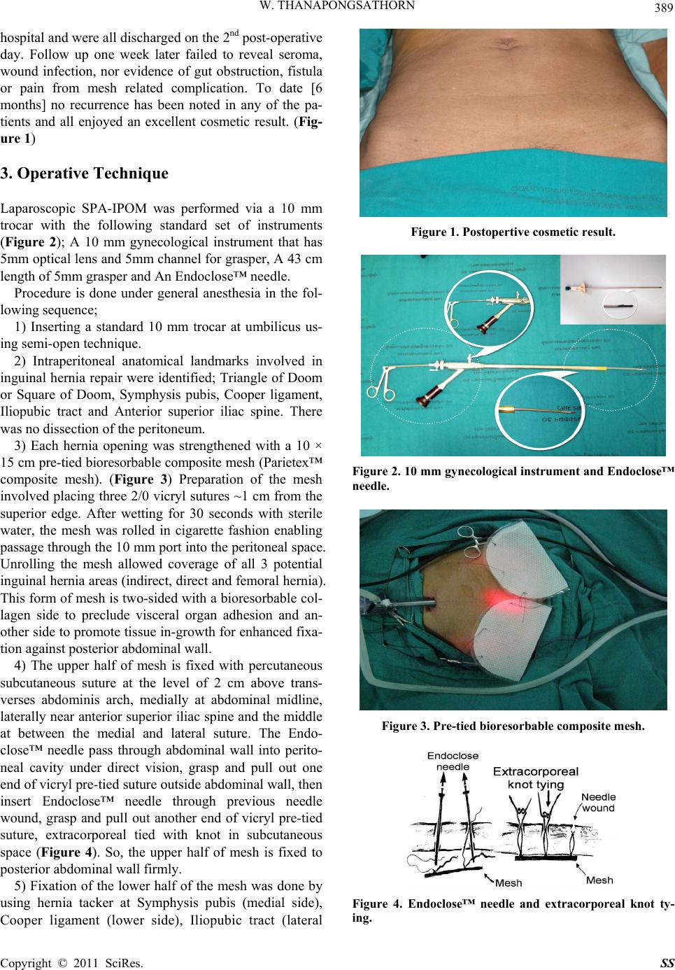 W. THANAPONGSATHORN 389 hospital and were all discharged on the 2nd post-operative day. Follow up one week later failed to reveal seroma, wound infection, nor evidence of gut obstruction, fistula or pain from mesh related complication. To date [6 months] no recurrence has been noted in any of the pa- tients and all enjoyed an excellent cosmetic result. (Fig- ure 1) 3. Operative Technique Laparoscopic SPA-IPOM was performed via a 10 mm trocar with the following standard set of instruments (Figure 2); A 10 mm gynecological instrument that has 5mm optical lens and 5mm channel for grasper, A 43 cm length of 5mm grasper and An Endoclose™ needle. Procedure is done under general anesthesia in the fol- lowing sequence; 1) Inserting a standard 10 mm trocar at umbilicus us- ing semi-open t echnique. 2) Intraperitoneal anatomical landmarks involved in inguinal hernia repair were identified; Triangle of Doom or Square of Doom, Symphysis pubis, Cooper ligament, Iliopubic tract and Anterior superior iliac spine. There was no dissection of the peritoneum. 3) Each hernia opening was strengthened with a 10 × 15 cm pre-tied bioresorbable composite mesh (Parietex™ composite mesh). (Figure 3) Preparation of the mesh involved placing three 2/0 vicryl sutures ~1 cm from the superior edge. After wetting for 30 seconds with sterile water, the mesh was rolled in cigarette fashion enabling passage through th e 10 mm port into the perito neal space. Unrolling the mesh allowed coverage of all 3 potential inguinal hernia areas (indirect, direct and femoral hernia). This form of mesh is two-sided with a bioresorbable col- lagen side to preclude visceral organ adhesion and an- other side to promote tissue in-growth for enhanced fixa- tion against posterior abdomin a l wall. 4) The upper half of mesh is fixed with percutaneous subcutaneous suture at the level of 2 cm above trans- verses abdominis arch, medially at abdominal midline, laterally near anterior superior iliac spine and the middle at between the medial and lateral suture. The Endo- close™ needle pass through abdominal wall into perito- neal cavity under direct vision, grasp and pull out one end of vicryl pre-tied suture outside abdominal wall, then insert Endoclose™ needle through previous needle wound, grasp and pull out another end of vicryl pre-tied suture, extracorporeal tied with knot in subcutaneous space (Figure 4). So, the upper half of mesh is fixed to posterior abdominal wall firmly. 5) Fixation of the lower half of the mesh was done by using hernia tacker at Symphysis pubis (medial side), Cooper ligament (lower side), Iliopubic tract (lateral Figure 1. Postopertive cosmetic result. Figure 2. 10 mm gynecological instrument and Endoclose™ needle. Figure 3. Pre-tied bioresorbable composite mesh. Figure 4. Endoclose™ needle and extracorporeal knot ty- ing. Copyright © 2011 SciRes. SS 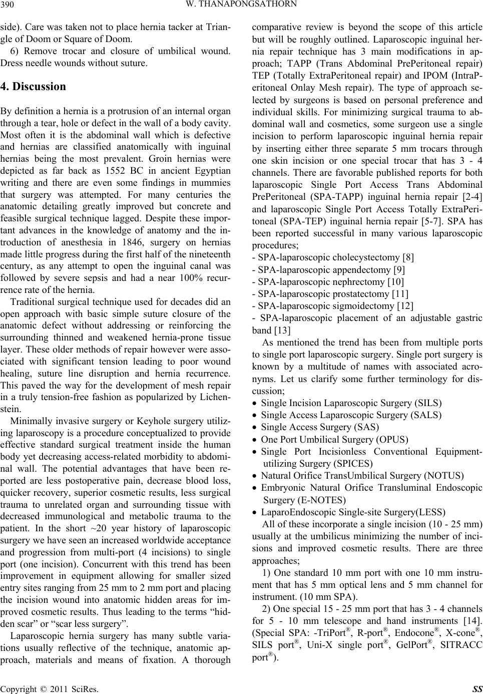 390 W. THANAPONGSATHORN side). Care was taken not to place hernia tacker at Trian- gle of Doom or Square of Doom. 6) Remove trocar and closure of umbilical wound. Dress needle wounds without suture. 4. Discussion By definition a hernia is a protrusion of an internal organ through a tear, hole or defect in the wall of a body cavity. Most often it is the abdominal wall which is defective and hernias are classified anatomically with inguinal hernias being the most prevalent. Groin hernias were depicted as far back as 1552 BC in ancient Egyptian writing and there are even some findings in mummies that surgery was attempted. For many centuries the anatomic detailing greatly improved but concrete and feasible surgical technique lagged. Despite these impor- tant advances in the knowledge of anatomy and the in- troduction of anesthesia in 1846, surgery on hernias made little progress during the first half of the nineteenth century, as any attempt to open the inguinal canal was followed by severe sepsis and had a near 100% recur- rence rate of the hernia. Traditional surgical technique used for decades did an open approach with basic simple suture closure of the anatomic defect without addressing or reinforcing the surrounding thinned and weakened hernia-prone tissue layer. These older methods of repair however were asso- ciated with significant tension leading to poor wound healing, suture line disruption and hernia recurrence. This paved the way for the development of mesh repair in a truly tension-free fashion as popularized by Lichen- stein. Minimally invasive surgery or Keyhole surgery utiliz- ing laparoscopy is a pro cedure conceptualized to provide effective standard surgical treatment inside the human body yet decreasing access-related morbidity to abdomi- nal wall. The potential advantages that have been re- ported are less postoperative pain, decrease blood loss, quicker recovery, superior cosmetic results, less surgical trauma to unrelated organ and surrounding tissue with decreased immunological and metabolic trauma to the patient. In the short ~20 year history of laparoscopic surgery we have seen an increased worldwide acceptance and progression from multi-port (4 incisions) to single port (one incision). Concurrent with this trend has been improvement in equipment allowing for smaller sized entry sites ranging from 25 mm to 2 mm port and placing the incision wound into anatomic hidden areas for im- proved cosmetic results. Thus leading to the terms “hid- den scar” or “scar less surgery”. Laparoscopic hernia surgery has many subtle varia- tions usually reflective of the technique, anatomic ap- proach, materials and means of fixation. A thorough comparative review is beyond the scope of this article but will be roughly outlined. Laparoscopic inguinal her- nia repair technique has 3 main modifications in ap- proach; TAPP (Trans Abdominal PrePeritoneal repair) TEP (Totally ExtraPeritoneal repair) and IPOM (IntraP- eritoneal Onlay Mesh repair). The type of approach se- lected by surgeons is based on personal preference and individual skills. For minimizing surgical trauma to ab- dominal wall and cosmetics, some surgeon use a single incision to perform laparoscopic inguinal hernia repair by inserting either three separate 5 mm trocars through one skin incision or one special trocar that has 3 - 4 channels. There are favorable published reports for both laparoscopic Single Port Access Trans Abdominal PrePeritoneal (SPA-TAPP) inguinal hernia repair [2-4] and laparoscopic Single Port Access Totally ExtraPeri- toneal (SPA-TEP) inguinal hernia repair [5-7]. SPA has been reported successful in many various laparoscopic procedures; - SPA-laparoscopic cholecystectomy [8] - SPA-laparoscopic appendectomy [9] - SPA-laparoscopic nephrectomy [10] - SPA-laparoscopic prostatectomy [11] - SPA-laparoscopic sigmoidectomy [12] - SPA-laparoscopic placement of an adjustable gastric band [13] As mentioned the trend has been from multiple ports to single port laparo scopic surg ery. Single port surger y is known by a multitude of names with associated acro- nyms. Let us clarify some further terminology for dis- cussion; Single Incision Lapa rosco pi c Sur gery (SILS) Single Access Laparoscopic Surgery (SALS) Single Access Surgery (SAS) One Port Umbilical Surgery (OPUS) Single Port Incisionless Conventional Equipment- utilizing Surg ery (SPICES) Natural Orifice TransUmbilical Surgery (NOTUS) Embryonic Natural Orifice Transluminal Endoscopic Surgery (E-NOTES) LaparoEndoscopi c Si n gle-si te Surgery(LESS) All of these incorporate a single incision (10 - 25 mm) usually at the umbilicus minimizing the number of inci- sions and improved cosmetic results. There are three approaches; 1) One standard 10 mm port with one 10 mm instru- ment that has 5 mm optical lens and 5 mm channel for instrument. (10 mm SPA). 2) One special 15 - 25 mm port that has 3 - 4 channels for 5 - 10 mm telescope and hand instruments [14]. (Special SPA: -TriPort®, R-port®, Endocone®, X-cone®, SILS port®, Uni-X single port®, GelPort®, SITRACC port®). Copyright © 2011 SciRes. SS 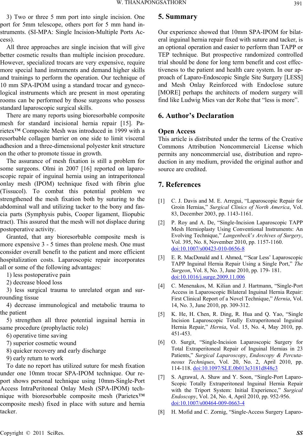 W. THANAPONGSATHORN 391 3) Two or three 5 mm port into single incision. One port for 5mm telescope, others port for 5 mm hand in- struments. (SI-MPA: Single Incision-Multiple Ports Ac- cess). All three approaches are single incision that will give better cosmetic results than multiple incision procedure. However, specialized trocars are very expensive, require more special hand instruments and demand higher skills and trainings to perform the operation. Our technique of 10 mm SPA-IPOM using a standard trocar and gyneco- logical instruments which are present in most operating rooms can be performed by those surgeons who possess standard laparoscopic surgical skills. There are many reports using bioresorbable composite mesh for standard incisional hernia repair [15]. Pa- rietex™ Composite Mesh was introduced in 1999 with a resorbable collagen barrier on one side to limit visceral adhesion and a three-dimensional polyester knit structure on the other to promote tissue in growth. The assurance of mesh fixation is still a problem for some surgeons. Olmi in 2007 [16] reported on laparo- scopic repair of inguinal hernia using an intraperitoneal onlay mesh (IPOM) technique fixed with fibrin glue (Tissucol). To combat this potential problem we strengthened the mesh fixation both by suturing to the abdominal wall and utilizing tacker to the bony and fas- cia parts (Symphysis pubis, Cooper ligament, Iliopubic tract). This assured that the mesh will not displace during postoperative activity. Granted, that any bioresorbable composite mesh is more expensive 3 - 5 times than prolene mesh. One must consider overall benefit to the patient and more efficient hospitalization costs. Laparoscopic repair incorporates all or some of the following advantages: 1) less postope rat i ve pai n 2) decrease blood loss 3) less surgical trauma to unrelated organ and sur- rounding tissue 4) decrease immunological and metabolic trauma to the patient 5) strengthen all three potential inguinal hernia in same procedure (prophylactic ro le) 6) operative time saving 7) superior cosmetic wound 8) quicker recovery and early discharge 9) early return to work To date no report has utilized suture for mesh fixation under one 10mm trocar SPA-IPOM technique. Our re- port shows personal technique using 10mm-Single-Port Access IntraPeritoneal Onlay Mesh (SPA-IPOM) tech- nique with bioresorbable composite mesh (Parietex™ composite mesh) fixed in place with suture and hernia tacker. 5. Summary Our experience showed that 10mm SPA-IPOM for bilat- eral inguinal hernia repair fixed with su ture and tacker, is an optional operation and easier to perform than TAPP or TEP technique. But prospective randomized controlled trial should be done for long term benefit and cost effec- tiveness to the patient and health care system. In our ap- proach of Laparo-Endoscopic Single Site Surgery [LESS] and Mesh Onlay Reinforced with Endoclose suture [MORE] perhaps the architects of modern surgery will find like Ludwig Mies van der Rohe that “less is more”. 6. Author’s Declaration Open Access This article is distributed under the terms of the Creative Commons Attribution Noncommercial License which permits any noncommercial use, distribution and repro- duction in any medium, provided the original author and source are credited. 7. References [1] C. J. Davis and M. E. Arregui, “Laparoscopic Repair for Groin Hernias,” Surgical Clinics of North America, Vol. 83, December 2003, pp. 1143-1161. [2] P. Roy and A. De, “Single-Incision Laparoscopic TAPP Mesh Hernioplasty Using Conventional Instruments: An Evolving Technique,” Langenbeck’s Archives of Surgery, Vol. 395, No. 8, November 2010, pp. 1157-1160. doi:10.1007/s00423-010-0656-8 [3] E. R. MacDonald and I. Ahmed, “‘Scar Less’ Laparoscopic TAPP Inguinal Hernia Repair Using a Single Port,” The Surgeon, Vol. 8, No. 3, June 2010, pp. 179- 181. doi:10.1016/j.surge.2009.11.006 [4] C. Menenakos, M. Kilian and J. Hartmann, “Single-Port Access in Laparoscopic Bilateral Inguinal Hernia Repair: First Clinical Report of a Novel Technique,” Hernia, Vol. 14, No. 3, June 2010, pp. 309-312. [5] K. He, H. Chen, R. Ding, R. Hua and Q. Yao, “Single Incision Laparoscopic Totally Extraperitoneal Inguinal Hernia Repair,” Hernia, Vol. 15, No. 4, May 2010, pp. 451-453. [6] O. Surgit, “Single-Incision Laparoscopic Surgery for Total Extraperitoneal Repair of Inguinal Hernias in 23 Patients,” Surgical Laparoscopy, Endoscopy & Percuta- neous Techniques, Vol. 20, No. 2, April 2010, pp. 114-118. doi:10.1097/SLE.0b013e3181d848c3 [7] S. Agrawal, A. Shaw and Y. Soon, “Single-Port Laparo- Scopic Totally Extraperitoneal Inguinal Hernia Repair with the Triport System: Initial Experience,” Surgical Endoscopy, Vol. 24, No. 4, April 2010, pp. 952-956. doi:10.1007/s00464-009-0663-4 [8] H. Mofid and C . Zornig, “Single-Access Surgery Laparo- Copyright © 2011 SciRes. SS  W. THANAPONGSATHORN Copyright © 2011 SciRes. SS 392 scopic Cholecystectomy and Appendectomy,” Surgical Technology International, Vol. 19, April 2010, pp. 61-64. http://www.springerlink.com/content/q26131ut15503w15/ [9] T. H. Hong, H. L. Kim, Y. S. Lee, J. J. Kim, K. H. Lee, Y. K. You, S. J. Oh and S. M. Park, “Transumbillical Sin- gle-Port Laparoscopic Appendectomy,” Journal of Laparo- endoscopic & Advanced Surgical Techniques. Part A, Vol. 19, No. 1, February 2009, pp. 75-78. doi:10.1089/lap.2008.0338 [10] M. Desai, “Single-Port Surgery for Donor Nephrectomy: A New Era in Laparoscopic Surgery?” Nature Clinical Practice Urology, Vol. 6, No. 1, January 2009, p. 1. doi:10.1038/ncpuro1278 [11] J. H. Kaouk, R. K. Goel, G. P. Haber, S. Crouzet, M. M. Desai and I. S. Gill, “Single-Port Laparoscopic Radical Prostatectomy,” Urology, Vol. 72, No. 6, December 2008, pp. 1190-1193. [12] J. Leroy, R. A. Cahil, M. Asakuma, B. Dallemagne and J. Marescaux, “Single-Access Laparoscopic Sigmoidectomy as Definitive Surgical Management of Prior Diverticulitis in a Human Patient,” Archives of Surgery, Vol. 144, No. 2, February 2009, pp. 173-179. doi:10.1001/archsurg.2008.562 [13] J. Teixeira, K. McGill, S. Binenbaum and G. Forrester, “Laparoscopic Single-Site Surgery for Placement of an Adjustable Gastric Band: Initial Experience,” Surgical Endoscopy, Vol. 23, No. 6, June 2009, pp. 1409-1414. doi:10.1007/s00464-009-0411-9 [14] M. G. Neto, A. Ramos and J. Campos, “Single Port Laparoscopic Access Surgery,” Techniques in Gastroin- testinal Endoscopy, Vol. 11, No. 2, April 2009, pp. 84-93. doi:10.1016/j.tgie.2009.06.007 [15] A. Moreno-Egea, J. A. Bustos, E. Girela and J. L. Aguayo-Albasini, “Long-Term Results of Laparoscopic Repair of Incisional Hernias Using an Intraperitoneal Composite Mesh,” Surgical Endoscopy, Vol. 24, No. 2, February 2010, pp. 359-365. doi:10.1007/s00464-009-0573-5 [16] S. Olmi, A. Scaini, L. Erba, A. Bertolini and E. Croce, “Laparoscopic Repair of Inguinal Hernias Using an In- traperitoneal Onlay Mesh Technique and a Parietex Composite Mesh Fixed with Fibrin Glue (Tissucol). Per- sonal Technique and Preliminary Results,” Surgical En- doscopy, Vol. 21, No. 11, November 2007, pp. 1961- 1964. doi:10.1007/s00464-007-9355-0 |