

Paper Menu >>
Journal Menu >>
 Surgical Science, 2011, 2, 366-368 doi:10.4236/ss.2011.27080 Published Online September 2011 (http://www.SciRP.org/journal/ss) Copyright © 2011 SciRes. SS A Rare Combınation of Gastric Inflammatory Fibroid Polyp and Hiatal Hernıa Coskun Polat, Murat Burc Yazicioglu*, Serkan Turel, Mehmet Nuri Kosar, Yuksel Arikan Departme nt of Surgery, Afyon Kocatepe University, School of Medicine, Afyonkarahisar, Turkey E-mail: *[email protected] Received March 8, 2011; revised May 13, 2011; accepted August 10, 2011 Abstract Inflammatory fibroid polyp (IFP) is a rare solitary gastrointestinal lesion of unknown etiology. It generally presents as polypoid mass in the gastric antrum. Mostly affects adults at average age of 60 years. IFP can cause different symptoms such as abdominal pain, gastrointestinal bleeding, intestinal obstruction or intussusception. IFP is a bening lesion and it may rarely mimic the submucosal tumor. The diagnosis can be possible after removal of the detected lession either by laparoscopic or open approach. In the present paper, we report a patient with gastric IFP and hiatal hernia. IFP was excised throughout the gastrotomy and following hiatal hernia was repaired. According to our best knowledge, this may be the first case report of gastric IFP concomitance with a hiatal hernia. Keywords: Gastric Inflamatuary Fibroid Tumor, Vanek, Diagnosis and Treatment 1. Introduction Inflammatory fibroid polyp is a relatively rare disorder which is thougt to b e clin ically an d histo logically ben ig n, and was first described as “polypoid fibroma” in 1920 by Kojetzny [1]. It may occu r throughou t the digestiv e tr act , but is most often seen in the stomach (approximately 80%) and usually occured either in the antrum or prepyloric region [2]. It is slightly more common in women (female:male ratio 1.6:1) [3]. It can be encoun- tered in all age group but peak incid ence is between sixth and seventh decades [4]. IFP originates from submucosa and grows as a solitary polypoid or sessile mass which may macroscopically mimic gastric cancer. When it is in the stomach, most frequently seen symptoms are vomiting, epigastric pain and bleeding. The lesion lo- cated in the small bowel can cause intussusception and obstruction. The diagnosis of IFP remains difficult be- cause of all available methods such as barium swallow or even endoscopic with endo scop ic ultrasonog raphy (EUS ) provide only nonspesific, insufficent information. In the case of submucosal lesions, standard biopsies are in- sufficent in obtaining adequate tissue. So that endo- scopic tumor excision or laparotomy is recommended. In this paper, we aimed to report a patient with IFP and hiatal hernia and to review the literature. 2. Case Report A 75 year-old man was referred to our hospital with complaints of nausea, abdominal pain and distention which was known for three years. Endoscopic exami- nation had been performed from the same complaints three years ago. En doscopy revealed a hernia poch at 7th cm below the Z line, and a 5 - 6 cm diameter of hiatal hernia and stomach was torsionized from this bare area. We could not perform endoscopic biopsy, because of the severe elevation in his arterial tension. A barium swallow was recommended, but he accepted neither barium swallow nor treatment. In his last admission, endoscopic examination showed a giant hiatal hernia, chronic gastritis and a mass in the bulbus; then multiple biopsy was obtained. On histhopathological examination, chronic inflammation and erosion were seen. Computed tomo- graphy (CT) revealed a giant hiatal hernia and a 2 × 7 cm hypodense image in hernia bowel which was close to liver at the right side (Figure 1). In his history, he was operated for Benign Prostate Hipertrophy and right inguinal hernia. No spesific family history was identified. On laboratory examination, WBC was 13,800/mm3. We have decided to perform an exploratory lapa- rotomy and Nissen fundoplication, because of he had a 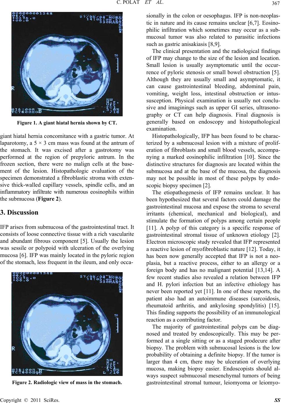 C. POLAT ET AL. 367 Figure 1. A giant hiatal hernia shown by CT. giant hiatal hernia concomita nce with a gastric tumor. At laparotomy, a 5 × 3 cm mass was found at the antrum of the stomach. It was excised after a gastrotomy was performed at the region of prepyloric antrum. In the frozen section, there were no malign cells at the base- ment of the lesion. Histopathologic evaluation of the specimen demonstrated a fibroblastic stroma with exten- sive thick-walled capillary vessels, spindle cells, and an inflammatory infiltrate with numerous eosinophils within the submucosa (Figure 2). 3. Discussion IFP arises from submucosa of the gastrointestinal tract. It consists of loose connective tissue with a rich vascularite and abundant fibrous component [5]. Usually the lesion was sessile or polypoid with ulceration of the overlying mucosa [6]. IFP was mainly located in the pyloric region of the stomach, less frequent in the ileum, and only occa- Figure 2. Radiologic view of mass in the stomach. sionally in the colon or oesophagus. IFP is non-neoplas- tic in nature and its cause remains unclear [6,7]. Eosino- philic infiltration which sometimes may occur as a sub- mucosal tumor was also related to parasitic infections such as gastric anisakiasis [8,9]. The clinical presentation and the radiological findings of IFP may change to the size of the lesion and location. Small lesion is usually asymptomatic until the occur- rence of pyloric stenosis or small bowel obstruction [5]. Although they are usually small and asymptomatic, it can cause gastrointestinal bleeding, abdominal pain, vomiting, weight loss, intestinal obstruction or intus- susception. Physical examination is usually not conclu- sive and imaginings such as upper GI series, ultrasono- graphy or CT can help diagnosis. Final diagnosis is generally based on endoscopy and histopathological examination. Histopathologically, IFP has been found to be charac- terized by a submucosal lesion with a mixture of prolif- eration of fibroblasts and small blood vessels, accompa- nying a marked eosinophilic infiltration [10]. Since the distinctive structures for diagnosis are located within the submucosa and at the base of the mucosa, the diagnosis may not be possible in most of these polyps by endo- scopic biopsy specimen [2]. The etiopathogenesis of IFP remains unclear. It has been hypothesized that several factors could damage the gastrointestinal mucosa and expose the stroma to several irritants (chemical, mechanical and biological), and stimulate the formation of polyps among certain people [11]. A polyp of this category is a specific response of gastrointestinal stromal tissue of unknown etiology [2]. Electron microscopic study revealed that IFP represented a reactive lesion of myofibroblastic nature [12]. Tod ay, it has been now generally accepted that IFP is not a neo- plasia, but a reactive process, either to an allergy or a foreign body and has no malignant potential [13,14]. A few recent studies also revealed a relation between IFP and H. pylori infection but an infective ethiology has never been reported yet [11]. In one of these reports, the patient also had an autoimmune diseases (sarcoidosis, rheumatoid arthritis, and ankylosing spondylitis) [15]. This finding supports the possibility of an immunological reaction as a contributing factor. The majority of gastrointestinal polyps can be diag- nosed and treated by endoscopically. This may be per- formed at a single sitting or as a staged prodecure after biopsy. The problem with submucosal lesions is the low probability of obtaining a definite biopsy. If the tumor is larger than 4 cm, there may be ulceration of overlying mucosa, making biopsy easier. Endoscopists should al- ways suspect submucosal mesenchymal tumors of being gastrointestinal stromal tumour, leiomyoma or leiomyo- Copyright © 2011 SciRes. SS 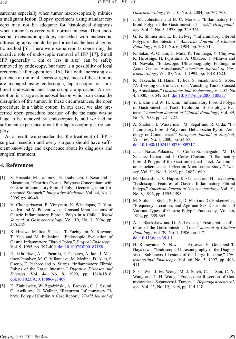 C. POLAT ET AL. Copyright © 2011 SciRes. SS 368 sarcoma especially when tumor macroscopically mimics a malignant lesion. Biopsy specimens using standart for- ceps may not be adequate for histological diagnosis when tumor is covered with normal mucosa. Then endo- scopic excision/polipectomy preceded with endoscopic ultrasonography should be performed as the best diagnos- tic method [6]. There were some reports concerning the curative role of endoscopic removal of IFP [17]. Small IFP (generally 1 cm or less in size) can be safely removed by endoscopy, but there is a possibility of local recurrence after operation [16]. But with increasing ex- perience in minimal access surgery, most of these tumors are managed using endoscopic, laparoscopic, or com- bined endoscopic and laparoscopic approache. An ex- ception is a large submucosal lesion which can cause the disruption of the tumor. In these circumstances, the open procedure is a viable option. In our case, we also pre- ferred open procedure because of the the mass was so huge to be removed by endoscopically and we had no sufficient experience about the laparoscopic gastric sur- gery. As a result, we consider that the treatment of IFP is surgical resection and every surgeon should have suffi- cient knowledge and experience about its diagnosis and surgical treatment. 4. References [1] S. Hırasaki, M. Tanimizu, E. Tsubouchı, J. Nasu and T. Masumoto, “Gastritis Cystica Polyposa Concomitant with Gastric Inflammatory Fibroid Polyp Occurring in an Un- operated Stomach,” Integrative Medicine, Vol. 44, No. 1, 2005, pp. 46-49. [2] V. Chongsrisawat, P. Yimyeam, N. Wisedopas, D. Vira- vaidya and Y. Poovorawan, “Unusual Manifestations of Gastric Inflammatory Fibroid Polyp in a Child,” World Journal of Gastroenterology, Vol. 10, No. 3, 2004, pp. 460-462. [3] K. Hizawa, M. Iida, S. Tada, T. Fuchigami, Y. Kuwano, T. Yao and M. Fujishima, “Endoscopic Evaluation of Gastric Inflammatory Fibroid Polyp,” Surgical Endoscopy, Vol. 9, 1995, pp. 397-400. doi:10.1007/BF00187158 [4] R. de la Plaza, A. L. Pıcardo, R. Cuberes, A. Jara, I. Mar- tinez-Penalver, M. C. Villanueva, M. Medina, D. Alias, S. Osorio, E. Pacheco and A. Suarez, “Inflammatory Fibroid Polyps of the Large Intestine,” Digestive Diseases and Sciences, Vol. 44, No. 9, 1999, pp. 1810-1816. doi:10.1023/A:1018886421409 [5] K. Zinkiewicz, W. Zgodziñski, A. Browski, O. J. Szumi, G. Awik and G. Wallner, “Recurrent Inflammatory Fi- broid Polyp of Cardia: A Case Report,” World Journal of Gastroenterology, Vol. 10, No. 5, 2004, pp. 767-768. [6] J. M. Johnstone and B. C. Morson, “Inflammatory Fi- broid Polyp of the Gastrointestinal Tract,” Histopathol- ogy, Vol. 2, No. 5, 1978, pp. 349-361. [7] G. R. Shimer and E. B. Helwig, “Inflammatory Fibroid Polyps of the Intestine,” American Journal of Clinical Pathology, Vol. 81, No. 6, 1984, pp. 708-714. [8] K. Sakai, A. Ohtani, H. Muta, K. Tominaga, Y. Chijiiwa, K. Hiroshige, H. Fujishima, A. Ohkubo, T. Misawa and H. Nawata, “Endoscopic Ultrasonography Findings in Acute Gastric Anisakiasis,” American Journal of Gas- troenterology, Vol. 87, No. 11, 1992, pp. 1618-1623. [9] K. Takeuchi, H. Hanai, T. Iida, S. Suzuki and S. Isobe, “A Bleeding Gastric Ulcer on a Vanishing Tumor Caused by Anisakia sis,” Gastrointestinal Endoscopy, Vol. 52, No. 4, 2000, pp. 549-551. doi:10.1067/mge.2000.108527 [10] Y. I. Kim and W. H. Kim, “Inflammatory Fi broid Polyps of Gastrointestinal Tract. Evolution of Histologic Pat- terns,” American Journal of Clinical Pathology, Vol. 89, No. 6, 1988, pp. 721-727. [11] A. Shalom, I. Wasserman, M. Segal and R. Orda, “In- flammatory Fibroid Polyp and Helicobacter Pylori. Aeti- ology or Coincidence?” European Journal of Surgical, Vol. 166, No. 1, 2000, pp. 54-57. doi:10.1080/110241500750009717 [12] J. J. Navas-Palacios, F. Colina-Ruizdelgado, M. D. Sanchez- Larrea and J. Cortes-Cansino, “Inflammatory Fibroid Polyps of the Gastrointestinal Tract. An Immu- nohistochemical and Electron Microscopic Study,” Can- cer, Vol. 51, No. 9, 1983, pp. 1682-1690. [13] M. Matsushita, K. Hajiro, K. Okazaki and H. Takakuwa, “Endoscopic Features of Gastric Inflammatory Fibroid Polyps,” American Journal of Gastroenterology, Vol. 91, No. 8, 1996, pp. 1595-1598. [14] M. Stolte, T. Sticht, S. Eidt, D. Ebert and G. Finkenzeller, “Frequency, Location, and Age and Sex Distribution of Various Types of Gastric Polyp,” Endoscopy, Vol. 26, 1994, pp. 659-665. [15] A. J. Blackshaw and D. A. Levison, “Eosinophilic Infil- trates of the Gastrointestinal Tract,” Journal of Clinical Pathology, Vol. 39, No. 1, 1986, pp. 1-7. doi:10.1136/jcp.39.1.1 [16] H. Kameyama, Y. Niwa, T. Arisawa, H. Goto and T. Hayakawa, “Endoscopic Ultrasonography in the Diagno- sis of Submucosal Lesions of the Large Intestine,” Gas- trointestinal Endoscopy, Vol. 46, No. 5, 1997, pp. 406- 411. [17] S. C. Wei, J. M. Wong, M. J. Shieh, C. T. Sun, C. Y. Wang and T. H. Wang, “Endoscopic Resection of Gas- trointestinal Submucosal Tumors,” Hepatogastroenterol- ogy, Vol. 45, No. 19, 1998, pp. 114-118. |