

Paper Menu >>
Journal Menu >>
 Open Journal of Nephrology, 2011, 1, 1-4 doi:10.4236/ojneph.2011.11001 Published Online September 2011 (http://www.SciRP.org/journal/ojneph) Copyright © 2011 SciRes. OJNeph Hyponatremia during a Severe Pneumococcal Meningitis with Renal Salt Wasting: A Cerebral Salt Wasting Syndrome? —Diagnosing a Cerebral Salt Wasting Syndrome Jean-Philippe Delabre, Stephane Pommet, Laurent Amigues, Olivier Jonquet, Kada Klouche Department of Intensive Care Unit Lapeyronie University Hospital, Montpellier, France E-mail: [email protected] Received September 11, 2011; revised September 19, 2011; accepted September 26, 2011 Abstract Background: The existence and prevalence of cerebral salt wasting and its differentiation from syndrome of inappropriate antidiuretic hormone have been controversial. This controversy stems from overlapping clini- cal and laboratory findings and an inability to assess the volume status of these patients. Objectives:To pre- sent a case of a cerebral salt wasting syndrome secondary to a bacterial mengitis and to emphasize the diffi- culty to assess the diagnosis. Case report: A 51-year-old male admitted to the ICU for a severe bacterial meningitis who developed, four days later, hyponatremia associated with hypovolemia due to a renal salt wasting. Clinical and biological parameters were collected; electrolytes balances and salt clearances were calculated to best investigate this hydroelectrolytic disorder. Patient's volume status and improvement after fluid and sodium replacement highly suggests the diagnosis of a cerebral salt wasting syndrome. Conclusion: A thorough analysis of this case showed that occurrence of hyponatremia and renal salt wasting after brain agression is not univocal underlying the fact that the diagnosis of cerebral salt wasting should be assessed only after a careful examination of the different determinants of the hydroelectrolytical balance. Keywords: Bacterial Meningitis, Cerebral Salt Wasting, Hyponatremia, Natriuresis 1. Introduction Hyponatremia is a common electrolyte disturbance in patients after central nervous system disease [1,2]. The syndrome of inappropriate secretion of antidiuretic hor- mone (SIADH) is the main attributed cause to this dis- order [3]. In some few cases, however, hyponatremia may be due to a renal salt wasting and diagnosed as a cerebral salt wasting syndrome (CSWS). CSWS, first described by Peters in 1950, is defined as the occurrence of an elevated diuresis and natriuresis after a cerebral injury, leading to hyponatremia and hypovolemia [4]. Despite substantial evidence in animal experiments, its pathophysiology and pertinency remains a question of debate in clinical settings [5]. We report herein the case of a patient who developped hyponatremia with renal salt wasting associated with hypovolemia following a severe bacterial meningitis suggestive of CSWS. Since many different mechanisms can lead to hyponatremia and a diagnosis of CSWS is seldom univocal, we thoroughly analyzed electrolytes balances, and salt clearances to assess the diagnosis. 2. Case Report A 51 year old male was admitted in our ICU with fever, headache and drowsiness evolving since 24 hours. At admission, the computed tomography (CT) scann was normal. The lumbar puncture was purulent with 7000 leucocytes per mm3, 95% being polynuclears. Cefotaxi- me and vancomycin in association with dexamethasone were administered to the patient. At day 1, he required mechanical ventilation because its consciousness dete- riorated. Cerebro spinal flu id was positiv e with p enicillin - sensitive streptococcus pneumoniae and antibiotherapy was switched to amoxicillin. At day 5, the patient signi- ficantly improved and was weaned of mechanical venti- lation with a good neurological status. 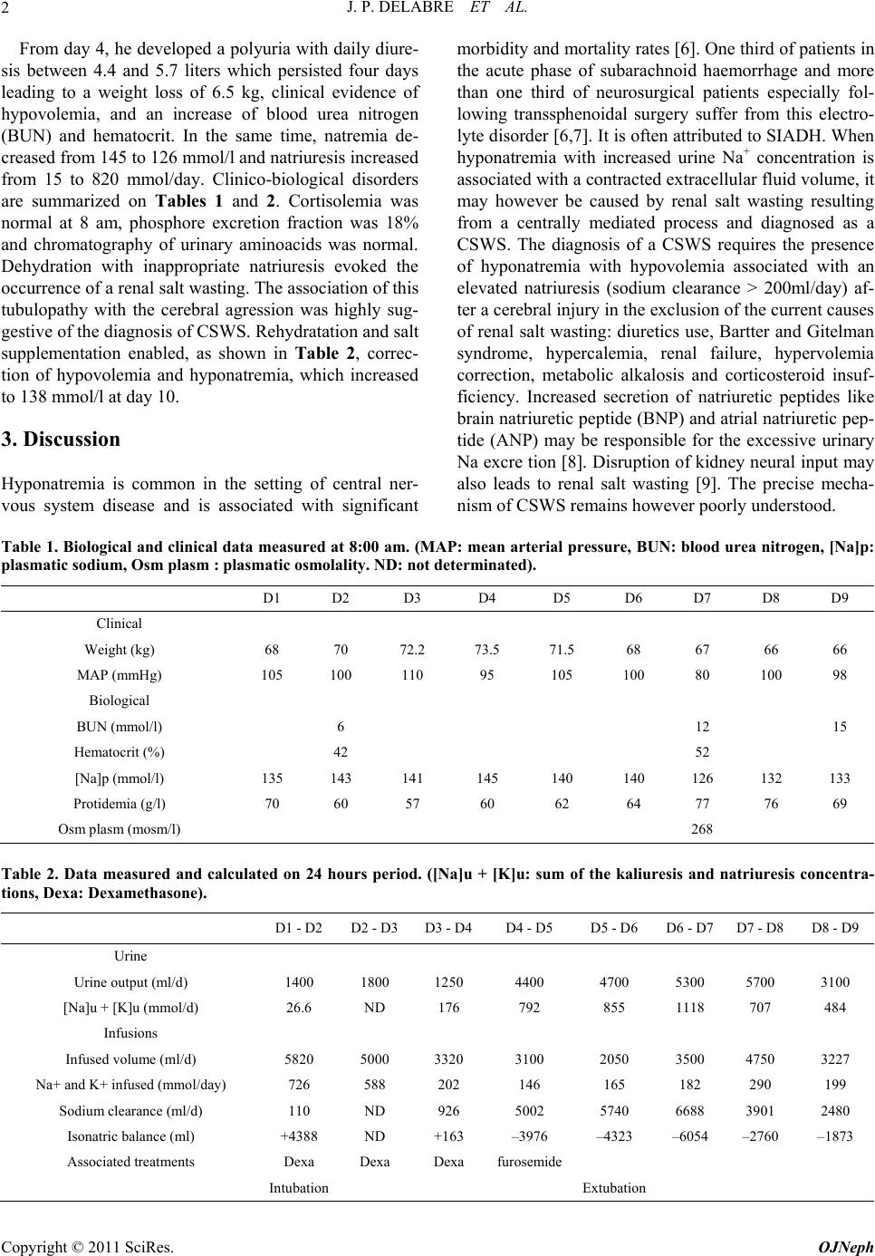 J. P. DELABRE ET AL. 2 From day 4, he developed a polyuria with daily diure- sis between 4.4 and 5.7 liters which persisted four days leading to a weight loss of 6.5 kg, clinical evidence of hypovolemia, and an increase of blood urea nitrogen (BUN) and hematocrit. In the same time, natremia de- creased from 145 to 126 mmol/l and natriuresis increased from 15 to 820 mmol/day. Clinico-biological disorders are summarized on Tables 1 and 2. Cortisolemia was normal at 8 am, phosphore excretion fraction was 18% and chromatography of urinary aminoacids was normal. Dehydration with inappropriate natriuresis evoked the occurrence of a renal salt wasting. The association of this tubulopathy with the cerebral agression was highly sug- gestive of the diagnosis of CSWS. Rehydratation and salt supplementation enabled, as shown in Table 2, correc- tion of hypovolemia and hyponatremia, which increased to 138 mmol/l at day 10. 3. Discussion Hyponatremia is common in the setting of central ner- vous system disease and is associated with significant morbidity and mortality rates [6]. One third of patients in the acute phase of subarachnoid haemorrhage and more than one third of neurosurgical patients especially fol- lowing transsphenoidal surgery suffer from this electro- lyte disorder [6,7 ]. It is often attributed to SIADH. When hyponatremia with increased urine Na+ concentration is associated with a contracted extracellular fluid volume, it may however be caused by renal salt wasting resulting from a centrally mediated process and diagnosed as a CSWS. The diagnosis of a CSWS requires the presence of hyponatremia with hypovolemia associated with an elevated natriuresis (sodium clearance > 200ml/day) af- ter a cerebral injury in the exclusion of the curren t causes of renal salt wasting: diuretics use, Bartter and Gitelman syndrome, hypercalemia, renal failure, hypervolemia correction, metabolic alkalosis and corticosteroid insuf- ficiency. Increased secretion of natriuretic peptides like brain natriuretic peptide (BNP) and atrial natriuretic pep- tide (ANP) may be responsible for the excessive urinary Na excre tion [8]. Disruption of kidney neural input may also leads to renal salt wasting [9]. The precise mecha- nism of CSWS remains however poorly under s tood. Table 1. Biological and clinical data measured at 8:00 am. (MAP: mean arterial pressure, BUN: blood urea nitrogen, [Na]p: plasmatic sodium, Osm plasm : plasmatic osmolality. ND: not determinated). D1 D2 D3 D4 D5 D6 D7 D8 D9 Clinical Weight (kg) 68 70 72.2 73.5 71.5 68 67 66 66 MAP (mmHg) 105 100 110 95 105 100 80 100 98 Biological BUN (mmol/l) 6 12 15 Hematocrit (%) 42 52 [Na]p (mmol/l) 135 143 141 145 140 140 126 132 133 Protidemia (g/l) 70 60 57 60 62 64 77 76 69 Osm plasm (mosm/l) 268 Table 2. Data measured and calculated on 24 hours period. ([Na]u + [K]u: sum of the kaliuresis and natriuresis concentra- tions, Dexa: Dexamethasone). D1 - D2 D2 - D3D3 - D4D4 - D5 D5 - D6 D6 - D7 D7 - D8 D8 - D9 Urine Urine output (ml/d) 1400 1800 1250 4400 4700 5300 5700 3100 [Na]u + [K]u (mmol/d) 26.6 ND 176 792 855 1118 707 484 Infusions Infused volume (ml/d) 5820 5000 3320 3100 2050 3500 4750 3227 Na+ and K+ infused (m mol/day) 726 588 202 146 165 182 290 199 Sodium clearance (ml/d) 110 ND 926 5002 5740 6688 3901 2480 Isonatric balance (ml) +4388 ND +163 –3976 –4323 –6054 –2760 –1873 Associated treatments Dexa Dexa Dexa furosem ide Intubation Extubation Copyright © 2011 SciRes. OJNeph  J. P. DELABRE ET AL. Copyright © 2011 SciRes. OJNeph 3 CSWS is yet unrecognized and its incidence is varia- bly estimated among authors [1-3,10]. In the study of Sherlock, hyponatremia after subarachnoid haemorrhage was induced by a CSWS in 6.5% of cases and by SIADH in 69% of cases [10]. A few observations of CSWS after infectious meningitis have been published and they were mainly tuberculous or viral origin [11-13]. Yet, at the best of our knowledge, only two cases of bacterial me- ningitis with a CSWS has been reported [14,15]. This report adds a new observation of a CSWS after bacterial meningitis, but also underlines the difficulty to assess this diagnosis particularly in ICU settings. Indeed, our patient developed at day 7 of a severe pneumococcal me- ningitis a clinical disorder associating dehydration, hy- ponatremia and renal salt wasting. Significant weight loss, increase of BUN and hematocrit support the fact that he was hypovolemic. Moreover, the occurrence of polyuria and correction of the disorder by saline infusion ruled out the diagnosis of SIADH. Main etiologies of tubulopathy have been also ruled out (calcemia, kaliemia, acido-basic status, cortisolemia and renal function were normals) but multiple factors that may play a role must be taken into acco unt to secure the diagnosis. Firstly, the patient exhibited three days before occurrence of hypo- natremia (at day 4), an opposite situation associating hypernatremia and hypervolemia as a result of a net gain of isonatric volume (infusion of isotonic saline) and the occurrence of a low clearance of sodium (Table 2). De- creased urinary Na excretion was secondary to the asso- ciation of corticotherapy and positive pressure ventila- tion. Secondarly, natriuresis and diuresis increased after day 4 leading to an increase of Na clearance and a dra- matic decrease of isonatric balance to day 8 as displayed in Table 2. This results probably from the occurrence of a CSWS but also, for a part, from positive pression ven- tilation interruption, and discontinuation of dexametha- sone (Table 2). Unless integrating all these elements, it seems therefore somewhat difficult to assess definitively the diagnosis of CSWS and this should be a general rule in hyponatremia diagnosis in similar circumstances. Nonetheless, rehydration and salt loading, that is thera- peutic management of CSWS, best corrected the disor- der. 4. Conclusions The case of our patient is an illustration of the problem in diagnosing a CSWS: the appearance of a renal salt wast- ing syndrome with hypovolemia and hyponatremia after a cerebral injury is insufficient to claim the presence of a CSWS. In fact, such patients generally benefit from se- veral treatments affecting the hydroelectrolytical balance: isotonic saline, positive pressure ventilation, corticoste- roid s and man nitol. Even af ter ex cluding major cause s of tubulopath y, the presence of a CSWS should be assessed only afte r a careful examinati on of the different determi- nants of the hydroelectrolytical balance, and considering cessation of treatments. 5. References [1] M. Cerda-Esteve, A. Ruiz-González, M. Gudelis, A. Go- day, J. Trujillano and E. Cuadrado, et al., “Incidence of Hyponatremia and Its Causes in Neurological Patients,” Endocrinol Nutrition, Vol. 5, No. 57, 2010, pp.182-186. doi:10.1016/j.endonu.2010.02.009 [2] M. G. Betjes, “Hyponatremia in Acute Brain Disease: The Cerebral Salt Wasting Syndrome,” European Journal of Internal Medicine, Vol. 1, No 13, 2002, pp. 9-14. doi:10.1016/S0953-6205(01)00192-3 [3] N. Moro, Y. Katayama, T. Igarashi, T. Mori, T. Kawa- mata and J. Kojima, “Hyponatremia in Patients with Traumatic Brain Injury: Incidence, Mechanism, and Re- sponse to Sodium Supplementation or Retention Therapy with Hydrocortisone,” Surgery Neurological, Vol. 4, No. 68, 2007, pp. 387-393. doi:10.1016/j.surneu.2006.11.052 [4] J. P. Peters, G. Weltg, E. A. H. Sims, J. Orloff and J. Needham, “A Salt-Wasting Syndrome Associated with Cerebral Disease,” Transactions of the Association of American Physicians, No. 63, 1950, pp. 57-64. [5] J. Kojima, Y. Katayama, N. Moro, H. Kawai, M. Yoneko and T. Mori, “Cerebral Salt Wasting in Subarachnoid Hemorrhage Rats: Model, Mechanism, and Tool,” Life Science, Vol. 20, No. 76, 2005, pp. 2361-2370. [6] B. Zheng, Y. Qiu, H. Jin, L. Wang, X. Chen and C. Shi, et al., “A Predictive Value of Hyponatremia for Poor Outcome and Cerebral Infarction in High-Grade Aneu- rysmal Subarachnoid Haemorrhage Patients,” Journal of Neurol Neurosurg Psychiatry, Vol. 2, No. 82, 2011, pp. 213-217. doi:10.1136/jnnp.2009.180349 [7] R. A. Kristof, M. Rother, G. Neuloh and D. Klingmüller, “Incidence, Clinical Manifestations, and Course of Water and Electrolyte Metabolism Disturbances Following Transsphenoidal Pituitary Adenoma Surgery: A Prospec- tive Observational Study,” Journal of Neurosurg, Vol. 3, No. 111, 2009, pp. 555-562. doi:10.3171/2008.9.JNS08191 [8] E. Berendes, M. Walter, P. Cullen, T. Prien, H. Van Aken and J. Horsthemke, et al., “Secretion of Brain Natriuretic Peptide in Patients with Aneurysmal Subarachnoid Haemorrhage,” Lancet, Vol. 349, No. 9047, 1997, pp. 245-249. doi:10.1016/S0140-6736(96)08093-2 [9] B. F. Palmer, “Hyponatremia in Patients with Central Nervous System Disease: SIADH Versus CSW,” Trends Endocrinol Metab, Vol. 4, No. 14, 2003, pp. 182-187. doi:10.1016/S1043-2760(03)00048-1 [10] M. Sherlock, E. O’Sullivan, A. Agha, L. A. Behan, D. Rawluk and P. Brennan, et al., “The Incidence and Patho- physiology of Hyponatraemia After Subarachnoid Haem-  J. P. DELABRE ET AL. 4 orrhage,” Clinical Endocrinol, Vol. 3, No. 64, 2006, pp. 250-254. doi:10.1111/j.1365-2265.2006.02432.x [11] L. K. Ti, S. C. Kang, and K. F. Cheong, “Acute Hypona- traemia Secondary to Cerebral Salt-Wasting Syndrome in a Patient with Tuberculous Meningitis,” Anaesth Inten- sive Care, Vol. 4, No.26, 1998, pp. 420-423. [12] U. S. Celik, D. Alabaz, D. Yildizdas, E. Alhan, E. Koca- bas and S. Ulutan, “Cerebral Salt-Wasting in Tuberculous Meningitis: Treatment with Fludrocortisone,” Ann Trop Paediatr, Vol. 4, No. 25, 2005, pp. 297-302. doi:10.1179/146532805X72458 [13] M. J. Brookes and T. H. Gould, “Cerebral Salt-Wasting Syndrome in Meningoencephalitis: A Case Report,” Journal of Neurol Neurosurg Psychiatry, Vol. 2, No. 74, 2003, pp. 277. doi:10.1136/jnnp.74.2.277 [14] H. Attout, S. Guez and C. Seriès, “Cerebral Salt-Wasting Syndrome in Bacterial Meningitis,” Ann Endocrinol, Vol. 5, No. 68, 2007, pp. 395-397. [15] M. H. Benito, P. Iglesias, P. Guevara, R. Sánchez Hernán-Dez and M. J. Fernández-Reyes, “Hyponatremia Secondary to Cerebral Salt-Wasting Syndrome Associ- ated to Bacterial Meningitis,” Nefrologia, Vol. 5, No. 28, 2008, pp. 570-571. Copyright © 2011 SciRes. OJNeph |