

Paper Menu >>
Journal Menu >>
 Materials Sciences and Applicatio ns, 2011, 2, 1194-1198 doi:10.4236/msa.2011.29161 Published Online September 2011 (http://www.SciRP.org/journal/msa) Copyright © 2011 SciRes. MSA Synthesis and Evaluation of Calcium-Deficient Hydroxyapatite with SiO2 Atsushi Nakahira1,2, Kentaro Nakata1, Chiya Numako3, Hidenobu Murata4, Katsuyuki Matsunaga4 1Osaka Prefecture University, Sakai, Japan; 2Osaka Center, IMR, Tohoku University, Sendai, Japan; 3The University of Tokushima, Tokushima, Japan; 4Nagoya University, Chikusa-ku, Japan. Email: [email protected] Received May 23rd, 2011; revised June 2nd, 2011; accepted June 9th, 2011. ABSTRACT Effect of SiO2 addition on the microstructures of calcium-deficient hydroxyapatite prepared by the heat-treatments was examined. Obtained hydroxyapatites substituted with Si ion were characterized by XRD, FT-IR and XAFS measure- ments. XANES results of Si K-edge and P K-edge in these modified hydroxyapatites indicated the shift of peaks of P K-edge with the SiO2 contents, although no change of Si local structures. In this study, the effect of Si ion on micro- and local structure of hydroxyapatite with SiO2 addition was mainly clarified. Keywords: Hydroxyapatite, SiO2, XAFS, Local Structure 1. Introduction It is well-known that hydroxyapatite (HAP) is a major inorganic component of bone and teeth. Since HAP pos- sesses high bioactivity, osteoconductivity and bio-com- patibility, it is extremely useful as a biomaterial for arti- ficial implant parts, bone fillers, and bone-cements etc. [1-6]. On the contrary, HAP has also both the high ability of ion exchange against cations and anions and adsorbent for various amino acids and proteins among other inor- ganic materials [5-10]. In bone, HAP is usually calcium- deficient below Ca/P of 1.67, not stoichiometry “(Ca10(PO4)6(OH)2) with Ca/P of 1.67”, for the achieve- ment of doping metal ions and control of the solubility. As a consequence, nonstoichiomeric HAP with Ca/P of less than 1.67 possesses the high ion-exchange ability of various cations. Therefore, calcium-deficient HAP is expected to be applicable as an ion-exchange media for solving environmental problems, such as the purification for water and soil polluted with heavy metals as well as biomedical applications [10,11]. Recently, the syntheses of HAP doped with functional elements (Zn and Fe) are also attempted for developing high performance bioce- ramics for biomedical application and the clarification of effects of functional elements on the properties and mi- crostructures is carried out in order to develop the high performance bioceramics. The syntheses of HAP substituted with other metals have been eagerly attempted by various synthetic pro- cesses, such as hydrothermal treatments, ion-exchange treatments, and normal heat-treatments at high tempera- ture in order to enhance the ion exchange ability and bioactivity of HAP [9-15]. For example, Fe substituted HAP i.e. abbreviated by “HAP modified with Fe ions”, synthesized by hydrothermal treatment indicated the good exchange ability against As and Pb etc., compared to monolithic HAP. However, in fact, monolithic HAP doped with metal ions is significantly difficult to synthe- size without any other phase. Since Bonefield reported that HAP containing with SiO2 indicated high bioactivity and osteoconductivity [14], the synthesis of HAP with SiO2 and tricalcium phosphate (TCP) with SiO2 have been performed by many researchers. However, HAP with SiO2 addition often results in the formation of reaction phase between HAP and SiO2, such as calcium silicate, and phosphate glass etc, and unreacted SiO2 residue. Thus, the SiO2 doped HAP without reaction phases and SiO2 residue is difficult to synthesize by the conventional methods. However, the successful synthesis of SiO2 doped HAP will lead to understanding effect of SiO2 element on the high bioactivity and osteoconductivity for SiO2 doped HAP. In this study, the main purpose is to synthesize SiO2 doped HAP. As a strategy, the incorporation of Si ions 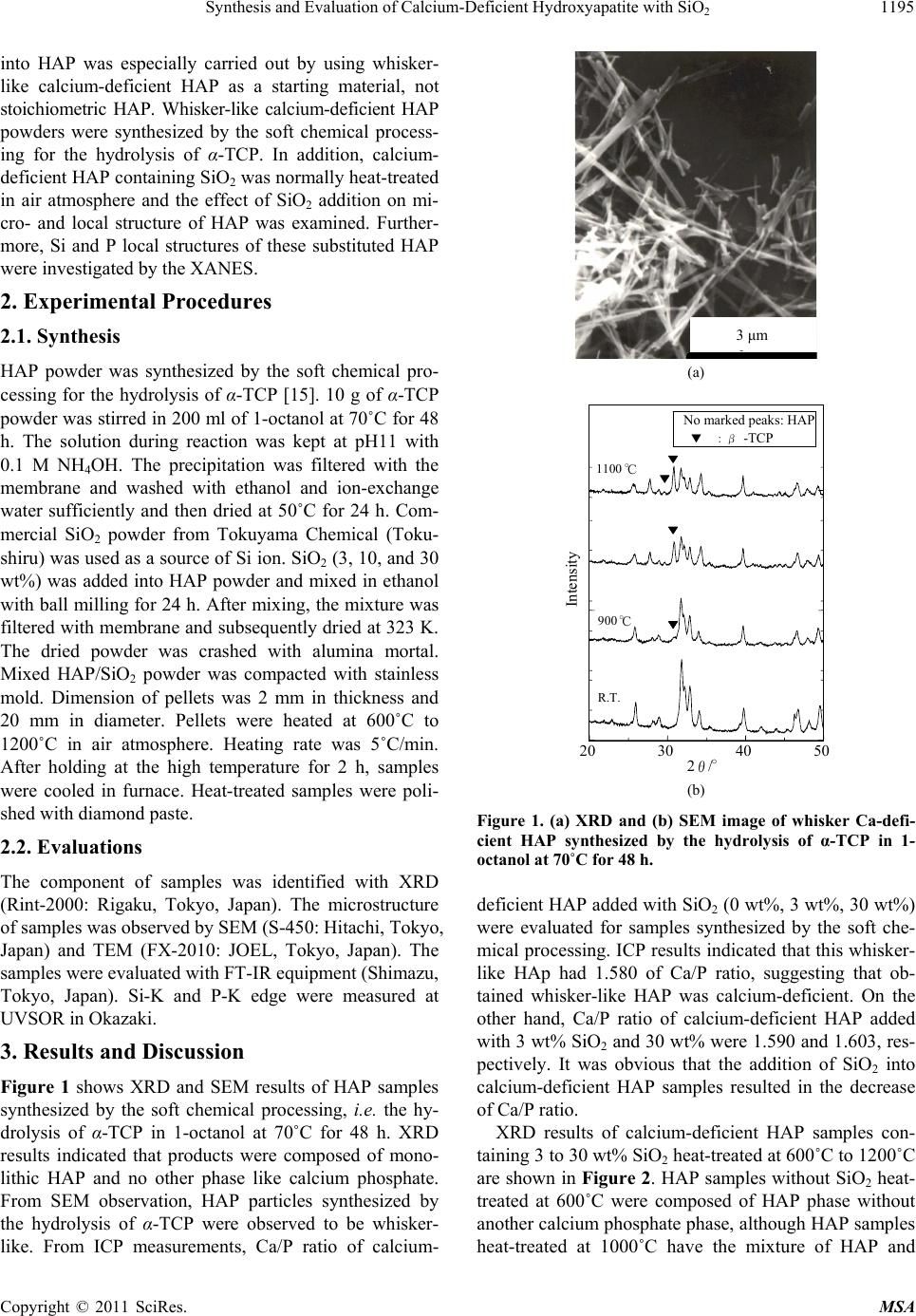 Synthesis and Evaluation of Calcium-Deficient Hydroxyapatite with SiO1195 2 into HAP was especially carried out by using whisker- like calcium-deficient HAP as a starting material, not stoichiometric HAP. Whisker-like calcium-deficient HAP powders were synthesized by the soft chemical process- ing for the hydrolysis of α-TCP. In addition, calcium- deficient HAP containing SiO2 was normally heat-treated in air atmosphere and the effect of SiO2 addition on mi- cro- and local structure of HAP was examined. Further- more, Si and P local structures of these substituted HAP were investigated by the XANES. 2. Experimental Procedures 2.1. Synthesis HAP powder was synthesized by the soft chemical pro- cessing for the hydrolysis of α-TCP [15]. 10 g of α-TCP powder was stirred in 200 ml of 1-octanol at 70˚C for 48 h. The solution during reaction was kept at pH11 with 0.1 M NH4OH. The precipitation was filtered with the membrane and washed with ethanol and ion-exchange water sufficiently and then dried at 50˚C for 24 h. Com- mercial SiO2 powder from Tokuyama Chemical (Toku- shiru) was used as a source of Si ion. SiO2 (3, 10, and 30 wt%) was added into HAP powder and mixed in ethanol with ball milling for 24 h. After mixing, the mixture was filtered with membrane and subsequently dried at 323 K. The dried powder was crashed with alumina mortal. Mixed HAP/SiO2 powder was compacted with stainless mold. Dimension of pellets was 2 mm in thickness and 20 mm in diameter. Pellets were heated at 600˚C to 1200˚C in air atmosphere. Heating rate was 5˚C/min. After holding at the high temperature for 2 h, samples were cooled in furnace. Heat-treated samples were poli- shed with diamond paste. 2.2. Evaluations The component of samples was identified with XRD (Rint-2000: Rigaku, Tokyo, Japan). The microstructure of samples was observed by SEM (S-450: Hitachi, Tokyo, Japan) and TEM (FX-2010: JOEL, Tokyo, Japan). The samples were evaluated with FT-IR equipment (Shimazu, Tokyo, Japan). Si-K and P-K edge were measured at UVSOR in Okazaki. 3. Results and Discussion Figure 1 shows XRD and SEM results of HAP samples synthesized by the soft chemical processing, i.e. the hy- drolysis of α-TCP in 1-octanol at 70˚C for 48 h. XRD results indicated that products were composed of mono- lithic HAP and no other phase like calcium phosphate. From SEM observation, HAP particles synthesized by the hydrolysis of α-TCP were observed to be whisker- like. From ICP measurements, Ca/P ratio of calcium- 3μm 3 μm (a) 20 30 40 50 R.T. 900 ℃ 1100 ℃ Intensity 2θ/° No marked peaks: HAP ▼:β -TCP (b) Figure 1. (a) XRD and (b) SEM image of whisker Ca-defi- cient HAP synthesized by the hydrolysis of α-TCP in 1- octanol at 70˚C for 48 h. deficient HAP added with SiO2 (0 wt%, 3 wt%, 30 wt%) were evaluated for samples synthesized by the soft che- mical processing. ICP results indicated that this whisker- like HAp had 1.580 of Ca/P ratio, suggesting that ob- tained whisker-like HAP was calcium-deficient. On the other hand, Ca/P ratio of calcium-deficient HAP added with 3 wt% SiO2 and 30 wt% were 1.590 and 1.603, res- pectively. It was obvious that the addition of SiO2 into calcium-deficient HAP samples resulted in the decrease of Ca/P ratio. XRD results of calcium-deficient HAP samples con- taining 3 to 30 wt% SiO2 heat-treated at 600˚C to 1200˚C are shown in Figure 2. HAP samples without SiO2 heat- treated at 600˚C were composed of HAP phase without another calcium phosphate phase, although HAP samples heat-treated at 1000˚C have the mixture of HAP and Copyright © 2011 SciRes. MSA 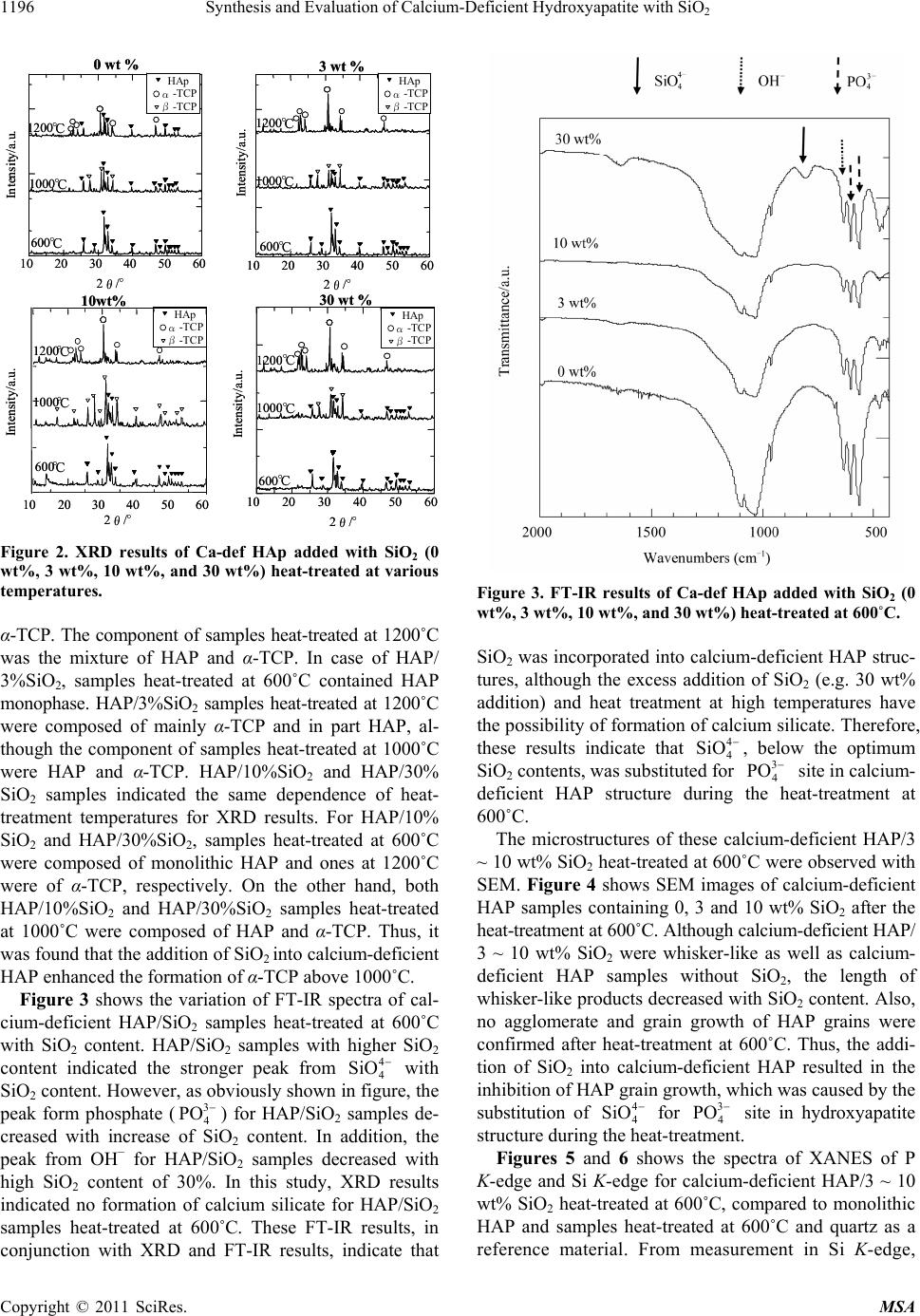 Synthesis and Evaluation of Calcium-Deficient Hydroxyapatite with SiO 1196 2 30 wt % 3 wt % 0 wt % 10wt% 10 20 30 40 50 60 2θ/° Intensity/a. u. 600℃ 1000℃ 1200℃ HAp α-TCP β-TCP 10 20 3040 5060 2θ/° 600℃ 1000℃ 1200℃ 1020 30 4050 60 600 ℃ 1000 ℃ 1200 ℃ 10 2030 40 50 60 2θ/° 600℃ 1000℃ 1200℃ HAp α-TCP β-TCP HAp α-TCP β-TCP HAp α-TCP β-TCP Intensity/a. u. Intensity/a.u. Intensity/a.u. 2θ/° 30 wt % 3 wt % 0 wt % 10wt% 10 20 30 40 50 60 2θ/° Intensity/a. u. 600℃ 1000℃ 1200℃ HAp α-TCP β-TCP HAp α-TCP β-TCP 10 20 3040 5060 2θ/° 600℃ 1000℃ 1200℃ 1020 30 4050 60 600 ℃ 1000 ℃ 1200 ℃ 10 2030 40 50 60 2θ/° 600℃ 1000℃ 1200℃ HAp α-TCP β-TCP HAp α-TCP β-TCP HAp α-TCP β-TCP HAp α-TCP β-TCP HAp α-TCP β-TCP HAp α-TCP β-TCP Intensity/a. u. Intensity/a.u. Intensity/a.u. 2θ/° Figure 2. XRD results of Ca-def HAp added with SiO2 (0 wt%, 3 wt%, 10 wt%, and 30 wt%) heat-treated at various temperatures. α-TCP. The component of samples heat-treated at 1200˚C was the mixture of HAP and α-TCP. In case of HAP/ 3%SiO2, samples heat-treated at 600˚C contained HAP monophase. HAP/3%SiO2 samples heat-treated at 1200˚C were composed of mainly α-TCP and in part HAP, al- though the component of samples heat-treated at 1000˚C were HAP and α-TCP. HAP/10%SiO2 and HAP/30% SiO2 samples indicated the same dependence of heat- treatment temperatures for XRD results. For HAP/10% SiO2 and HAP/30%SiO2, samples heat-treated at 600˚C were composed of monolithic HAP and ones at 1200˚C were of α-TCP, respectively. On the other hand, both HAP/10%SiO2 and HAP/30%SiO2 samples heat-treated at 1000˚C were composed of HAP and α-TCP. Thus, it was found that the addition of SiO2 into calcium-deficient HAP enhanced the formation of α-TCP above 1000˚C. Figure 3 shows the variation of FT-IR spectra of cal- cium-deficient HAP/SiO2 samples heat-treated at 600˚C with SiO2 content. HAP/SiO2 samples with higher SiO2 content indicated the stronger peak from 4 4 SiO with SiO2 content. However, as obviously shown in figure, the peak form phosphate () for HAP/SiO2 samples de- creased with increase of SiO2 content. In addition, the peak from OH− for HAP/SiO2 samples decreased with high SiO2 content of 30%. In this study, XRD results indicated no formation of calcium silicate for HAP/SiO2 samples heat-treated at 600˚C. These FT-IR results, in conjunction with XRD and FT-IR results, indicate that 3 4 PO Figure 3. FT-IR results of Ca-def HAp added with SiO2 (0 wt%, 3 wt%, 10 wt%, and 30 wt%) heat-treated at 600˚C. SiO2 was incorporated into calcium-deficient HAP struc- tures, although the excess addition of SiO2 (e.g. 30 wt% addition) and heat treatment at high temperatures have the possibility of formation of calcium silicate. Therefore, these results indicate that , below the optimum SiO2 contents, was substituted for site in calcium- deficient HAP structure during the heat-treatment at 600˚C. 4 4 SiO 3 4 PO The microstructures of these calcium-deficient HAP/3 ~ 10 wt% SiO2 heat-treated at 600˚C were observed with SEM. Figure 4 shows SEM images of calcium-deficient HAP samples containing 0, 3 and 10 wt% SiO2 after the heat-treatment at 600˚C. Although calcium-deficient HAP/ 3 ~ 10 wt% SiO2 were whisker-like as well as calcium- deficient HAP samples without SiO2, the length of whisker-like products decreased with SiO2 content. Also, no agglomerate and grain growth of HAP grains were confirmed after heat-treatment at 600˚C. Thus, the addi- tion of SiO2 into calcium-deficient HAP resulted in the inhibition of HAP grain growth, which was caused by the substitution of 4 4 SiO for site in hydroxyapatite structure during the heat-treatment. 3 4 PO Figures 5 and 6 shows the spectra of XANES of P K-edge and Si K-edge for calcium-deficient HAP/3 ~ 10 wt% SiO2 heat-treated at 600˚C, compared to monolithic HAP and samples heat-treated at 600˚C and quartz as a reference material. From measurement in Si K-edge, Copyright © 2011 SciRes. MSA 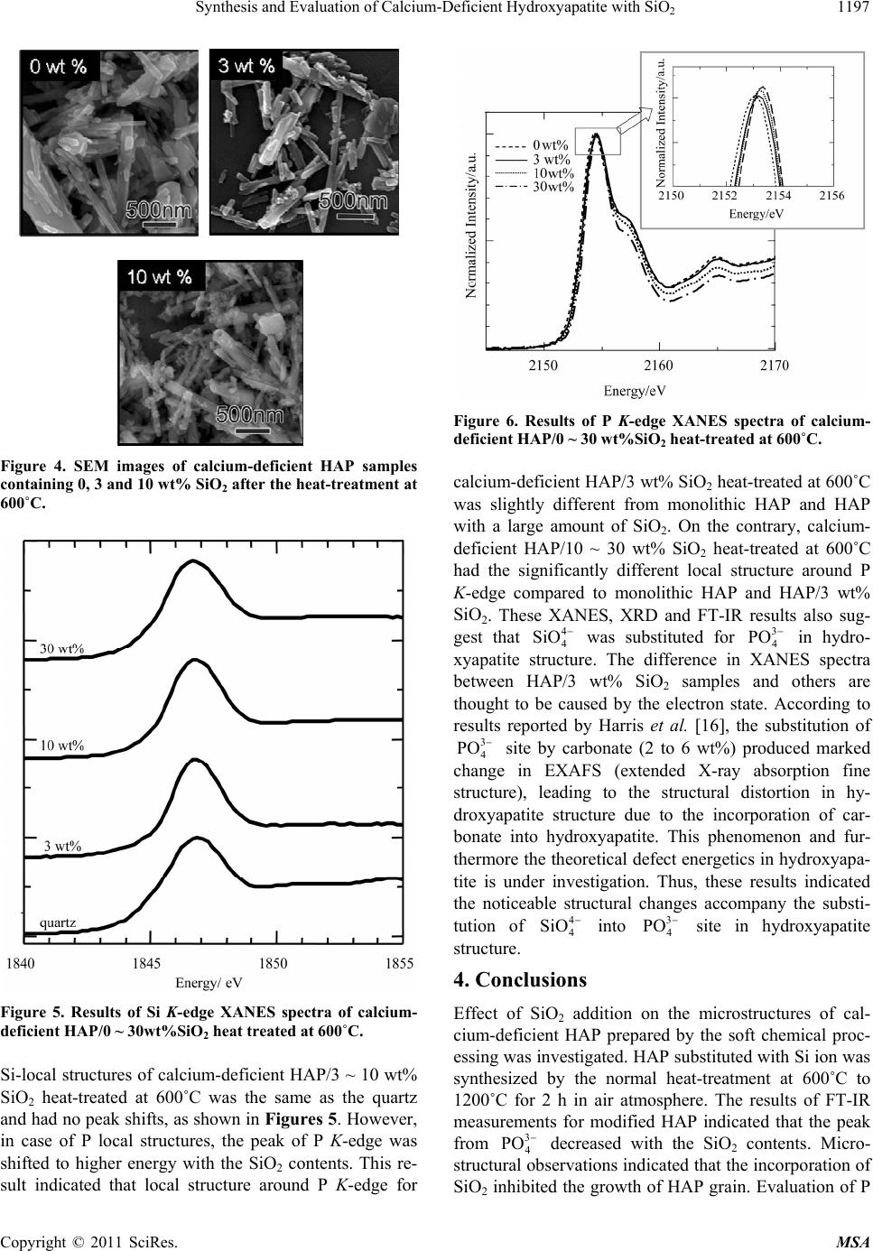 Synthesis and Evaluation of Calcium-Deficient Hydroxyapatite with SiO1197 2 Figure 4. SEM images of calcium-deficient HAP samples containing 0, 3 and 10 wt% SiO2 after the heat-treatment at 600˚C. Figure 5. Results of Si K-edge XANES spectra of calcium- deficient HAP/0 ~ 30wt%SiO2 heat treated at 600˚C. Si-local structures of calcium-deficient HAP/3 ~ 10 wt% SiO2 heat-treated at 600˚C was the same as the quartz and had no peak shifts, as shown in Figures 5. However, in case of P local structures, the peak of P K-edge was shifted to higher energy with the SiO2 contents. This re- sult indicated that local structure around P K-edge for Figure 6. Results of P K-edge XANES spectra of calcium- deficient HAP/0 ~ 30 wt%SiO2 heat-treated at 600˚C. calcium-deficient HAP/3 wt% SiO2 heat-treated at 600˚C was slightly different from monolithic HAP and HAP with a large amount of SiO2. On the contrary, calcium- deficient HAP/10 ~ 30 wt% SiO2 heat-treated at 600˚C had the significantly different local structure around P K-edge compared to monolithic HAP and HAP/3 wt% SiO2. These XANES, XRD and FT-IR results also sug- gest that 4 4 SiO was substituted for in hydro- xyapatite structure. The difference in XANES spectra between HAP/3 wt% SiO2 samples and others are thought to be caused by the electron state. According to results reported by Harris et al. [16], the substitution of 3 4 PO 3 4 PO site by carbonate (2 to 6 wt%) produced marked change in EXAFS (extended X-ray absorption fine structure), leading to the structural distortion in hy- droxyapatite structure due to the incorporation of car- bonate into hydroxyapatite. This phenomenon and fur- thermore the theoretical defect energetics in hydroxyapa- tite is under investigation. Thus, these results indicated the noticeable structural changes accompany the substi- tution of 4 4 SiO into site in hydroxyapatite structure. 3 4 PO 4. Conclusions Effect of SiO2 addition on the microstructures of cal- cium-deficient HAP prepared by the soft chemical proc- essing was investigated. HAP substituted with Si ion was synthesized by the normal heat-treatment at 600˚C to 1200˚C for 2 h in air atmosphere. The results of FT-IR measurements for modified HAP indicated that the peak from 3 4 PO decreased with the SiO2 contents. Micro- structural observations indicated that the incorporation of SiO2 inhibited the growth of HAP grain. Evaluation of P Copyright © 2011 SciRes. MSA 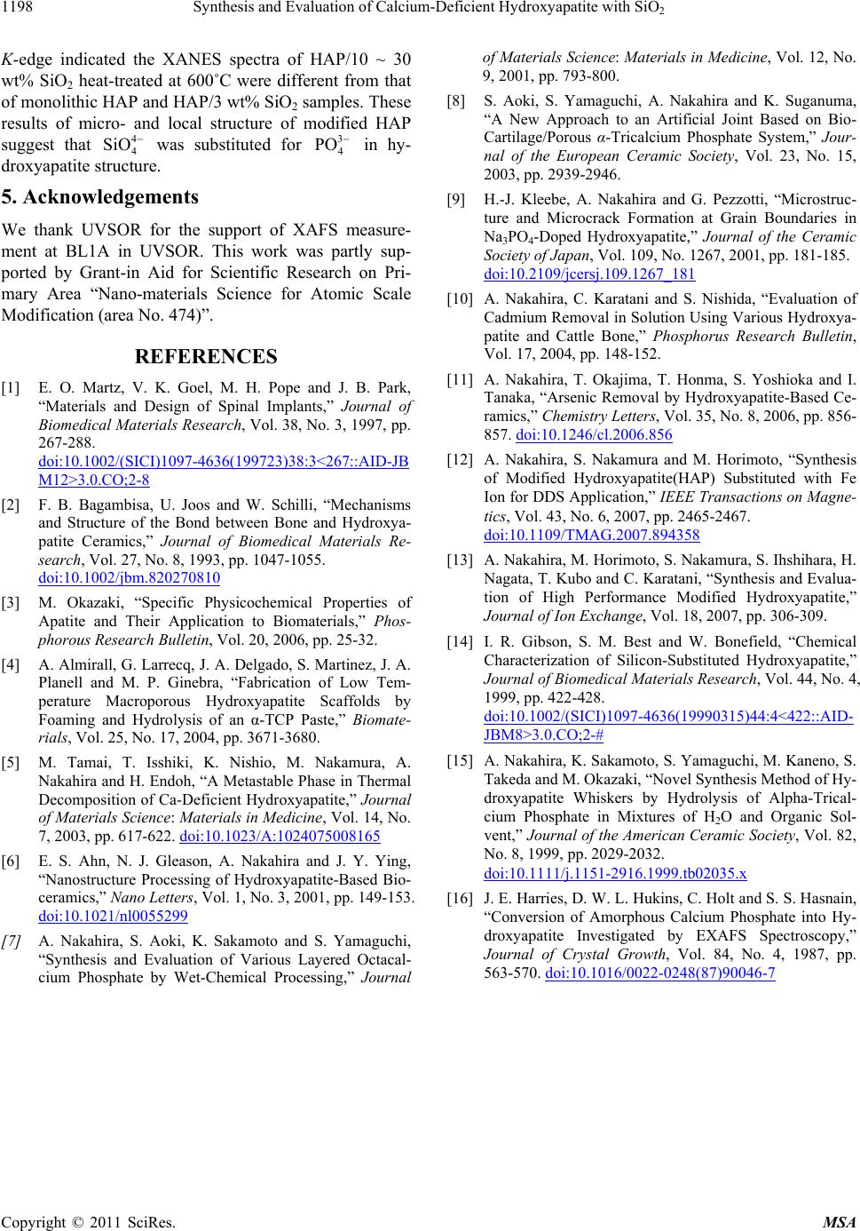 Synthesis and Evaluation of Calcium-Deficient Hydroxyapatite with SiO2 Copyright © 2011 SciRes. MSA 1198 K-edge indicated the XANES spectra of HAP/10 ~ 30 wt% SiO2 heat-treated at 600˚C were different from that of monolithic HAP and HAP/3 wt% SiO2 samples. These results of micro- and local structure of modified HAP suggest that was substituted for 4 4 SiO3 4 PO in hy- droxyapatite structure. 5. Acknowledgements We thank UVSOR for the support of XAFS measure- ment at BL1A in UVSOR. This work was partly sup- ported by Grant-in Aid for Scientific Research on Pri- mary Area “Nano-materials Science for Atomic Scale Modification (area No. 474)”. REFERENCES [1] E. O. Martz, V. K. Goel, M. H. Pope and J. B. Park, “Materials and Design of Spinal Implants,” Journal of Biomedical Materials Research, Vol. 38, No. 3, 1997, pp. 267-288. doi:10.1002/(SICI)1097-4636(199723)38:3<267::AID-JB M12>3.0.CO;2-8 [2] F. B. Bagambisa, U. Joos and W. Schilli, “Mechanisms and Structure of the Bond between Bone and Hydroxya- patite Ceramics,” Journal of Biomedical Materials Re- search, Vol. 27, No. 8, 1993, pp. 1047-1055. doi:10.1002/jbm.820270810 [3] M. Okazaki, “Specific Physicochemical Properties of Apatite and Their Application to Biomaterials,” Phos- phorous Research Bulletin, Vol. 20, 2006, pp. 25-32. [4] A. Almirall, G. Larrecq, J. A. Delgado, S. Martinez, J. A. Planell and M. P. Ginebra, “Fabrication of Low Tem- perature Macroporous Hydroxyapatite Scaffolds by Foaming and Hydrolysis of an α-TCP Paste,” Biomate- rials, Vol. 25, No. 17, 2004, pp. 3671-3680. [5] M. Tamai, T. Isshiki, K. Nishio, M. Nakamura, A. Nakahira and H. Endoh, “A Metastable Phase in Thermal Decomposition of Ca-Deficient Hydroxyapatite,” Journal of Materials Science: Materials in Medicine, Vol. 14, No. 7, 2003, pp. 617-622. doi:10.1023/A:1024075008165 [6] E. S. Ahn, N. J. Gleason, A. Nakahira and J. Y. Ying, “Nanostructure Processing of Hydroxyapatite-Based Bio- ceramics,” Nano Letters, Vol. 1, No. 3, 2001, pp. 149-153. doi:10.1021/nl0055299 [7] A. Nakahira, S. Aoki, K. Sakamoto and S. Yamaguchi, “Synthesis and Evaluation of Various Layered Octacal- cium Phosphate by Wet-Chemical Processing,” Journal of Materials Science: Materials in Medicine, Vol. 12, No. 9, 2001, pp. 793-800. [8] S. Aoki, S. Yamaguchi, A. Nakahira and K. Suganuma, “A New Approach to an Artificial Joint Based on Bio- Cartilage/Porous α-Tricalcium Phosphate System,” Jour- nal of the European Ceramic Society, Vol. 23, No. 15, 2003, pp. 2939-2946. [9] H.-J. Kleebe, A. Nakahira and G. Pezzotti, “Microstruc- ture and Microcrack Formation at Grain Boundaries in Na3PO4-Doped Hydroxyapatite,” Journal of the Ceramic Society of Japan, Vol. 109, No. 1267, 2001, pp. 181-185. doi:10.2109/jcersj.109.1267_181 [10] A. Nakahira, C. Karatani and S. Nishida, “Evaluation of Cadmium Removal in Solution Using Various Hydroxya- patite and Cattle Bone,” Phosphorus Research Bulletin, Vol. 17, 2004, pp. 148-152. [11] A. Nakahira, T. Okajima, T. Honma, S. Yoshioka and I. Tanaka, “Arsenic Removal by Hydroxyapatite-Based Ce- ramics,” Chemistry Letters, Vol. 35, No. 8, 2006, pp. 856- 857. doi:10.1246/cl.2006.856 [12] A. Nakahira, S. Nakamura and M. Horimoto, “Synthesis of Modified Hydroxyapatite(HAP) Substituted with Fe Ion for DDS Application,” IEEE Transactions on Magne- tics, Vol. 43, No. 6, 2007, pp. 2465-2467. doi:10.1109/TMAG.2007.894358 [13] A. Nakahira, M. Horimoto, S. Nakamura, S. Ihshihara, H. Nagata, T. Kubo and C. Karatani, “Synthesis and Evalua- tion of High Performance Modified Hydroxyapatite,” Journal of Ion Exchange, Vol. 18, 2007, pp. 306-309. [14] I. R. Gibson, S. M. Best and W. Bonefield, “Chemical Characterization of Silicon-Substituted Hydroxyapatite,” Journal of Biomedical Materials Research, Vol. 44, No. 4, 1999, pp. 422-428. doi:10.1002/(SICI)1097-4636(19990315)44:4<422::AID- JBM8>3.0.CO;2-# [15] A. Nakahira, K. Sakamoto, S. Yamaguchi, M. Kaneno, S. Takeda and M. Okazaki, “Novel Synthesis Method of Hy- droxyapatite Whiskers by Hydrolysis of Alpha-Trical- cium Phosphate in Mixtures of H2O and Organic Sol- vent,” Journal of the American Ceramic Society, Vol. 82, No. 8, 1999, pp. 2029-2032. doi:10.1111/j.1151-2916.1999.tb02035.x [16] J. E. Harries, D. W. L. Hukins, C. Holt and S. S. Hasnain, “Conversion of Amorphous Calcium Phosphate into Hy- droxyapatite Investigated by EXAFS Spectroscopy,” Journal of Crystal Growth, Vol. 84, No. 4, 1987, pp. 563-570. doi:10.1016/0022-0248(87)90046-7 |