

Paper Menu >>
Journal Menu >>
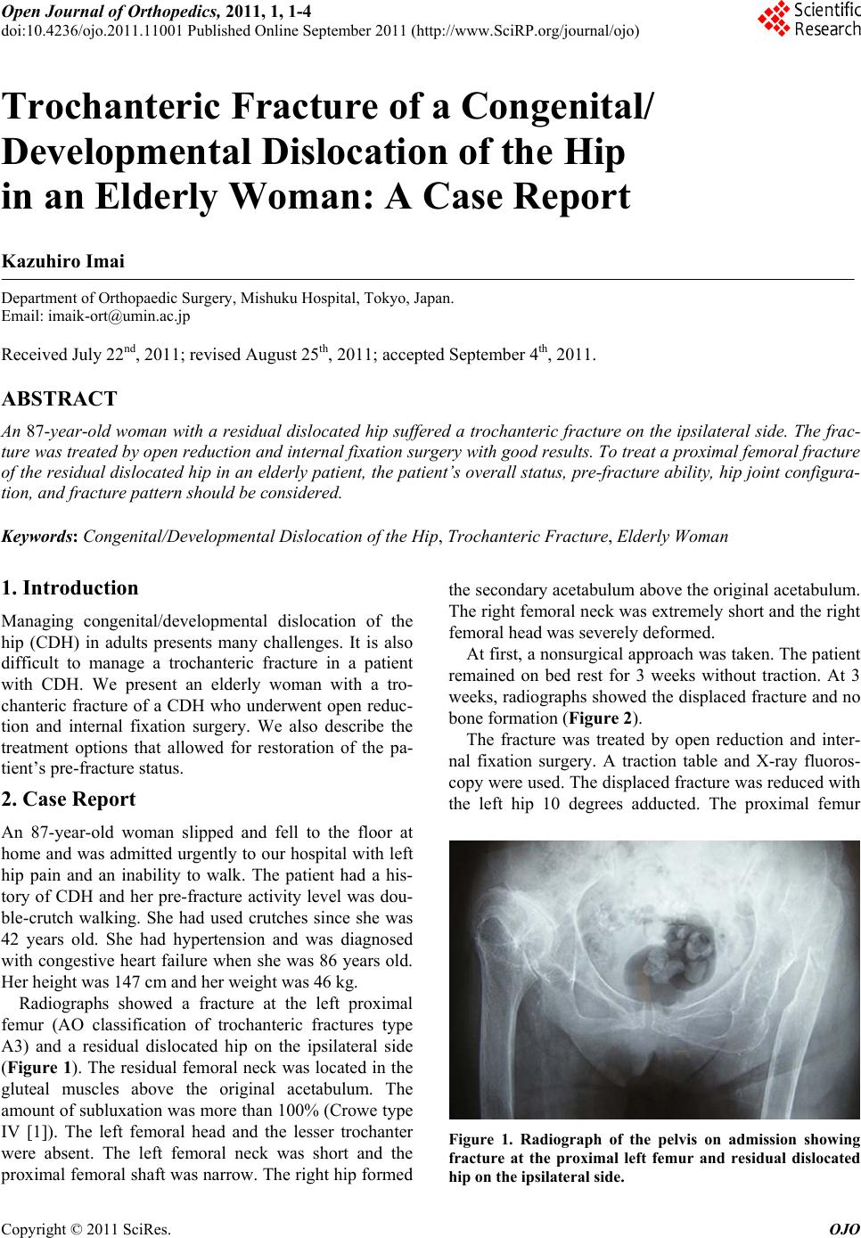 Open Journal of Orthopedics, 2011, 1, 1-4 doi:10.4236/ojo.2011.11001 Published Online September 2011 (http://www.SciRP.org/journal/ojo) Copyright © 2011 SciRes. OJO 1 Trochanteric Fracture of a Congenital/ Developmental Dislocation of the Hip in an Elderly Woman: A Case Report Kazuhiro Imai Department of Orthopaedic Surgery, Mishuku Hospital, Tokyo, Japan. Email: [email protected] Received July 22nd, 2011; revised August 25th, 2011; accepted September 4th, 2011. ABSTRACT An 87-year-old woman with a residual dislocated hip suffered a trochanteric fracture on the ipsilateral side. The frac- ture was treated by open reductio n and intern al fixation surgery with good results. To treat a proxima l femoral fracture of the residual dislocated hip in an elderly patient, the patient’s overall sta tus, pre-fracture ability, hip joint configura- tion, and fracture pattern should be considered. Keywords: Congenital/Developmental Dislocation of the Hip, Trochanteric Fracture, Elderly Woman 1. Introduction Managing congenital/developmental dislocation of the hip (CDH) in adults presents many challenges. It is also difficult to manage a trochanteric fracture in a patient with CDH. We present an elderly woman with a tro- chanteric fracture of a CDH who underwent open reduc- tion and internal fixation surgery. We also describe the treatment options that allowed for restoration of the pa- tient’s pre-fracture status. 2. Case Report An 87-year-old woman slipped and fell to the floor at home and was admitted urgently to our hospital with left hip pain and an inability to walk. The patient had a his- tory of CDH and her pre-fracture activity level was dou- ble-crutch walking. She had used crutches since she was 42 years old. She had hypertension and was diagnosed with congestive heart failure when she was 86 years old. Her height was 147 cm and her weight was 46 kg. Radiographs showed a fracture at the left proximal femur (AO classification of trochanteric fractures type A3) and a residual dislocated hip on the ipsilateral side (Figure 1). The residual femoral neck was located in the gluteal muscles above the original acetabulum. The amount of subluxation was more than 100% (Crowe type IV [1]). The left femoral head and the lesser trochanter were absent. The left femoral neck was short and the proximal femoral shaft was narrow. The right hip formed the secondary acetabulum above the original acetabulum. The right femoral neck was extremely short and the right femoral head was severely deformed. At first, a nonsurgical approach was taken. The patient remained on bed rest for 3 weeks without traction. At 3 weeks, radiographs showed the displaced fracture and no bone format ion (Figure 2). The fracture was treated by open reduction and inter- nal fixation surgery. A traction table and X-ray fluoros- copy were used. The displaced fracture was reduced with the left hip 10 degrees adducted. The proximal femur Figure 1. Radiograph of the pelvis on admission showing fracture at the proximal left femur and residual dislocated hip on the ipsilateral side. 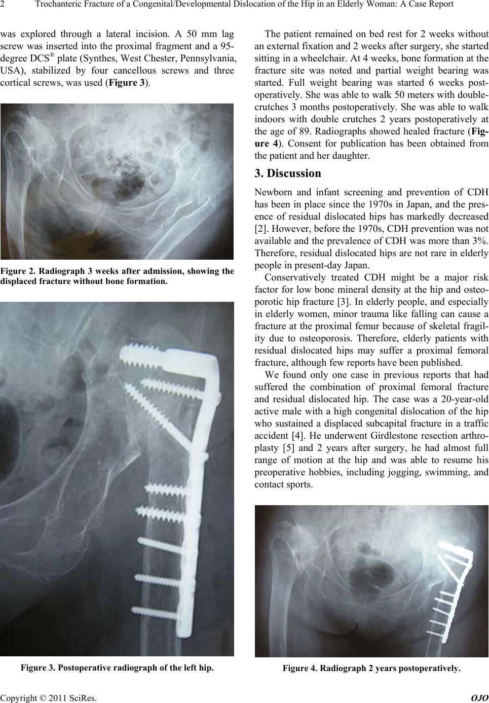 Trochanteric Fracture of a Congenital/Developmental Dislocation of the Hip in an Elderly Woman: A Case Report 2 was explored through a lateral incision. A 50 mm lag screw was inserted into the proximal fragment and a 95- degree DCS® plate (Synthes, West Ch ester, Pennsylvan ia, USA), stabilized by four cancellous screws and three cortical screws, was used (Figure 3). Figure 2. Radiograph 3 weeks after admission, showing the displaced fracture without bone formation. Figure 3. Postoperative radiograph of the left hip. The patient remained on bed rest for 2 weeks without an extern a l f ixatio n and 2 week s af ter sur g er y, sh e st ar ted sitting in a wheelchair. At 4 weeks, bone formation at the fracture site was noted and partial weight bearing was started. Full weight bearing was started 6 weeks post- operatively. She was able to walk 50 meters with double- crutches 3 months postoperatively. She was able to walk indoors with double crutches 2 years postoperatively at the age of 89. Radiographs showed healed fracture (Fig- ure 4). Consent for publication has been obtained from the patient and her daughter. 3. Discussion Newborn and infant screening and prevention of CDH has been in place since the 1970s in Japan, and the pres- ence of residual dislocated hips has markedly decreased [2]. Howev er, before th e 1970s, CDH prev ention was no t available and the prevalence of CDH was more than 3%. Therefore, residual dislocated hips are not rare in elderly people in present-day Japan. Conservatively treated CDH might be a major risk factor for low bone mineral density at the hip and osteo- porotic hip fracture [3]. In elderly people, and especially in elderly women, minor trauma like falling can cause a fracture at the proximal femur because of skeletal fragil- ity due to osteoporosis. Therefore, elderly patients with residual dislocated hips may suffer a proximal femoral fracture, althoug h few reports have been pub lishe d. We found only one case in previous reports that had suffered the combination of proximal femoral fracture and residual dislocated hip. The case was a 20-year-old active male with a high congenital dislocation of the hip who sustained a displaced subcapital fracture in a traffic accident [4]. He underwent Girdlestone resection arthro- plasty [5] and 2 years after surgery, he had almost full range of motion at the hip and was able to resume his preoperative hobbies, including jogging, swimming, and contact sports. Figure 4. Radiograph 2 years postoperatively. Copyright © 2011 SciRes. OJO 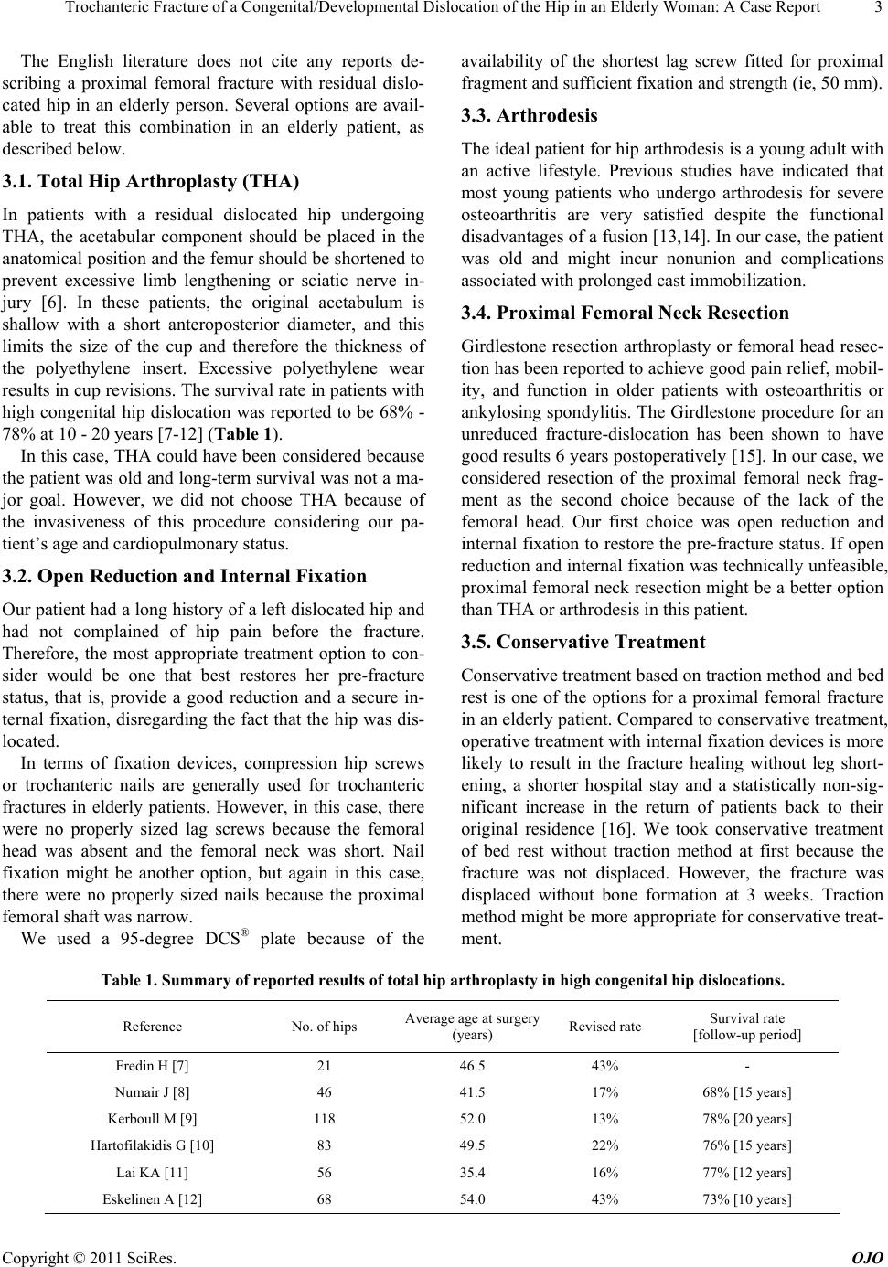 Trochanteric Fracture of a Congenital/Developmental Dislocation of the Hip in an Elderly Woman: A Case Report Copyright © 2011 SciRes. OJO 3 The English literature does not cite any reports de- scribing a proximal femoral fracture with residual dislo- cated hip in an elderly person. Several options are avail- able to treat this combination in an elderly patient, as described below. 3.1. Total Hip Arthroplasty (THA) In patients with a residual dislocated hip undergoing THA, the acetabular component should be placed in the anatomical position and the femur should be shortened to prevent excessive limb lengthening or sciatic nerve in- jury [6]. In these patients, the original acetabulum is shallow with a short anteroposterior diameter, and this limits the size of the cup and therefore the thickness of the polyethylene insert. Excessive polyethylene wear results in cup revisions. The surv ival rate in patients with high congenital hip dislocation was reported to be 68% - 78% at 10 - 20 years [7-12] (Table 1). In this case, THA could have been considered because the patient was old and long-term survival was not a ma- jor goal. However, we did not choose THA because of the invasiveness of this procedure considering our pa- tient’s age and cardiopulmonary status. 3.2. Open Reduction and Internal Fixation Our patient had a long h istory of a left dislocated hip and had not complained of hip pain before the fracture. Therefore, the most appropriate treatment option to con- sider would be one that best restores her pre-fracture status, that is, provide a good reduction and a secure in- ternal fixation, disregarding the fact that the hip was dis- located. In terms of fixation devices, compression hip screws or trochanteric nails are generally used for trochanteric fractures in elderly patients. However, in this case, there were no properly sized lag screws because the femoral head was absent and the femoral neck was short. Nail fixation might be another option, but again in this case, there were no properly sized nails because the proximal femoral shaft was narrow. We used a 95-degree DCS® plate because of the availability of the shortest lag screw fitted for proximal fragment and suf fi c i ent fi xat i on and st ren gth (ie, 50 mm ). 3.3. Arthrodesis The ideal patient for hip arthrodesis is a young adult with an active lifestyle. Previous studies have indicated that most young patients who undergo arthrodesis for severe osteoarthritis are very satisfied despite the functional disadvantages of a fusion [13,14]. In our case, the patient was old and might incur nonunion and complications associated with prolonged cast immobilization. 3.4. Proximal Femoral Neck Resection Girdlestone resection arthroplasty or femoral head resec- tion has been reported to achieve good pain relief, mobil- ity, and function in older patients with osteoarthritis or ankylosing spondylitis. Th e Girdlestone procedure for an unreduced fracture-dislocation has been shown to have good results 6 years postoperatively [15]. In our case, we considered resection of the proximal femoral neck frag- ment as the second choice because of the lack of the femoral head. Our first choice was open reduction and internal fixation to restore the pr e-fracture status. If open reduction and internal fixation was technically unf easible, proximal femoral neck resection might be a better option than THA or arthrodesis in this patient. 3.5. Conservative Treatment Conservative treatment based on traction method and bed rest is one of the options for a proximal femoral fracture in an elderly patient. Compared to conservative treatment, operative treatment with internal fixation devices is more likely to result in the fracture healing without leg short- ening, a shorter hospital stay and a statistically non-sig- nificant increase in the return of patients back to their original residence [16]. We took conservative treatment of bed rest without traction method at first because the fracture was not displaced. However, the fracture was displaced without bone formation at 3 weeks. Traction method might be more appropriate for conservative treat- ment. Table 1. Summary of reported results of total hip arthroplasty in high c ong enital hip disloc ations. Reference No. of hips Average age at surgery (years) Revised rateSurvival rate [follow-up period] Fredin H [7] 21 46.5 43% - Numair J [8] 46 41.5 17% 68% [15 years] Kerboull M [9] 118 52.0 13% 78% [20 years] Hartofilakidis G [10] 83 49.5 22% 76% [15 years] Lai KA [11] 56 35.4 16% 77% [12 years] Eskelinen A [12] 68 54.0 43% 73% [10 years] 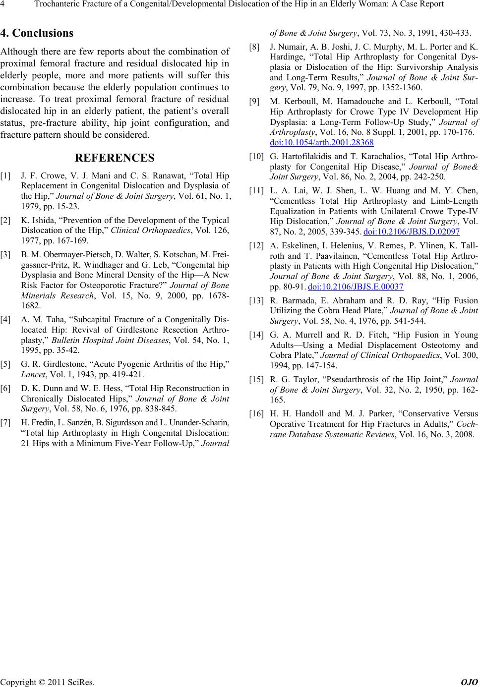 Trochanteric Fracture of a Congenital/Developmental Dislocation of the Hip in an Elderly Woman: A Case Report 4 4. Conclusions Although there are few reports about the combination of proximal femoral fracture and residual dislocated hip in elderly people, more and more patients will suffer this combination because the elderly population continues to increase. To treat proximal femoral fracture of residual dislocated hip in an elderly patient, the patient’s overall status, pre-fracture ability, hip joint configuration, and fracture pattern should be considered. REFERENCES [1] J. F. Crowe, V. J. Mani and C. S. Ranawat, “Total Hip Replacement in Congenital Dislocation and Dysplasia of the Hip,” Journal of Bone & Joint Surgery, Vol. 61, No. 1, 1979, pp. 15-23. [2] K. Ishida, “Prevention of the Development of the Typical Dislocation of the Hip,” Clinical Orthopaedics, Vol. 126, 1977, pp. 167-169. [3] B. M. Obermayer-Pietsch, D. Walter, S. Kotschan, M. Frei- gassner-Pritz, R. Windhager and G. Leb, “Congenital hip Dysplasia and Bone Mineral Density of the Hip—A New Risk Factor for Osteoporotic Fracture?” Journal of Bone Minerials Research, Vol. 15, No. 9, 2000, pp. 1678- 1682. [4] A. M. Taha, “Subcapital Fracture of a Congenitally Dis- located Hip: Revival of Girdlestone Resection Arthro- plasty,” Bulletin Hospital Joint Diseases, Vol. 54, No. 1, 1995, pp. 35-42. [5] G. R. Girdlestone, “Acute Pyogenic Arthritis of the Hip,” Lancet, Vol. 1, 1943, pp. 419-421. [6] D. K. Dunn and W. E. Hess, “Total Hip Reconstruction in Chronically Dislocated Hips,” Journal of Bone & Joint Surgery, Vol. 58, No. 6, 1976, pp. 838-845. [7] H. Fredin , L . S an zén , B . S igurds so n and L . Un and er- Sch ari n, “Total hip Arthroplasty in High Congenital Dislocation: 21 Hips with a Minimum Five-Year Follow-Up,” Journal of Bone & Joint Surgery, Vol. 73, No. 3, 1991, 430-433. [8] J. Numair, A. B. Joshi, J. C. Murphy , M. L. Porter and K. Hardinge, “Total Hip Arthroplasty for Congenital Dys- plasia or Dislocation of the Hip: Survivorship Analysis and Long-Term Results,” Journal of Bone & Joint Sur- gery, Vol. 79, No. 9, 1997, pp. 1352-1360. [9] M. Kerboull, M. Hamadouche and L. Kerboull, “Total Hip Arthroplasty for Crowe Type IV Development Hip Dysplasia: a Long-Term Follow-Up Study,” Journal of Arthroplasty, Vol. 16, No. 8 Suppl. 1, 2001, pp. 170-176. doi:10.1054/arth.2001.28368 [10] G. Hartofilakidis and T. Karachalios, “Total Hip Arthro- plasty for Congenital Hip Disease,” Journal of Bone& Joint Surgery, Vol. 86, No. 2, 2004, pp. 242-250. [11] L. A. Lai, W. J. Shen, L. W. Huang and M. Y. Chen, “Cementless Total Hip Arthroplasty and Limb-Length Equalization in Patients with Unilateral Crowe Type-IV Hip Dislocation,” Journal of Bone & Joint Surgery, Vol. 87, No. 2, 2005, 339-345. doi:10.2106/JBJS.D.02097 [12] A. Eskelinen, I. Helenius, V. Remes, P. Ylinen, K. Tall- roth and T. Paavilainen, “Cementless Total Hip Arthro- plasty in Patients with High Congenital Hip Dislocation,” Journal of Bone & Joint Surgery, Vol. 88, No. 1, 2006, pp. 80-91. doi:10.2106/JBJS.E.00037 [13] R. Barmada, E. Abraham and R. D. Ray, “Hip Fusion Utilizing the Cobra Head Plate,” Journal of Bone & Joint Surgery, Vol. 58, No. 4, 1976, pp. 541-544. [14] G. A. Murrell and R. D. Fitch, “Hip Fusion in Young Adults—Using a Medial Displacement Osteotomy and Cobra Plate,” Journal of Clinical Orthopaedics, Vol. 300, 1994, pp. 147-154. [15] R. G. Taylor, “Pseudarthrosis of the Hip Joint,” Journal of Bone & Joint Surgery, Vol. 32, No. 2, 1950, pp. 162- 165. [16] H. H. Handoll and M. J. Parker, “Conservative Versus Operative Treatment for Hip Fractures in Adults,” Coch- rane Database Systematic Reviews, Vol. 16, No. 3, 2008. Copyright © 2011 SciRes. OJO |