

Paper Menu >>
Journal Menu >>
 World Journal of Neuroscience, 2011, 1, 28-30 WJNS doi:10.4236/wjns.2011.12004 Published Online August 2011 (http://www.SciRP.org/journal/wjns/). Published Online August 2011 in SciRes. http://www.scirp.org/journal/WJNS A minor perimesencephalic subarachnoid hemorrhage on CT changes Yeting Zhou1, Daoming Tong2 1Medical Evaluation Unit, Shuyang People’Hospital, Shuyang, China; 2Departments of Neurology, Shuyang People’Hospital, Shuyang, China. Email: [email protected] Received 2 July 2011; revised 25 July 2011; accepted 4 August 2011. ABSTRACT Despite the increasing number of reports of patients with perimesencephalic subarachnoid hemorrhage (PMSAH), a minor or atypical PMSAH on CT changes has not been reported. We present the first described case of a minor subarachnoid hemorrhage located in the right perimesencephalic cistern on CT 4 h after headache onset. Twenty-six hours after headache on- set, another CT of the head showed that blood dis- persion. On the third day in hospital, examination of cerebrospinal fluid revealed xanthochromia. Minor PMSAH is rare type of SAH, and can been missed if there is a delay in CT imaging of the head. Any pa- tient with a suspected minor PMSAH or equivocal results on CT should undergo routine lumbar punc- ture. Keywords: Headache; Perimesencephalic Subarachnoid Hemorrhage; CT Imaging; Lumbar Puncture; Angiography 1. INTRODUCTION Perimesencephalic subarachnoid hemorrhage (PMSAH) appears to have an etiology and natural history distinct from aneurysm rupture [1]. The radiographic pattern of PMSAH is relatively dis- tinct. It involves hemorrhage centered anterior to the midbrain or pons (with or without extension of blood around the brainstem) into the sup rasellar cistern, or into the proximal Sylvian fissure [2]. Headache from PMSAH is a common presenting chief complaint. Despite the increasing number of reports of patients with PMSAH [1, 3,4], a minor or atypical PMSAH has not been reported. Here, we detail the rare case of CT changes in the brain of a patient with headac he fr o m minor PMSAH. 2. CASE REPORT A 36-year-old female was admitted to an affiliated hos- pital of university because of headache. Five hours be- fore hospital admission, she suddenly began to have a headache without nausea or vomiting. Four hours after the onset of headache, CT of the head revealed a minor or equivocal subarachnoid hemorrhage (SAH) located in the right perimesencephalic cistern (Figures 1(a) and (b)). On physical examination, the blood pressure was 150/ 72 mmHg. She was alert and oriented, with mild nuchal rigidity. The results of the remainder of the physical ex- amination were normal. Laboratory examinations, in- cluding complete blood count, serum electrolytes, liver Figure 1. Four hours after headache onset, CT of the head revealed a minor or equivocal subarachnoid hemorrhage ((a) and (b), arrow). Twenty-six hours after headache onset, CT of the head showed that extravasated blood dispersed quickly ((c) and (d)). 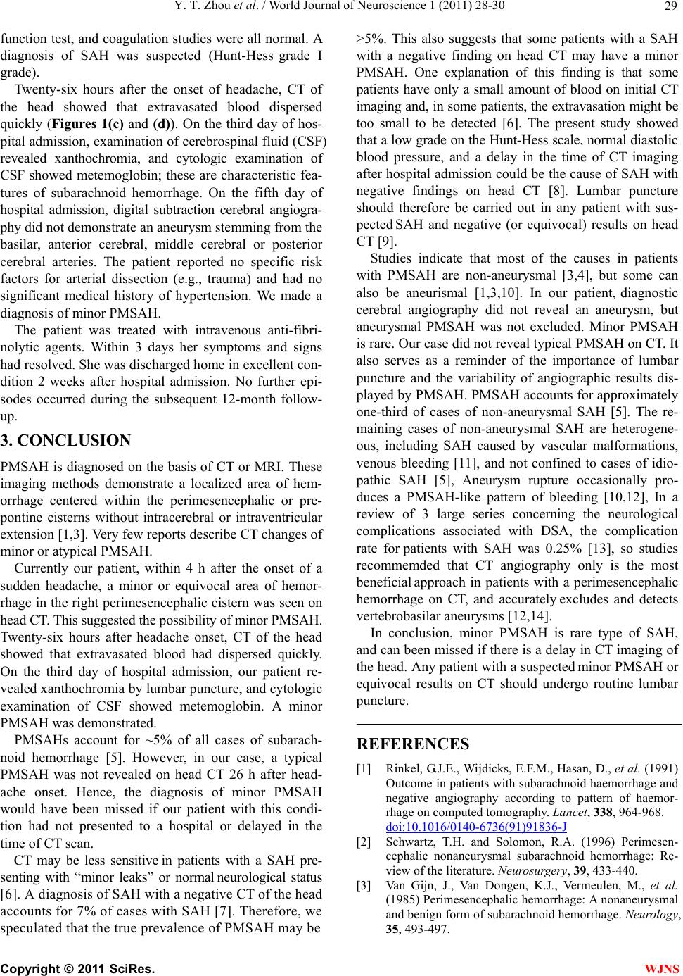 Y. T. Zhou et al. / World Journal of Neuroscience 1 (2011) 28-30 29 function test, and coagu lation studies were all normal. A diagnosis of SAH was suspected (Hunt-Hess grade I grade). Twenty-six hours after the onset of headache, CT of the head showed that extravasated blood dispersed quickly (Figures 1(c) and (d)). On the third day of hos- pital admission, examination of cerebrospinal fluid (CSF) revealed xanthochromia, and cytologic examination of CSF showed metemoglobin; these are characteristic fea- tures of subarachnoid hemorrhage. On the fifth day of hospital admission, digital subtraction cerebral angiogra- phy did not demonstrate an aneurysm stemming from the basilar, anterior cerebral, middle cerebral or posterior cerebral arteries. The patient reported no specific risk factors for arterial dissection (e.g., trauma) and had no significant medical history of hypertension. We made a diagnosis of minor PMSAH. The patient was treated with intravenous anti-fibri- nolytic agents. Within 3 days her symptoms and signs had resolved. She was discharged home in excellent con- dition 2 weeks after hospital admission. No further epi- sodes occurred during the subsequent 12-month follow- up. 3. CONCLUSION PMSAH is diagnosed on the basis of CT or MRI. These imaging methods demonstrate a localized area of hem- orrhage centered within the perimesencephalic or pre- pontine cisterns without intracerebral or intraventricular extension [1,3]. Very few reports describe CT changes of minor or atypical PMSAH. Currently our patient, within 4 h after the onset of a sudden headache, a minor or equivocal area of hemor- rhage in the right perimesencephalic cistern was seen on head CT. This suggested the possibility of minor PMSAH. Twenty-six hours after headache onset, CT of the head showed that extravasated blood had dispersed quickly. On the third day of hospital admission, our patient re- vealed xanthochromia by lumbar puncture, and cytologic examination of CSF showed metemoglobin. A minor PMSAH was demonstrated. PMSAHs account for ~5% of all cases of subarach- noid hemorrhage [5]. However, in our case, a typical PMSAH was not revealed on head CT 26 h after head- ache onset. Hence, the diagnosis of minor PMSAH would have been missed if our patient with this condi- tion had not presented to a hospital or delayed in the time of CT scan. CT may be less sensitive in patients with a SAH pre- senting with “minor leaks” or normal neurological status [6]. A diagnosis of SAH with a negative CT of the head accounts for 7% of cases with SAH [7]. Therefore, we speculated that the true prevalence of PMSAH may be >5%. This also suggests that some patients with a SAH with a negative finding on head CT may have a minor PMSAH. One explanation of this finding is that some patients have only a small amount of blood on initial CT imaging and, in some patients, the extravasation might be too small to be detected [6]. The present study showed that a low grade on the Hunt-Hess scale, normal d iast olic blood pressure, and a delay in the time of CT imaging after hospital admission could be the cause of SAH with negative findings on head CT [8]. Lumbar puncture should therefore be carried out in any patient with sus- pected SAH and negative (or equivocal) results on head CT [9]. Studies indicate that most of the causes in patients with PMSAH are non-aneurysmal [3,4], but some can also be aneurismal [1,3,10]. In our patient, diagnostic cerebral angiography did not reveal an aneurysm, but aneurysmal PMSAH was not excluded. Minor PMSAH is rare. Our case did not reveal typical PMSAH on CT. It also serves as a reminder of the importance of lumbar puncture and the variability of angiographic results dis- played by PMSAH. PMSAH accounts for approximately one-third of cases of non-aneurysmal SAH [5]. The re- maining cases of non-aneurysmal SAH are heterogene- ous, including SAH caused by vascular malformations, venous bleeding [11], and not confined to cases of idio- pathic SAH [5], Aneurysm rupture occasionally pro- duces a PMSAH-like pattern of bleeding [10,12], In a review of 3 large series concerning the neurological complications associated with DSA, the complication rate for patients with SAH was 0.25% [13], so studies recommemded that CT angiography only is the most beneficial approach in patients with a perimesencephalic hemorrhage on CT, and accurately excludes and detects vertebrobasilar aneurysms [12,14]. In conclusion, minor PMSAH is rare type of SAH, and can been missed if there is a delay in CT imaging of the head. Any patient with a suspected minor PMSAH or equivocal results on CT should undergo routine lumbar puncture. REFERENCES [1] Rinkel, G.J.E., Wijdicks, E.F.M., Hasan, D., et al. (1991) Outcome in patients with subarachnoid haemorrhage and negative angiography according to pattern of haemor- rhage on computed tomography. Lancet, 338, 964-968. doi:10.1016/0140-6736(91)91836-J [2] Schwartz, T.H. and Solomon, R.A. (1996) Perimesen- cephalic nonaneurysmal subarachnoid hemorrhage: Re- view of the literature. Neurosurgery, 39, 433-440. [3] Van Gijn, J., Van Dongen, K.J., Vermeulen, M., et al. (1985) Perimesencephalic hemorrhage: A nonaneurysmal and benign form of subarachnoid hemorrhage. Neurology, 35, 493-497. C opyright © 2011 SciRes. WJNS  Y. T. Zhou et al. / World Journal of Neuroscience 1 (2011) 28-30 Copyright © 2011 SciRes. WJNS 30 [4] Hui, F.K., Tumialán, L.M., Tanaka, T., et al. (2009) Cli- nical differences between angiographically negative, dif- fuse subarachnoid hemorrhage and perimesencephalic subarachnoid hemorrhage. Neurocrit Care, 11, 64-70. doi:10.1007/s12028-009-9203-2 [5] Flaherty, M.L., Haverbusch, M., Kissela, B., et al. (2005) Perimesencephalic subarachnoid hemorrhage: Incidence, risk factors,and outcoma. Journal of Stroke Cerebrovas- cular Diseases, 14, 267-71. doi:10.1016/j.jstrokecerebrovasdis.2005.07.004 [6] Van der Wee, N., Rinkel, G.J.E., Hasan, D., et al. (1995) Detection of subarachnoid haemorrhage on early CT: Is lumbar puncture still needed after a negative scan? Jour- nal of Neurology, Neurosurgery & Psychiatry, 58, 357- 359. doi:10.1136/jnnp.58.3.357 [7] Richard, L.B, William, R.M, Natalie, S., Gelareh, Z.G., Shanna, F. and Larry, J.B. (2008) Sensitivity of noncon- trast cranial computed tomography for the emergency department diagnosis of subarachnoid hemorrhage. An- nals of Emergency Medicine, 51, 697-703. doi:10.1016/j.annemergmed.2007.10.007 [8] Tong, D.M. and Chou, Y.T. (2010) Predictors of the su- barachnoid hemorrhage of a negative CT scan. Stroke, 41, e566-e567. doi:10.1161/STROKEAHA.109.574038 [9] Suarez, J.I., Tarr, R.W. and Selman, W.R. (2006) Aneu- rysmal subarachnoid hemorrhage. New England Journal of Medici ne, 354, 387-396. doi:10.1056/NEJMra052732 [10] Schievink, W.I., Wijdicks, E.F.M., Piepgras, D.G., et al. (1994) Perimesencephalic subarachnoid hemorrhage: Additional perspectives from four cases. Stroke, 25, 1507-1511. doi:10.1161/01.STR.25.7.1507 [11] Van Der Schaaf, I.C., Velthuis, B.K., Gouw, A., et al. (2004) Venous drainage in perimesencephalic hemor- rhage. Stroke, 35, 1614-1618. doi:10.1161/01.STR.0000131657.08655.ce [12] Velthuis, B.K., Rinkel, G.J., Ramos, L.M., et al. (1999) Perimesencephalic hemorrhage: Exclusion of vertebro- basilar aneurysms with CT angiography. Stroke, 30, 1103-1109. doi:10.1161/01.STR.30.5.1103 [13] Cloft, H.J., Joseph, G.J. and Dion, J.E. (1999) Risk of cerebral angiography in patients with subarachnoid hem- orrhage, cerebral aneurysm, and arteriovenous malfor- mation: A meta-analysis. Stroke, 30, 317-320. doi:10.1161/01.STR.30.2.317 [14] Ruigrok, Y.M., Rinkel, G.J., Buskens, E., et al. (2000) Perimesencephalic hemorrhage and CT angiography: A decision analysis. Stro ke, 31, 2976-2983. doi:10.1161/01.STR.31.12.2976 |