

Paper Menu >>
Journal Menu >>
 Surgical Science, 2011, 2, 326-330 doi:10.4236/ss.2011.26069 Published Online August 2011 (http://www.SciRP.org/journal/ss) Copyright © 2011 SciRes. SS Validity of Ultrasound in Patients with Acute Pelvic Pain Related to Suspected Ovarian Torsion Leena Mawaldi1, Charu Gupta1, Hanadi Bakhsh1, Maissa Saadeh2, Mostafa A. Abolfotouh2* 1Department of Obstetrics and G ynaecology, King AbdulAziz Medical City, National Guard Health Affairs, Riyadh, Saudi Arabia 2King Abdullah International Medical Research Center (KAIMRC), King Saud Bin-Abdulaziz University fo r Health Sciences, National Guard Health Affairs, Riyadh, Saudi Arabia *E-mail: [email protected] m Received April 27, 2011; revised July 2, 2011; accepted July 18, 2011 Abstract Objective: Ultrasound has been proven to be useful in detecting underlying ovarian pathology. However, its role in the prediction of ovarian torsion has been controversial. The aim of the study was to assess the valid- ity of ultrasound in the prediction of ovarian torsion in patients with acute pelvic pain related to clinically suspected ovarian torsion. Methods: A retrospective observational study was conducted at the Ob/Gyn de- partment using a 10-year chart review of all female patients older than 11 years of age with highly suspected ovarian torsion who underwent clinical assessment and ultrasound prior to surgery (n = 62). The sensitivity and specificity of ultrasound were determined by cross-tabulation of the ultrasound and surgical findings. Results: Of the suspected cases, 54 (87.1%) were confirmed to be cases of ovarian torsion by surgery. The majority of the cases were suggestive of ovarian torsion, which was indicated by clinical examination (77.4%), ultrasound (77.4%), or pathological examination (79%). Almost one-half of the cases (46.8%) showed a pain score >6; two-thirds (62.9%) presented with vomiting and/or nausea; and more than one-third (38.7%) presented with leukocytosis. The estimated sensitivity and specificity of ultrasound were 0.74 and 0.0, respectively. The positive predictive value was 0.83. Ultrasound was significantly associated with both clinical examination (p = 0.039) and pain score (p = 0.008). Conclusion: The diagnosis of ovarian torsion cannot be exclusively based on ultrasound. Both clinical and sonographical evaluation of acute pelvic pain should be considered for the diagnosis. A definitive diagnosis remains challenging. Keywords: Acute Pelvic Pain, Ultrasound, Ovarian Torsion 1. Introduction Ovarian torsion refers to the twisting of the ovary on its ligamentous supports, which often results in an imped- ance of its blood supply. This condition is the fifth most common gynecological emergency and affects females of all ages [1]. Expedient diagnosis is important to pre- serve ovarian function and prevent adverse sequelae [2]; however, the diagnosis can be challenging because the symptoms are relatively nonspecific. The ovaries were examined in a large series of patients with surgically confirmed torsion-associated cysts in 48% and neopla- sms in 46%; the remainder occurred in normal-appearing ovaries [3]. Histopathology was benign in over 90% of patients [3-4]. Whereas anatomic factors usually account for ovarian torsion in adults, normal ovaries have been demonstrated in over 50% of ovarian torsion cases in children under the age of 15 years [5]. In adults, torsion has also been described following laparoscopic hyster- ectomy, suggesting that even release of the fulcrum, on which the ovaries usually twist, does not protect against torsion [6]. Strenuous exercise or a sudden increase in abdominal pressure also promotes torsion of the ovary around the vascular pedicle [7]. The right ovary is more likely than the left to underg o torsion, suggestin g that the sigmoid colon may help to prevent torsion [8]. Women who are pregnant [9-10] or are undergoing ovarian hyperstimulation during infertility treatment are at increased risk of ovarian torsion [11]. The overall in- cidence of torsion in pregnant women was reported as  L. MAWALDI ET AL.327 15% [12]. In association with pregnancy, torsion most commonly occurred between 10 and 17 weeks of gesta- tion and during the postpartum period. A much lower incidence was reported in another series of pregnant women [13-14]. Torsion was half as common as appen- dicitis during pregnancy [15]. The clinical presentation of ovarian torsion is nonspecific, and therefore, it is a challenge for the clinician to recognize this conditio n and differentiate it from other etiologies. The two most common presenting features of ovarian torsion are acute pelvic pain (83%) and an adnexal mass (72%) [16]. Other symptoms and findings include nausea and vomiting (70%), stabbing pain (70%), sudden and sharp pain in the lower abdomen (59%), pain radiating to the back, flank, or groin (51%), peritoneal signs (3%), fever (<2%), leukocytosis, and an increased level of In- terleukin-6 [17,18] . Ultrasound can detect adnexal lesions and ovarian enlargement. An enlarged, heterogeneous-appearing ovary is the most common ultrasound finding [19]; however, the presence of normal-appearing ovaries does not rule out the diagnosis [20]. Doppler ultrasound is also con- troversial [21] because it shows diminishing or absent ovarian vessel flow in two-dimensional color [21]. The aim of this study was to assess the valid ity of ultrasound in the prediction of ovarian torsion in patients with acute pelvic pain suggestive of ovarian torsion. 2. Methods A retrospective observational study was conducted in the Department of Obstetrics and Gynaecology, King Ab- dulAziz Medical City, Riyadh, Saudi Arabia. All female patients (n = 62) older than 11 years of age who pre- sented with acute pelvic pain with highly suspected ovarian torsion from January 2000 through December 2009 and for whom surgeries were performed within less than 6 hours of the ultrasound assessment were in cluded. Patients underwent a transabdominal scan using 3 - 5 MHz probes and a 5 - 7 MHz endovaginal transducer with both real-time gray-scale and color Doppler imag- ing. The data were collected by chart review for all pa- tients. All categorical variables (age, marital status, pregnancy, pain score, vomiting, white blood cell counts, abdomi- nal/pelvic exam, ultrasound, and surgery) were recorded, and their frequency distributi ons were measured. Sensitivity, specificity, and predictive values were de- termined by cross-tabulation of the results of ultrasound and the surgical findings of ovarian torsion as the gold standard. Based on this tabulation, the sensitivity, speci- ficity and positive predictive values were computed for ultrasound. The sensitivity of ultrasound diagnosis com- pared with the surgical diagnosis “gold standard” was determined by calculating how frequently the correct ultrasound diagnosis was made in each surgical diagnosis. The specificity of the ultrasound diagnosis was deter- mined by calculating how frequently the ultrasound di- agnosis was not made when the corresponding surgical diagnosis was not present. Positive predictability indi- cated how frequently the ultrasound diagnosis correctly reflected the surgical diagnosis. In addition, the level of agreement between the ultrasound diagnosis and the sur- gical diagnosis was determined by calculating the kappa coefficient. The data were analyzed using SPSS version 15.0. All of the statistical tests were considered significant at a P-value < 0.05 3. Results Table 1 shows a summary of the sociodemographic and clinical characteristics of the 62 cases of suspected ovar- ian torsion. Of these cases, 54 (87.1%) were confirmed as cases of ovarian torsion by surgery. The majority of the cases were suggestive of ovarian torsion, as deter- mined by clinical examination (77.4%), ultrasound (77.4%), or pathological examination (79%). Almost one-half of the cases (46.8%) were associated with a pain score > 6; two-thirds (62.9%) presented with vomiting and/or nausea; and more than one-third (38.7%) pre- sented with leukocytosis. Table 2 shows that when ultrasound is used for pre- dicting ovarian torsion, the sensitivity is 74%. That is, the ultrasound scan correctly diagnosed 74% of ovarian torsion cases and missed 26% of these cases (false nega- tives). However, free subjects were misclassified as ovarian torsion cases (false positives). Table 3 shows that ultrasound was significantly asso- ciated with clinical examination (p = 0.039) and pain score (p = 0.008). Table 4 shows that positive ultrasound alone is not predictive of ovarian torsion (PPV = 0.0). Additionally, examination and pain scores have 100% PPV each, yet the yield is only 13% and 3.7%, respectively. When re- lying upon ultrasound and one of these two techniques, the PPV reaches 87%, and the yield is 74%. 4. Discussion Ovarian torsion is an uncommon condition; however, it is the most common gynecological surgical emergency and has an overall incidence of 2.7% [22]. Awareness of its clinical and sonographical features may enable prompt treatment that can spare the ovary. The possible consequences of delayed diag nosis are serious, including Copyright © 2011 SciRes. SS 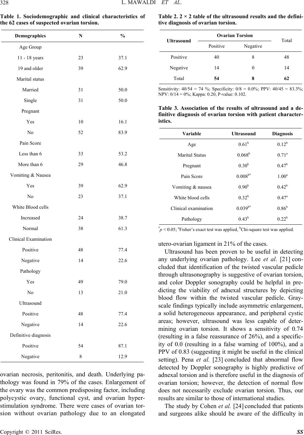 L. MAWALDI ET AL. 328 Table 1. Sociodemographic and clinical characteristics of the 62 cases of suspected ovarian torsion. Demographics N % Age Group 11 - 18 years 23 37.1 19 and older 39 62.9 Marital status Married 31 50.0 Single 31 50.0 Pregnant Yes 10 16.1 No 52 83.9 Pain Score Less than 6 33 53.2 More than 6 29 46.8 Vomiting & Nausea Yes 39 62.9 No 23 37.1 White Blood cells Increased 24 38.7 Normal 38 61.3 Clinical Examination Positive 48 77.4 Negative 14 22.6 Pathology Yes 49 79.0 No 13 21.0 Ultrasound Positive 48 77.4 Negative 14 22.6 Definitive diagnosis Positive 54 87.1 Negative 8 12.9 ovarian necrosis, peritonitis, and death. Underlying pa- thology was found in 79% of the cases. Enlargement of the ovary was the common predisposing factor, including polycystic ovary, functional cyst, and ovarian hyper- stimulation syndrome. There were cases of ovarian tor- sion without ovarian pathology due to an elongated Table 2. 2 × 2 table of the ultrasound results and the defini- tive diagnosis of ovarian torsion. Ovarian Torsion Ultrasound Positive Negative Total Positive 40 8 48 Negative 14 0 14 Total 54 8 62 Sensitivity: 40/54 = 74 %; Specificity: 0/8 = 0.0%; PPV: 40/45 = 83.3%; NPV: 0/14 = 0 %; Kappa: 0.20, P-value: 0.102. Table 3. Association of the results of ultrasound and a de- finitive diagnosis of ovarian torsion with patient character- istics. Variable Ultrasound Diagnosis Age 0.61b 0.12b Marital Status 0.068b 0.71a Pregnant 0.30b 0.47b Pain Score 0.008b* 1.00a Vomiting & nausea 0.90b 0.42b White blood cells 0.32b 0.47a Clinical examinat ion 0.039b* 0.86b Pathology 0.43b 0.22b *p < 0.05; aFisher’s exact test was applied, bChi-square test was applied. utero-ovarian ligament in 21% of the cases. Ultrasound has been proven to be useful in detecting any underlying ovarian pathology. Lee et al. [21] con- cluded that identification of the twisted vascular pedicle through ultrasonogr aphy is suggestive of ovarian tors ion, and color Doppler sonography could be helpful in pre- dicting the viability of adnexal structures by depicting blood flow within the twisted vascular pedicle. Gray- scale findings typically include asymmetric enlargement, a solid heterogeneous appearance, and peripheral cystic areas; however, ultrasound was less capable of deter- mining ovarian torsion. It shows a sensitivity of 0.74 (resulting in a false reassurance of 26%), and a specific- ity of 0.0 (resulting in a false warning of 100%), and a PPV of 0.83 (suggesting it might be useful in the clinical setting). Pena et al. [23] concluded that abnormal flow detected by Doppler sonography is highly predictive of adnexal torsion and is therefore useful in the diagnosis of ovarian torsion; however, the detection of normal flow does not necessarily exclude ovarian torsion. Thus, our results are similar to those of international studies. The study by Cohen et al. [24] concluded that patients and surgeons alike should be aware of the difficulty in Copyright © 2011 SciRes. SS 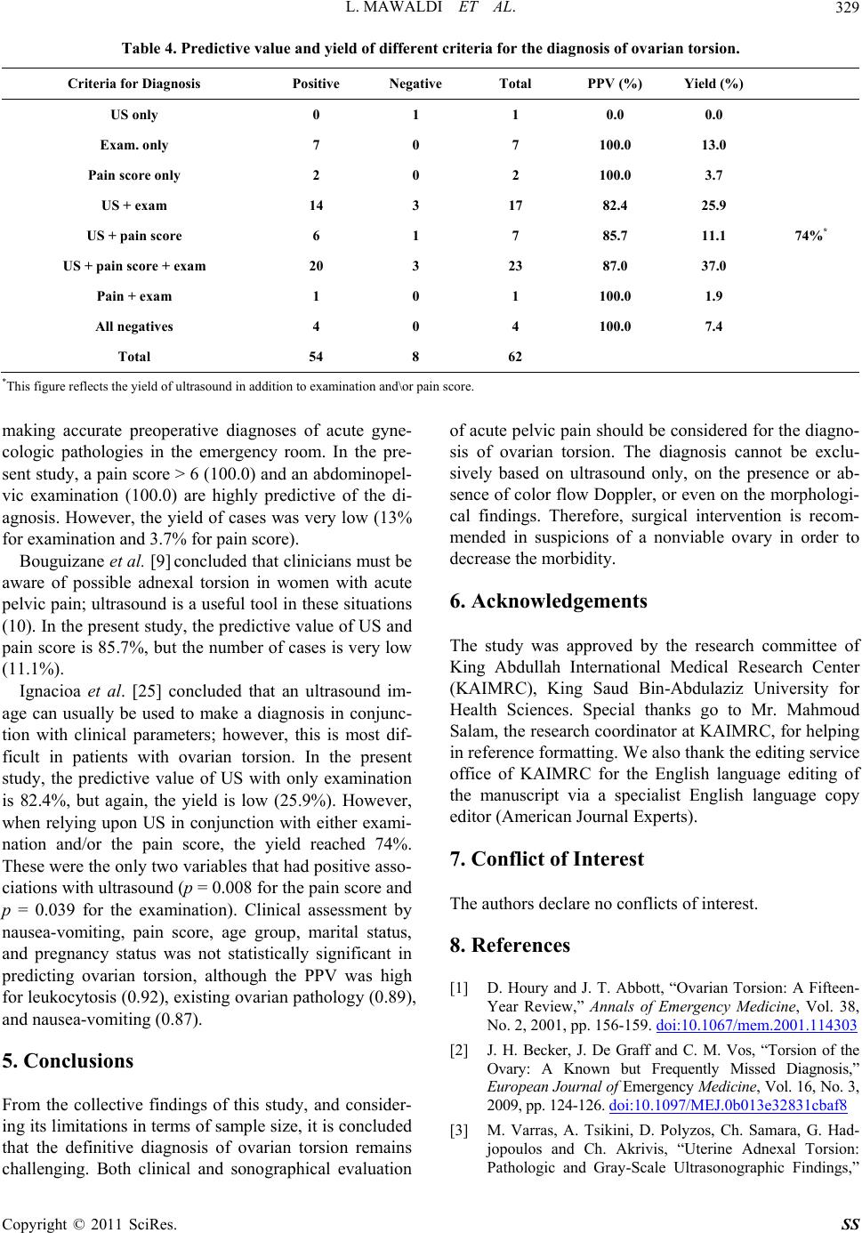 L. MAWALDI ET AL. Copyright © 2011 SciRes. SS 329 Table 4. Predictive value and yield of different criteria for the diagnosis of ovarian torsion. Criteria for Diagnosis Positive Negative Total PPV (%) Yield (%) US only 0 1 1 0.0 0 .0 Exam. only 7 0 7 100.0 13.0 Pain score only 2 0 2 100.0 3.7 US + exam 14 3 17 82.4 25.9 US + pain score 6 1 7 85.7 11.1 US + pain score + exam 20 3 23 87.0 37.0 74%* Pain + exam 1 0 1 100.0 1.9 All negatives 4 0 4 100.0 7.4 Total 54 8 62 *This figure reflects the yield of ultrasound in addition to examination and\or p ain score. making accurate preoperative diagnoses of acute gyne- cologic pathologies in the emergency room. In the pre- sent study, a pain score > 6 (100.0) and an abdominopel- vic examination (100.0) are highly predictive of the di- agnosis. However, the yield of cases was very low (13% for examination and 3.7% for pain score). Bouguizane et al. [9] concluded th at clinicians must be aware of possible adnexal torsion in women with acute pelvic pain; ultrasound is a useful tool in these situations (10). In the present stud y, the predictive value of US and pain score is 85.7%, but the number of cases is very low (11.1%). Ignacioa et al. [25] concluded that an ultrasound im- age can usually be used to make a diagnosis in conjunc- tion with clinical parameters; however, this is most dif- ficult in patients with ovarian torsion. In the present study, the predictive value of US with only examination is 82.4%, but again, the yield is low (25.9%). However, when relying upon US in conjunction with either exami- nation and/or the pain score, the yield reached 74%. These were the only two variables that h ad positive asso- ciations with ultrasound (p = 0.008 for the pain score and p = 0.039 for the examination). Clinical assessment by nausea-vomiting, pain score, age group, marital status, and pregnancy status was not statistically significant in predicting ovarian torsion, although the PPV was high for leukocytosis (0.92), existing ovarian pathology (0.89), and nausea-vomiting (0.87). 5. Conclusions From the collective findings of this study, and consider- ing its limitations in terms of sample size, it is concluded that the definitive diagnosis of ovarian torsion remains challenging. Both clinical and sonographical evaluation of acute pelvic pain should be considered for the diagno- sis of ovarian torsion. The diagnosis cannot be exclu- sively based on ultrasound only, on the presence or ab- sence of colo r flow Doppler, or even on the morp hologi- cal findings. Therefore, surgical intervention is recom- mended in suspicions of a nonviable ovary in order to decrease the morbidity. 6. Acknowledgements The study was approved by the research committee of King Abdullah International Medical Research Center (KAIMRC), King Saud Bin-Abdulaziz University for Health Sciences. Special thanks go to Mr. Mahmoud Salam, the research coordinator at KAIMRC, for helping in reference formatting. We also thank the editing service office of KAIMRC for the English language editing of the manuscript via a specialist English language copy editor (American Journal Experts). 7. Conflict of Interest The authors declare no conflicts of interest. 8. References [1] D. Houry and J. T. Abbott, “Ovarian Torsion: A Fifteen- Year Review,” Annals of Emergency Medicine, Vol. 38, No. 2, 2001, pp. 156-159. doi:10.1067/mem.2001.114303 [2] J. H. Becker, J. De Graff and C. M. Vos, “Torsion of the Ovary: A Known but Frequently Missed Diagnosis,” European Journal of Emergency Medicine , Vol. 16, No. 3, 2009, pp. 124-126. doi:10.1097/MEJ.0b013e32831cbaf8 [3] M. Varras, A. Tsikini, D. Polyzos, Ch. Samara, G. Had- jopoulos and Ch. Akrivis, “Uterine Adnexal Torsion: Pathologic and Gray-Scale Ultrasonographic Findings,”  L. MAWALDI ET AL. 330 Clinical & Experimental Obstetrics & Gynecology, Vol. 31, No. 1, 2004, pp. 34-38. [4] M. Sommerville, D. A. Grimes, P. P. Koonings and K. Campbell, “Ovarian Neoplasms and the Risk of Adnexal Torsion,” American Journal of Obstetrics & Gynecology, Vol. 164, No. 2, 1991, pp. 577-578. [5] J. F. Anders and E. C. Powell, “Urgency of Evaluation and Outcome of Acute Ovarian Torsion in Pediatric Pa- tients,” Archives of Pediatrics & Adolescent Medicine, Vol. 159, No. 6, 2005, pp. 532-535. doi:10.1001/archpedi.159.6.532 [6] R. Mashiach, M. Canis, K. Jardon, G. Mage, J. L. Pouly and A. Wattiez, “Adenexal Torsion after Laparoscopic Hysterectomy: Description of Seven Cases,” The Journal of the American Association of Gynecologic Laparo- scopists, Vol. 11, No. 3, 2004, pp. 336-339. doi:10.1016/S1074-3804(05)60046-1 [7] E. D. Littman, J. Rydfors and A. A. Milki, “Exer- cise-Induced Ovarian Torsion in the Cycle Following Gonadotrophin Therapy: Case Report,” Oxford Journals: Human Reproduction, Vol. 18, No. 8, 2003, pp. 1641-1642. doi:10.1093/humrep/deg338 [8] M. Beaunoyer, J. Chapdelaine, S. Bochard and A. Ouimet, “Asynchronous Bilateral Ovarian Torsion,” Journal of Pediatric Surgery, Vol. 39, No. 5, 2004, pp. 746-749. doi:10.1016/j.jpedsurg.2004.01.037 [9] S. Bouguizane, H. Bibi, Y. Farhat, S. Dhifallah, F. Darraji, S. Hidar, L. Lassoued, A. Chaieb and H. Khairi, “Adnexal Torsion: A Report of 1 35 Cases,” Journal de Gynécologie, Obstétrique et Biologie de l a Reproductio n, Vol. 32, No. 6, 2003, pp. 535-540. [10] C. Born, S. Wirth, A. Stabler and M. Reiser, “Diagnosis of Adnexal Torsion in the Third Trimester of Pregnancy: A Case Report,” Abdominal Imaging Journal, Vol. 29, No. 1, 2004, pp. 123-127. [11] H. Gorkemli, M. Camus and K. Clasen, “Adnexal Tor- sion after Gonadotrophin Ovulation Induction for IVF or ICSI and Its Conservative Treatment,” Archives of Gy- necology and Obstetrics, Vol. 267, No. 1, 2002, pp. 4-6. doi:10.1007/s00404-001-0251-x [12] C. F. Yen, S. L. Lin, W. Murk, C.-J. Wang, C.-L. Lee, Y. K. Soong, M. Phil and A. Arici, “Risk Analysis of torsion an d Malignancy for Adnexal Masses during Pregnancy,” Fertility Sterility Home, Vol. 91, No. 5, 2009, pp. 1895- 1902. doi:10.1016/j.fertnstert.2008.02.014 [13] B. Bromley and B. Benacerraf, “Adnexal Masses during Pregnancy: Accuracy of Sonographic Diagnosis and Outcome,” Journal of Ultrasound in Medicine, Vol. 16, No. 7, 1997, pp. 447-452. [14] K. M. Schmeler, W. W. Mayo-Smith, J. F. Peipert, S. Weitzen, M. D. Manuel and M. E. Gordinier, “Adnexal Masses in Pregnancy: Surgery Compared with Observa- tion,” The American College of Obstetricians and Gyne- cologists, Vol. 105, No. 5, 2005, pp. 1098-1103. [15] T. R. Johanson Jr. and J. D. Woodruff, “Surgical Emer- gencies of the Uterine Adnexae during Pregnancy,” In- ternational Journal of Gynecology & Obstetrics, Vol. 24, No. 5, 1986, pp. 331-335. doi:10.1016/0020-7292(86)90150-5 [16] A. I. Bayer and A. K. Wiskind, “Adnexal Torsion: Can the Adnexa Be Saved?” American Journal of Obstetrics & Gynecology, Vol. 171, No. 6, 1994, pp. 1506-1510. [17] A. B. Pinto, V. S. Ratts, D. B. Williams, S. L. Keller and R. R. Odem, “Reduction of Ovarian Torsion 1 Week after Embryo Transfer in a Patient with Bilateral Hype- rstimu- lated Ovaries,” Fertility Sterility Home, Vol. 76, No. 2, 2001, pp. 403-406. doi:10.1016/S0015-0282(01)01910-0 [18] S. B. Cohen, A. Wattiez, D. Stockheim, D. S. Seidman, A. L. Lidor, S. Mashiach and M. Goldenberg, “The Accu- racy of Serum Interleukin-6 and Tumour Necrosis Factor as Markers for Ovarian Torsion,” Oxford Journals: Hu- man Reproduction, Vol. 16, No. 10, 2001, pp. 2195-2197. doi:10.1093/humrep/16.10.2195 [19] S. Servaes, D. Zurakowski, M. R. Laufer, N. Feins and J. S. Chow, “Sonographic findings of ovarian torsion in children,” Journal of Pediatric Radiology, Vol. 37, No. 5, 2007, pp. 446-451. doi:10.1007/s00247-007-0429-x [20] M. Ben-Ami, Y. Perlitz and S. Haddad, “The Effective- ness of Spectral and Color Doppler in Predicting Ovarian Torsion. A Prospective Study,” European Journal of Ob- stetrics & Gynecology and Reproductive Biology, Vol. 104, No. 1, 2002, pp. 64-66. doi:10.1016/S0301-2115(02)00056-8 [21] E .J. Lee, H. C. Kwon, H. J. Joo, J. H. Suh and A. C. Fleischer, “Diagnosis of Ovarian Torsion with Color Doppler Sonography: Depiction of Twisted Vascular Pedicle,” Journal of Ultrasound in Medicine, Vol. 17, No. 2, 1998, pp. 83-89. [22] L. T. Hibbard, “Adnexal Torsion,” American Journal of Obstetrics & Gynecology, Vol. 152, No. 4, 1985, pp. 456-461. [23] J. E. Pena, D. Ufberg, N. Coony and A. L. Denis, “Use- fulness of Doppler Sonography in the Diagnosis of Ovar- ian Torsion,” Fertility Sterility Home, Vol. 73, No. 5, 2000, pp. 1047-1050. doi:10.1016/S0015-0282(00)00487-8 [24] S. B. Cohen, B. Weisz, D. S. Seidman, S. Mashiach, A. L. Lidor and M. Goldenberg, “Accuracy of the Preoperative Diagnosis in 100 Emergency Laparoscopies Performed Due to Acute Abdomen in Non-pregnant Women,” The Journal of the American A ssociation of Gynecolog ic Lapa- roscopist s, Vol. 8, No. 1, 2001, pp. 92-94. doi:10.1016/S1074-3804(05)60555-5 [25] E. A. Ignacio and M. C. Hill, “Ultrasound of the Acute Female Pelvis,” Ultrasound Quarterly, Vol. 19, No. 2, 2003, pp. 86-98. doi:10.1097/00013644-200306000-00004 Copyright © 2011 SciRes. SS |