

Paper Menu >>
Journal Menu >>
 Surgical Science, 2011, 2, 331-334 doi:10.4236/ss.2011.26070 Published Online August 2011 (http://www.SciRP.org/journal/ss) Copyright © 2011 SciRes. SS Titanium Rib Plate Technique for Huge Chest Wall Reconstruction Yong Han Yoon Department of Th oracic and Cardiovascular Surgery, Inha University College of Medicine, Incheon, Korea E-mail: [email protected] Received February 9, 2011; revised April 6, 2011; accepted August 2, 2011 Abstract Chest wall reconstruction after en-bloc tumor resection is very important to preserve functional mobility and to enhance the cosmetic effect. Because they are flexible and pliable, titanium plates are proposed to recon- struct the chest wall, even though such chest wall reconstruction has been performed in only a few cases worldwide. We present a case of a 49-year-old man with a chondrosarcoma arising from the left 1st rib, in- vading the manubrium, clavicle, 2nd and 3rd ribs, and the anterior segment of the left upper lobe. After wide resection, the chest wall was reconstructed using titanium rib plates and Marlex mesh—the Bovine pericar- dium sandwich type. The patient tolerated the pain well, and fourteen months after surgery, the chest wall was well preserved function mobility and improved pulmonary function test. Keywords: Chest Wall Reconstruction, Titanium Plate, Chondrosarcoma 1. Introduction Generally, chest wall defects larger than 5cm in size, in any location, and those more than 10cm in size, posterior or in close proximity to the scapula, must be recon- structed [1]. LeRoux and Shama [2], proposed that the ideal characteristics of a prosthetic material are rigidity to invalidate paradoxical chest wall motion, inertness to allow in-growth of fibrotic tissue and decrease the like hood to the appropriate shape at the time of operation, and radiolucency to allow radiographic follow-up the underlying problem. Techniques have been developed for thoracic reconstruction surgery that allows resection of chondrosarcomas. Wide excision surgical treatment is the best choice for chondrosarcoma, after which local- ized recurrence is the most common problem. Titanium plates have sufficient inertness to allow the in-growth of fibrous tissue [3]. Because rigid titanium plates have many advantages, we expect to use them widely for chest wall reconstruction. 2. Method Case Report A 48-year-old man was referred for evaluation of chest pain and a palpable chest wall mass. A chest computed tomography (CT) scan showed a large mass involving the left upper lobe, 1st rib, and chest wall, originating from a well-defined 12.6 × 10.1 cm chondrosarcoma in the anterior chest wall (Figures 1(a) and (b)). A local- ized chest wall biopsy showed a spindle cell sarcoma. The chest wall mass was resected. The wide excision was performed and included soft tissue, partial manu- brium, clavicle, the 1st-3rd ribs, and anterior segment of the left upper lobe without any problems. The surgical margin had to be free of tumor by frozen section before the reconstructive procedure was attempted. Skeletal reconstruction surgery was performed, including the manubrium, clavicle, and 1st-4th ribs. We used a latis- simus dorsi muscle flap associated with a split thickness skin graft. The pathology results revealed a dedifferenti- ated chondrosarcoma sarcoma, the 1st rib was a rhado- myosarcoma and cell necrosis extension into the inter- costal soft tissue invaded the lung parenchyma and manubrium that was 16.0 × 11.0 × 11.0 cm in size. The patient was discharged uneventfully on post-operative day 24 and underwent radiotherapy. 3. Surgical Tech nique Five rib plate surgery of the anterior chest wall was per- formed after resection of thechest wall chondrosarcoma, 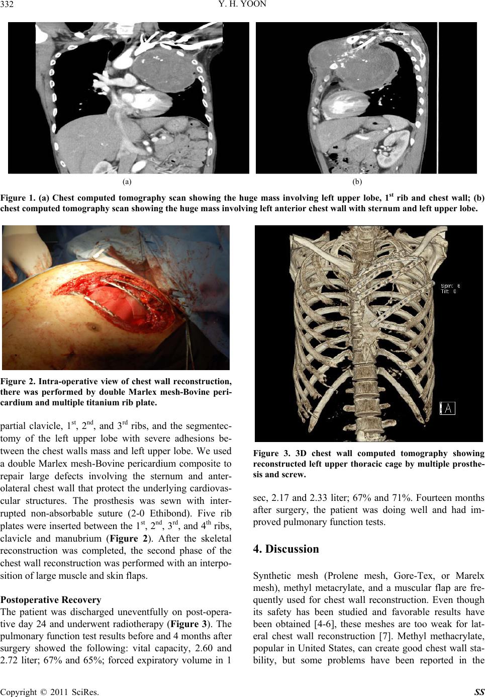 Y. H. YOON 332 (a) (b) Figure 1. (a) Chest computed tomography scan showing the huge mass involving left upper lobe, 1st rib and chest wall; (b) chest computed tomography scan showing the huge mass involving left anterior chest wall with sternum and left upper lobe. Figure 2. Intra-operative view of chest wall reconstruction, there was performed by double Marlex mesh-Bovine peri- cardium and multiple titanium rib plate. partial clavicle, 1st, 2nd, and 3rd ribs, and the segmentec- tomy of the left upper lobe with severe adhesions be- tween the chest walls mass and left upper lobe. We used a double Marlex mesh-Bovine pericardium composite to repair large defects involving the sternum and anter- olateral chest wall that protect the underlying cardiovas- cular structures. The prosthesis was sewn with inter- rupted non-absorbable suture (2-0 Ethibond). Five rib plates were inserted between the 1st, 2nd, 3rd, and 4th ribs, clavicle and manubrium (Figure 2). After the skeletal reconstruction was completed, the second phase of the chest wall reconstruction was performed with an interpo- sition of large muscle and skin flaps. Postoperative Recovery The patient was discharged uneventfully on post-opera- tive day 24 and underwent radiotherapy (Figure 3). The pulmonary function test results before and 4 months after surgery showed the following: vital capacity, 2.60 and 2.72 liter; 67% and 65%; forced expiratory volume in 1 Figure 3. 3D chest wall computed tomography showing reconstructed left upper thoracic cage by multiple prosthe- sis and screw. sec, 2.17 and 2.33 liter; 67% and 71%. Fourteen months after surgery, the patient was doing well and had im- proved pulmonary function tests. 4. Discussion Synthetic mesh (Prolene mesh, Gore-Tex, or Marelx mesh), methyl metacrylate, and a muscular flap are fre- quently used for chest wall reconstruction. Even though its safety has been studied and favorable results have been obtained [4-6], these meshes are too weak for lat- eral chest wall reconstruction [7]. Methyl methacrylate, popular in United States, can create good chest wall sta- bility, but some problems have been reported in the Copyright © 2011 SciRes. SS 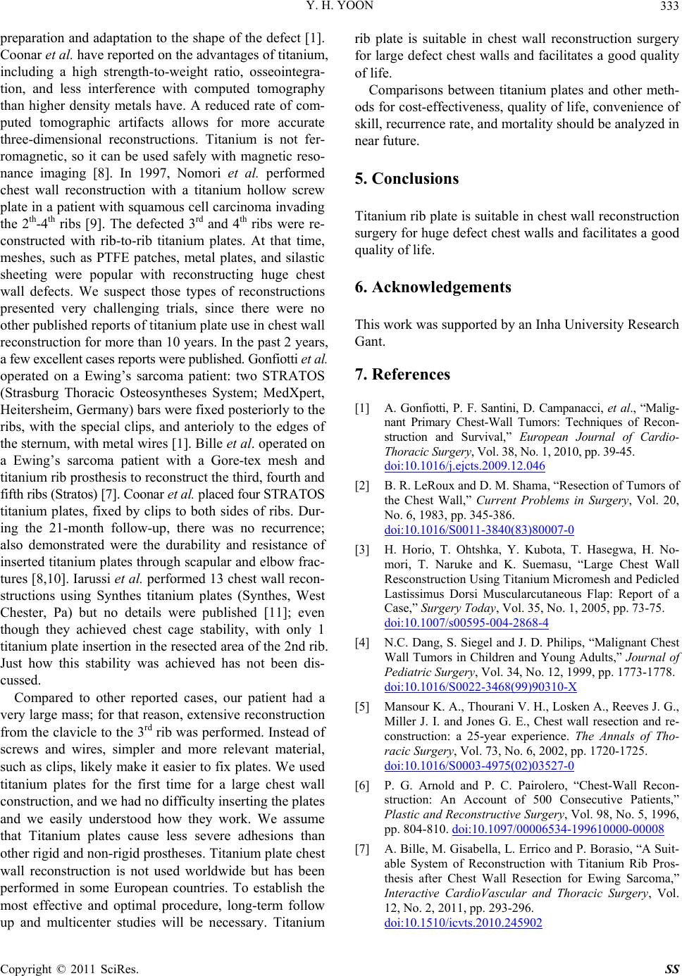 Y. H. YOON333 preparation and adaptation to the shape of the defect [1]. Coonar et al. have reported on the advan tages of titanium, including a high strength-to-weight ratio, osseointegra- tion, and less interference with computed tomography than higher density metals have. A reduced rate of com- puted tomographic artifacts allows for more accurate three-dimensional reconstructions. Titanium is not fer- romagnetic, so it can be used safely with magnetic reso- nance imaging [8]. In 1997, Nomori et al. performed chest wall reconstruction with a titanium hollow screw plate in a patient with squamous cell carcinoma invading the 2th-4th ribs [9]. The defected 3rd and 4th ribs were re- constructed with rib-to-rib titanium plates. At that time, meshes, such as PTFE patches, metal plates, and silastic sheeting were popular with reconstructing huge chest wall defects. We suspect those types of reconstructions presented very challenging trials, since there were no other published reports of titanium plate use in chest wall reconstruction for more than 10 years. In the past 2 years, a few excellent cases reports were published. Gonfiotti et al. operated on a Ewing’s sarcoma patient: two STRATOS (Strasburg Thoracic Osteosyntheses System; MedXpert, Heitersheim, Germany) bars were fixed posteriorly to the ribs, with the special clips, and anterioly to the edges of the sternum, with metal wires [1]. Bille et al. operated on a Ewing’s sarcoma patient with a Gore-tex mesh and titanium rib prosthesis to reconstruc t the third, fourth and fifth ribs (Stratos) [7]. Coonar et al. placed four STRATOS titanium plates, fixed by clips to both sides of ribs. Dur- ing the 21-month follow-up, there was no recurrence; also demonstrated were the durability and resistance of inserted titanium plates through scapular and elbow frac- tures [8,10]. Iarussi et al. performed 13 chest wall recon- structions using Synthes titanium plates (Synthes, West Chester, Pa) but no details were published [11]; even though they achieved chest cage stability, with only 1 titanium plate insertion in the resected area of the 2nd rib. Just how this stability was achieved has not been dis- cussed. Compared to other reported cases, our patient had a very large mass; for that reason, extensive reconstruction from the clavicle to the 3rd rib was performed. Instead of screws and wires, simpler and more relevant material, such as clips, likely make it easier to fix plates. We used titanium plates for the first time for a large chest wall construction, and we had no difficulty inserting the plates and we easily understood how they work. We assume that Titanium plates cause less severe adhesions than other rigid and non-rigid prostheses. Titanium plate chest wall reconstruction is not used worldwide but has been performed in some European countries. To establish the most effective and optimal procedure, long-term follow up and multicenter studies will be necessary. Titanium rib plate is suitable in chest wall reconstruction surgery for large defect chest walls and facilitates a good quality of life. Comparisons between titanium plates and other meth- ods for cost-effectiven ess, quality of life, convenience of skill, recurrence rate, and mortality sh ould be analyzed in near future. 5. Conclusions Titanium rib plate is suitable in chest wall reconstru ction surgery for huge defect chest walls and facilitates a good quality of life. 6. Acknowledgements This work was supported b y an Inha Univ ersity Research Gant. 7. References [1] A. Gonfiotti, P. F. Santini, D. Campanacci, et al., “Malig- nant Primary Chest-Wall Tumors: Techniques of Recon- struction and Survival,” European Journal of Cardio- Thoracic Su rgery , Vol. 38, No. 1, 2010, pp. 39-45. doi:10.1016/j.ejcts.2009.12.046 [2] B. R. LeRoux and D. M. Shama, “Resection of Tumors of the Chest Wall,” Current Problems in Surgery, Vol. 20, No. 6, 1983, pp. 345-386. doi:10.1016/S0011-3840(83)80007-0 [3] H. Horio, T. Ohtshka, Y. Kubota, T. Hasegwa, H. No- mori, T. Naruke and K. Suemasu, “Large Chest Wall Resconstruction Using Titanium Micromesh and Pedicled Lastissimus Dorsi Muscularcutaneous Flap: Report of a Case,” Surgery Today, Vol. 35, No. 1, 2005, pp. 73-75. doi:10.1007/s00595-004-2868-4 [4] N.C. Dang, S. Siegel and J. D. Philips, “Malignant Chest Wall Tumors in Children and Young Adults,” Journal of Pediatric Surgery, Vol. 34, No. 12, 1999, pp. 1773-1778. doi:10.1016/S0022-3468(99)90310-X [5] Mansour K. A., Thourani V. H., Losken A., Reeves J. G., Miller J. I. and Jones G. E., Chest wall resection and re- construction: a 25-year experience. The Annals of Tho- racic Surgery, Vol. 73, No. 6, 2002, pp. 1720-1725. doi:10.1016/S0003-4975(02)03527-0 [6] P. G. Arnold and P. C. Pairolero, “Chest-Wall Recon- struction: An Account of 500 Consecutive Patients,” Plastic and Reconstructive Surgery, Vol. 98, No. 5, 1996, pp. 804-810. doi:10.1097/00006534-199610000-00008 [7] A. Bille, M. Gisabella, L. Errico and P. Borasio, “A Suit- able System of Reconstruction with Titanium Rib Pros- thesis after Chest Wall Resection for Ewing Sarcoma,” Interactive CardioVascular and Thoracic Surgery, Vol. 12, No. 2, 2011, pp. 293-296. doi:10.1510/icvts.2010.245902 Copyright © 2011 SciRes. SS  Y. H. YOON Copyright © 2011 SciRes. SS 334 [8] A. S. Coonar, N. Qureshi, I. Smith, F. C. Wells and E. Reisberg and J.-M. Wihl m, “A Novel Titanium Ri b Bridge System for Chest Wall Reconstruction,” The Annals of Thoracic Su rgery , Vol. 87, No. 5, 2009, pp. e46-e48. doi:10.1016/j.athoracsur.2009.01.069 [9] H. Nomori, H. Horio and T. Hasegawa, “Chest Wall Re- construction Using Titanium Hollow Screw Reconstruc- tion Plate,” The Journal of Thoracic and Cardiovascular Surgery, Vol. 45, No. 1, 1997, pp. 35-37. doi:10.1055/s-2007-1013681 [10] A. S. Coonar, J. M. Wihlm, F. C. Wells and N. Qureshi, “Intermediate Outcome and Dynamic Computerized To- mography after Chest Wall Reconstruction with the STRTOS Titanium Rib Bridge System: Videodemonstra- tion of Preserved Bucket-Handle Rib Motion,” Interac- tive CardioVascular and Thoracic Surgery, Vol. 12, 2011, pp. 80-81. doi:10.1510/icvts.2010.249615 [11] T. Iarussi, A. Pardolesi, P. Camplese and R. Sacco, “Composite Chest Wall Reconstruction Using Titanium Plates and Mesh Preserves Chest Wall Function,” The Journal of Thoracic and Cardiovascular Surgery, Vol. 140, No. 2, 2010, pp. 476-7. doi:10.1016/j.jtcvs.2009.07.030 |