

Paper Menu >>
Journal Menu >>
 Surgical Science, 2011, 2, 297-302 doi:10.4236/ss.2011.26064 Published Online August 2011 (http://www.SciRP.org/journal/ss) Copyright © 2011 SciRes. SS Delayed Manifestation of Sacral Clear Cell Meningioma Distent Metastasis or Multifocal Disease? Habib Nouri1, Leila Abid2, Moez Ouertatani1, Dalenda Hentati3, Mondher Mestiri2, Habib Jaafoura2 1Department of “Adult” Surgery, Kassab’s Institute, Tunisia 2Department of “Pathology”, Kassab’s Institute, Tunisia 3Department of Radiat i o n Therapy, National Institute of Oncology, Tunisia E-mail: [email protected] Received December 6, 2010; revised December 26, 2010; accepted January 13, 2011 Abstract Study Design: the authors report on a clear cell meningioma (CCM) of the sacrum revealed 17 years after a removal of a spinal lesion. A review of 29 spinal CCM previously reported was done. Objective: To focus on clinical and biological behaviour of CCM. Summary of Background Data: CCM is a rare subtype charac- terrized by its inordinately aggressive clinical course despite its benign appearance. The tumour shows pro- clivity for the cerebellopontine angle and cauda equine region. Recurrence and metastasis have been com- monly reported. Method: a 26 year-old man presented with low back pain related to a lytic lesion of the sa- crum. He had a history of an intradural extramedullary meningioma of the cervical spine removed when he was 9 year-old. CT scan revealed a sacral osteolytic lesion with anterior and lateral extension. Open biopsy revealed a clear cell meningioma. That was the same pattern of the tumour removed 17 years before. Results: Our patient was managed conservatively by palliative radiation therapy. At 2 years follow-up, he experi- enced improvement of pain and walking. Conclusion: CCM is an aggressive tumour with a high risk of me- tastases through cerebrospinal fluid. A follow up with spinal MRI should be performed. Keywords: Clear Cell Meningioma, Spine, Cerebrospinal Fluid, Metastasis, Local Recurrence, Radiation Therapy, Pathology 1. Introduction Meningiomas are the most common primary tumours of the central nervous system. Approximately 90% to 95% of them are classified as grade I according to the World Health Organization (WHO) 2007 criteria [1], whereas 5 to 7% are graded II and 1 to 2.8% are graded III. Clear cell meningiomas (CCM) are rare and have been re- ported in only 0.2% of cases [2]. They are graded II or aggressive. The tumour shows proclivity for the cere- bellopontine angle and cauda equine region. Despite its benign appearance, CCM has an aggressive behaviour and recurrence and metastasis has been commonly re- ported. Here we report an unusual case of a metastatic clear cell meningioma to the sacrum, discovered 17 years after total resection of the primary cervical lesion. The clinical and pathological features are discussed. 2. Case Report A 26 year-old male was referred to our department in 2007 for a tumour of the sacrum. He underwent in 1990 when he was 9 year-old a resection of an intraspinal tu- mour. Cervical CT scan had revealed an intraduralex- tramedullary tumor measuring 2 cm at the level of C4-C5. The tumor was well-circumscribed, homogeneous, lo- cated posteriorly, and compressing the spinal cord. The tumor was excised totally. Pathological examination had revealed a particular meningioma characterized by the presence of mycrocystic cells and prominent interstitial collagen which separate or enclose the cellular aggre- gates. A Masson’s trichrome stain highlighted the abun- dant collagen component. The neoplastic cells were ar- ranged in sheets and forming a rare whorls. The stromal blood vessels have a thick hyalinized wall. The patient got no further therapy and follow-up was uneventful for 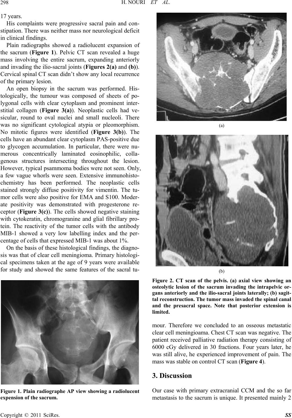 H. NOURI ET AL. 298 17 years. His complaints were progressive sacral pain and con- stipation. There was neither mass nor neurological deficit in clinical findings. Plain radiographs showed a radiolucent expansion of the sacrum (Figure 1). Pelvic CT scan revealed a huge mass involving the entire sacrum, expanding anteriorly and invading the ilio-sacral joints (Figures 2(a) and (b)). Cervical spinal CT scan didn’t show any local recurrence of the primary lesion. An open biopsy in the sacrum was performed. His- tologically, the tumour was composed of sheets of po- lygonal cells with clear cytoplasm and prominent inter- stitial collagen (Figure 3(a)). Neoplastic cells had ve- sicular, round to oval nuclei and small nucleoli. There was no significant cytological atypia or pleomorphism. No mitotic figures were identified (Figure 3(b)). The cells have an abundant clear cytoplasm PAS-positive due to glycogen accumulation. In particular, there were nu- merous concentrically laminated eosinophilic, colla- genous structures intersecting throughout the lesion. However, typical psammoma bodies were not seen. Only, a few vague whorls were seen. Extensive immunohisto- chemistry has been performed. The neoplastic cells stained strongly diffuse positivity for vimentin. The tu- mor cells were also positive for EMA and S100. Moder- ate positivity was demonstrated with progesterone re- ceptor (Figure 3(c)). The cells showed negative staining with cytokeratin, chromogranine and glial fibrillary pro- tein. The reactivity of the tumor cells with the antibody MIB-1 showed a very low labelling index and the per- centage of cells that expressed MIB-1 was about 1%. On the basis of these histological findings, the diagno- sis was that of clear cell meningioma. Primary histologi- cal specimens taken at the age of 9 years were available for study and showed the same features of the sacral tu- Figure 1. P lain radiographe AP view showing a radiolucent expension of the sacrum. (a) (b) Figure 2. CT scan of the pelvis. (a) axial view showing an osteolytic lesion of the sacrum invading the intrapelvic or- gans anteriorly and the ilio-sacral joints laterally; (b) sagit- tal reconstruction. The tumor mass invaded the spinal canal and the presacral space. Note that posterior extension is limited. mour. Therefore we concluded to an osseous metastatic clear cell meningioama. Chest CT scan was negative. The patient received palliative radiation therapy consisting of 6000 cGy delivered in 30 fractions. Four years later, he was still alive, he experienced improvement of pain. The mass was stable on control CT scan (Figure 4). 3. Discussion Our case with primary extracranial CCM and the so far metastasis to the sacrum is unique. It presented mainly 2 Copyright © 2011 SciRes. SS 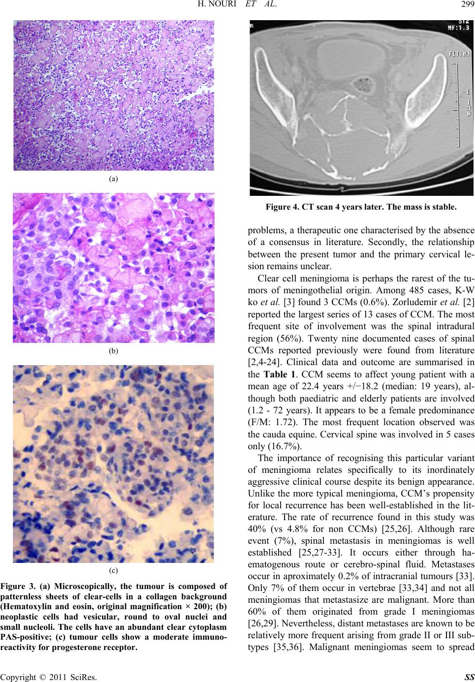 H. NOURI ET AL.299 (a) (b) (c) Figure 3. (a) Microscopically, the tumour is composed of patternless sheets of clear-cells in a collagen background (Hematoxylin and eosin, original magnification × 200); (b) neoplastic cells had vesicular, round to oval nuclei and small nucleoli. The cells have an abundant clear cytoplasm PAS-positive; (c) tumour cells show a moderate immuno- reactivity for progesterone receptor. Figure 4. CT scan 4 years later. The mass is stable. problems, a therapeutic one characterised by the absence of a consensus in literature. Secondly, the relationship between the present tumor and the primary cervical le- sion remains unclear. Clear cell meningioma is perhaps the rarest of the tu- mors of meningothelial origin. Among 485 cases, K-W ko et al. [3] found 3 CCMs (0.6%). Zorludemir et al. [2] reported the largest series of 13 cases of CCM. The most frequent site of involvement was the spinal intradural region (56%). Twenty nine documented cases of spinal CCMs reported previously were found from literature [2,4-24]. Clinical data and outcome are summarised in the Table 1. CCM seems to affect young patient with a mean age of 22.4 years +/−18.2 (median: 19 years), al- though both paediatric and elderly patients are involved (1.2 - 72 years). It appears to be a female predominance (F/M: 1.72). The most frequent location observed was the cauda equine. Cervical spine was involved in 5 cases only (16.7%). The importance of recognising this particular variant of meningioma relates specifically to its inordinately aggressive clinical course despite its benign appearance. Unlike the more typical meningioma, CCM’s propensity for local recurrence has been well-established in the lit- erature. The rate of recurrence found in this study was 40% (vs 4.8% for non CCMs) [25,26]. Although rare event (7%), spinal metastasis in meningiomas is well established [25,27-33]. It occurs either through ha- ematogenous route or cerebro-spinal fluid. Metastases occur in aproximately 0.2% of intracranial tumours [33]. Only 7% of them occur in vertebrae [33,34] and not all meningiomas that metastasize are malignant. More than 60% of them originated from grade I meningiomas [26,29]. Nevertheless, distant metastases are known to be relatively more frequent arising from grade II or III sub- types [35,36]. Malignant meningiomas seem to spread Copyright © 2011 SciRes. SS 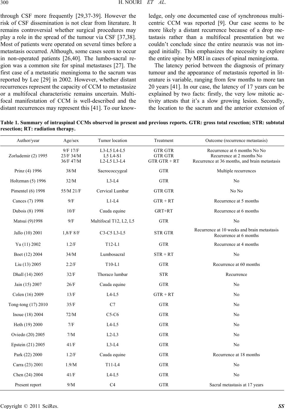 H. NOURI ET AL. Copyright © 2011 SciRes. SS 300 through CSF more frequently [29,37-39]. However the risk of CSF dissemination is not clear from literature. It remains controversial whether surgical procedures may play a role in the spread of the tumour via CSF [37,38]. Most of patients were operated on several times before a metastasis occurred. Although, some cases seem to occur in non-operated patients [26,40]. The lumbo-sacral re- gion was a common site for spinal metastases [27]. The first case of a metastatic meningioma to the sacrum was reported by Lee [29] in 2002. However, whether distant recurrences represent the capacity of CCM to metastasize or a multifocal characteristic remains uncertain. Multi- focal manifestation of CCM is well-described and the distant recurrences may represent this [41]. To our know- ledge, only one documented case of synchronous multi- centric CCM was reported [9]. Our case seems to be more likely a distant recurrence because of a drop me- tastasis rather than a multifocal presentation but we couldn’t conclude since the entire neuraxis was not im- aged initially. This emphasizes the necessity to explore the entire spine by MRI in cases of spinal meningioma. The latency period between the diagnosis of primary tumour and the appearance of metastasis reported in lit- erature is variable, ranging from few months to more tan 20 years [41]. In our case, the latency of 17 years can be explained by two facts: firstly, the very low mitotic ac- tivity attests that it’s a slow growing lesion. Secondly, the location to the sacrum and the anterior extension of Table 1. Summary of intraspinal CCMs observed in present and previous reports. GTR: gross total resection; STR: subtotal resection; RT: radiation therapy. Author/year Age/sex Tumor location Treatment Outcome (recurrence metastasis) Zorludemir (2) 1995 9/F 17/F 23/F 34/M 36/F 47/M L3-L5 L4-L5 L5 L4-S1 L2-L5 L3-L4 GTR GTR GTR GTR GTR GTR + RT Recurrence at 6 months No No Recurrence at 2 months No Recurrence at 36 months, and brain metastasis Prinz (4) 1996 38/M Sacrococcygeal GTR Multiple recurrences Holtzman (5) 1996 32/M L3-L4 GTR No Pimentel (6) 1998 55/M 21/F Cervical Lumbar GTR GTR No No Cances (7) 1998 9/F L1-L4 GTR + RT Recurrence at 5 months Dubois (8) 1998 10/F Cauda equine GRT+RT Recurrence at 6 months Matsui (9)1998 9/F Multifocal T12, L2, L5GTR No Jallo (10) 2001 1,8/F 8/F C3-C5 L3-L5 STR GTR Recurrence at 10 weeks and brain metastasis Recurrence at 6 months Yu (11) 2002 1.2/F T12-L1 GTR Recurrence at 4 months Boet (12) 2004 34/M Lumbosacral STR + RT No Liu (13) 2005 2.2/F T10-L1 GTR Recurrence at 60 months Dhall (14) 2005 32/F Thoraco lumbar STR Recurrence Jain (15) 2007 26/F Cauda equine GTR No Colen (16) 2009 13/F L4-L5 GTR + RT No Tong-tong (17) 2010 35/F C7 GTR No Inoue (18) 2004 72/M C5-C6 GTR No Heth (19) 2000 7/F L4-L5 GTR No Oviedo (20) 2005 7/M L2-L3 GTR No Epstein (21) 2005 41/F L3-L4 GTR No Park (22) 2000 1.2/F Cauda equine GTR Recurrence at 18 months Carra (23) 2001 1.9/M T11-L4 GTR No Chen (24) 2004 41/F L4-L5 GTR No Present report 9/M C4 GTR Sacral metastasis at 17 years 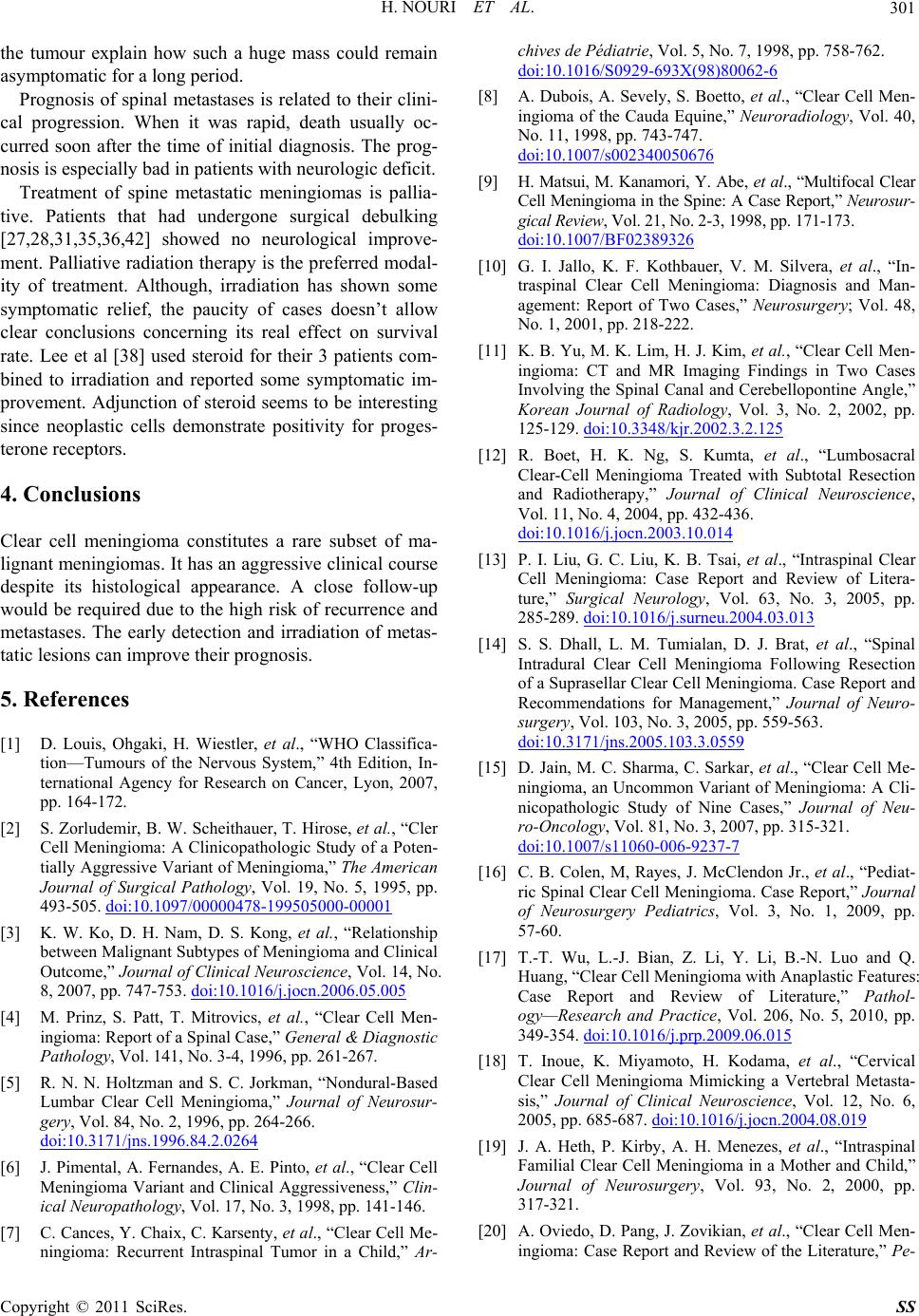 H. NOURI ET AL. Copyright © 2011 SciRes. SS 301 the tumour explain how such a huge mass could remain asymptomatic for a long period. Prognosis of spinal metastases is related to their clini- cal progression. When it was rapid, death usually oc- curred soon after the time of initial diagnosis. The prog- nosis is especially bad in patients with neurologic deficit. Treatment of spine metastatic meningiomas is pallia- tive. Patients that had undergone surgical debulking [27,28,31,35,36,42] showed no neurological improve- ment. Palliative radiation therapy is the preferred modal- ity of treatment. Although, irradiation has shown some symptomatic relief, the paucity of cases doesn’t allow clear conclusions concerning its real effect on survival rate. Lee et al [38] used steroid for their 3 patients com- bined to irradiation and reported some symptomatic im- provement. Adjunction of steroid seems to be interesting since neoplastic cells demonstrate positivity for proges- terone receptors. 4. Conclusions Clear cell meningioma constitutes a rare subset of ma- lignant meningiomas. It has an aggressive clinical course despite its histological appearance. A close follow-up would be required due to the high risk of recurrence and metastases. The early detection and irradiation of metas- tatic lesions can improve their prognosis. 5. References [1] D. Louis, Ohgaki, H. Wiestler, et al., “WHO Classifica- tion—Tumours of the Nervous System,” 4th Edition, In- ternational Agency for Research on Cancer, Lyon, 2007, pp. 164-172. [2] S. Zorludemir, B. W. Scheithauer, T. Hirose, et al., “Cler Cell Meningioma: A Clinicopathologic Study of a Poten- tially Aggressive Variant of Meningioma,” The American Journal of Surgical Pathology, Vol. 19, No. 5, 1995, pp. 493-505. doi:10.1097/00000478-199505000-00001 [3] K. W. Ko, D. H. Nam, D. S. Kong, et al., “Relationship between Malignant Subtypes of Meningioma and Clinical Outcome,” Journal of Clinical Neuroscience, Vol. 14, No. 8, 2007, pp. 747-753. doi:10.1016/j.jocn.2006.05.005 [4] M. Prinz, S. Patt, T. Mitrovics, et al., “Clear Cell Men- ingioma: Report of a Spinal Case,” General & Diagnostic Pathology, Vol. 141, No. 3-4, 1996, pp. 261-267. [5] R. N. N. Holtzman and S. C. Jorkman, “Nondural-Based Lumbar Clear Cell Meningioma,” Journal of Neurosur- gery, Vol. 84, No. 2, 1996, pp. 264-266. doi:10.3171/jns.1996.84.2.0264 [6] J. Pimental, A. Fernandes, A. E. Pinto, et al., “Clear Cell Meningioma Variant and Clinical Aggressiveness,” Clin- ical Neuropathology, Vol. 17, No. 3, 1998, pp. 141-146. [7] C. Cances, Y. Chaix, C. Karsenty, et al., “Clear Cell Me- ningioma: Recurrent Intraspinal Tumor in a Child,” Ar- chives de Pédiatrie, Vol. 5, No. 7, 1998, pp. 758-762. doi:10.1016/S0929-693X(98)80062-6 [8] A. Dubois, A. Sevely, S. Boetto, et al., “Clear Cell Men- ingioma of the Cauda Equine,” Neuroradiology, Vol. 40, No. 11, 1998, pp. 743-747. doi:10.1007/s002340050676 [9] H. Matsui, M. Kanamori, Y. Abe, et al., “Multifocal Clear Cell Meningioma in the Spine: A Case Report,” Neurosur- gical Review, Vol. 21, No. 2-3, 1998, pp. 171-173. doi:10.1007/BF02389326 [10] G. I. Jallo, K. F. Kothbauer, V. M. Silvera, et al., “In- traspinal Clear Cell Meningioma: Diagnosis and Man- agement: Report of Two Cases,” Neurosurgery; Vol. 48, No. 1, 2001, pp. 218-222. [11] K. B. Yu, M. K. Lim, H. J. Kim, et al., “Clear Cell Men- ingioma: CT and MR Imaging Findings in Two Cases Involving the Spinal Canal and Cerebellopontine Angle,” Korean Journal of Radiology, Vol. 3, No. 2, 2002, pp. 125-129. doi:10.3348/kjr.2002.3.2.125 [12] R. Boet, H. K. Ng, S. Kumta, et al., “Lumbosacral Clear-Cell Meningioma Treated with Subtotal Resection and Radiotherapy,” Journal of Clinical Neuroscience, Vol. 11, No. 4, 2004, pp. 432-436. doi:10.1016/j.jocn.2003.10.014 [13] P. I. Liu, G. C. Liu, K. B. Tsai, et al., “Intraspinal Clear Cell Meningioma: Case Report and Review of Litera- ture,” Surgical Neurology, Vol. 63, No. 3, 2005, pp. 285-289. doi:10.1016/j.surneu.2004.03.013 [14] S. S. Dhall, L. M. Tumialan, D. J. Brat, et al., “Spinal Intradural Clear Cell Meningioma Following Resection of a Suprasellar Clear Cell Meningioma. Case Report and Recommendations for Management,” Journal of Neuro- surgery, Vol. 103, No. 3, 2005, pp. 559-563. doi:10.3171/jns.2005.103.3.0559 [15] D. Jain, M. C. Sharma, C. Sarkar, et al., “Clear Cell Me- ningioma, an Uncommon Variant of Meningioma: A Cli- nicopathologic Study of Nine Cases,” Journal of Neu- ro-Oncology, Vol. 81, No. 3, 2007, pp. 315-321. doi:10.1007/s11060-006-9237-7 [16] C. B. Colen, M, Rayes, J. McClendon Jr., et al., “Pediat- ric Spinal Clear Cell Meningioma. Case Report,” Journal of Neurosurgery Pediatrics, Vol. 3, No. 1, 2009, pp. 57-60. [17] T.-T. Wu, L.-J. Bian, Z. Li, Y. Li, B.-N. Luo and Q. Huang, “Clear Cell Meningioma with Anaplastic Features: Case Report and Review of Literature,” Pathol- ogy—Research and Practice, Vol. 206, No. 5, 2010, pp. 349-354. doi:10.1016/j.prp.2009.06.015 [18] T. Inoue, K. Miyamoto, H. Kodama, et al., “Cervical Clear Cell Meningioma Mimicking a Vertebral Metasta- sis,” Journal of Clinical Neuroscience, Vol. 12, No. 6, 2005, pp. 685-687. doi:10.1016/j.jocn.2004.08.019 [19] J. A. Heth, P. Kirby, A. H. Menezes, et al., “Intraspinal Familial Clear Cell Meningioma in a Mother and Child,” Journal of Neurosurgery, Vol. 93, No. 2, 2000, pp. 317-321. [20] A. Oviedo, D. Pang, J. Zovikian, et al., “Clear Cell Men- ingioma: Case Report and Review of the Literature,” Pe-  H. NOURI ET AL. 302 diatric and Developmental Pathology, Vol. 8, No. 3, 2005, pp. 386-390. doi:10.1007/s10024-005-0119-3 [21] N. Epstein, S. Drexler and J. Shneider, “Clear cell Men- ingioma of the Cauda Equine in an Adult. Case Report and Literature Review,” Journal of Spinal Disorders & Tech- niques, Vol. 18, No. 6, 2005, pp. 539-543. doi:10.1097/01.bsd.0000173314.98401.b5 [22] H. C. Park, M. J. Sohn, E. Y. Kim, et al., “Spinal Clear Cell Meningiomas Presented with Progressive Parapresis in Infancy,” Child’s Nervous System, Vol. 16, No. 9, 2000, pp. 607-610. doi:10.1007/PL00007302 [23] S. Carra, P. Drigo, M. Gadriman, et al., “Clear Cell Me- ningioma in a 22 Months-Old Male; a Case Report and Literature Review,” Pediatric Neurosurgery, Vol. 34, No. 5, 2001, pp. 264-267. [24] M. H. Chen, S. J. Chen, S. M. Lin, et al., “A Lumbar Cleare Cell Meningioma with Foraminal Extension in a Renal Transplant Recipient,” Journal of Clinical Neuro- science, Vol. 11, No. 6, 2004, pp. 665-667. doi:10.1016/j.jocn.2003.10.024 [25] S. K. Ludwin and F. K. Conley, “Malignant Meningioma Metastatizing through the Cerebrospinal Pathways,” Journal of Neurology, Neurosurgery & Psychiatry, Vol. 38, No. 2, 1975, pp. 136-142. doi:10.1136/jnnp.38.2.136 [26] P. M. Son, M. Sacher, S. W. Strenger, et al., “Benign Metastasizing Meningiomas,” American Journal of Neu- roradiology, Vol. 8, No. 1, 1987, pp. 127-130. [27] A. T. kimura, T. Orita, O. Hayashida, et al., “Malignant Meningiomas Metastatizing through the Cerebrospinal Pathway,” Acta Neurologica Scandinavica, Vol. 85, No. 5, 1992, pp. 368-371. doi:10.1111/j.1600-0404.1992.tb04063.x [28] K. Kamiya, T. Inagawa and R. Nagasako, “Malignant Intraventricular Meningioma with Spinal Metastasis through the Cerebrospinal Fluid,” Surgical Neurology, Vol. 32, No. 3, 1989, pp. 213-218. doi:10.1016/0090-3019(89)90181-X [29] Y. Y. Lee, W. W. Hsu, T. J. Huang, et al., “Metastatic Meningioma in the Sacrum: A Case Report,” Spine, Vol. 27, No. 4, 2002, pp. E100-E103. doi:10.1097/00007632-200202150-00022 [30] W. C. Peh and Y. W. Fan, “Case Report: Intraventricular Meningioma with Cerebelopontine Angle and Drop Me- tastases,” The British Journal of Radiology, Vol. 68, No. 808, 1995, pp. 428-430. doi:10.1259/0007-1285-68-808-428 [31] C. G. Riley, “Metastatizing Meningeal Tumour Case Report,” The New Zealand Medical Journal, Vol. 73, 1974, pp. 210-214. [32] F. J. Wippold II, J. G. Smimoitopoulos and T. K. Pilgram, “Lesions of the Cauda Equine: A Clinical Pathology Re- view from the Armed Forces Institute of Pathology,” Clinical Neurology and Neurosurgery, Vol. 9, No. 4, 1997, pp. 229-234. doi:10.1016/S0303-8467(97)00095-4 [33] J. L Karasik and S. Mullens, “A Survey of Metastatic Meningiomas,” Journal of Neurosurgery, Vol. 40, No. 2, 1974, pp. 206-212. doi:10.3171/jns.1974.40.2.0206 [34] R. Wasserkurg, E. Peyser and C. Lichtig, “Extracranial Bone Metastases from Intracranial Meningiomas,” Surgical Neurology, Vol. 12, No. 6, 1979, pp. 480-484. [35] M. D. Conrad, C. Schonauer, I. Pelissou-Guyotat, et al., “Recurrent Lumbosacral Metastases from Intracranial Meningioma. Report of a Case and Review of the Litera- ture,” Acta Neurochirurgica, Vol. 143, No. 9, 2001, pp. 935-937. doi:10.1007/s007010170024 [36] S. A. Enam, S. Abdulrauf, B. Mehta, et al., “Metastasis in Meningioma,” Acta Neurochirurgica, Vol. 138, No. 10, 1996, pp. 1172-1178. doi:10.1007/BF01809747 [37] M. C. Chamberlain and M. J. Glantz, “Cerebrospinal Fluid- Dessiminated Meningioma,” Cancer, Vol. 103, No. 7, 2005, pp. 1427-1430. doi:10.1002/cncr.20926 [38] T. T. Lee and H. J. Landy, “Spinal Metastases of Malig- nant Intracranial Meningioma,” Surgical Neurology, Vol. 50, No. 5, 1998, pp. 437-441. doi:10.1016/S0090-3019(97)00348-0 [39] P. D. Delgado-Lopez, V. Martin-Velasco, J. M. Castilla- Diez, et al., “Metastatic Meningioma to the Eleventh Dorsal Vertebal Body: Total En Bloc Spondylectomy. Case Report and Review of the Literature,” Neur- ocirur- gia, Vol. 17, No. 3, 2006, pp. 240-249. [40] S. W. Strenger, Y. P. Huang and V. P. Sachdez, “Malig- nant Meningioma within the Third Ventricle: A Case Report,” Neurosurgery, Vol. 20, No. 3, 1978, pp. 465-468. doi:10.1227/00006123-198703000-00019 [41] W. Lee, K. H. Chang, G. Choe, et al., “MR Imaging Fea- tures of Clear Cell Meningioma with Diffuse Lep- to- meningeal Seeding,” American Journal of Neuror- adiology, Vol. 21, No. 1, 2000, pp. 130-132. [42] S. Fuentes, P. Metellus, C. Bouvier, et al., “Metastatic Meningioma to the First Thoracic Vertebral Body. A Case Report and Review of the Literature,” Neurochirur- gie, Vol. 48, No. 1, 2002, pp. 53-56. Copyright © 2011 SciRes. SS |