

Paper Menu >>
Journal Menu >>
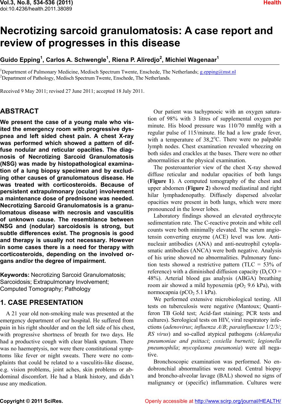 Vol.3, No.8, 534-536 (2011) Health doi:10.4236/health.2011.38089 Copyright © 2011 SciRes. Openly accessible at http://www.scirp.org/journal/HEALTH/ Necrotizing sarcoid granulomatosis: A case report and review of progresses in this disease Guido Epping1, Carlos A. Schwengle1, Riena P. Aliredjo2, Michiel Wagenaar 1 1Department of Pulmonary Medicine, Medisch Spectrum Twente, Enschede, The Netherlands; [email protected] 2Department of Pathology, Medisch Spectrum Twente, Enschede, The Netherlands. Received 9 May 2011; revised 27 June 2011; accepted 18 July 2011. ABSTRACT We present the case of a young male who vis- ited the emergency room with progressive dys- pnea and left sided chest pain. A chest X-ray was performed which showed a pattern of dif- fuse nodular and reticular opacities. The diag- nosis of Necrotizing Sarcoid Granulomatosis (NSG) was made by histopathological examina- tion of a lung biopsy specimen and by exclud- ing other causes of granulomatous disease. He was treated with corticosteroids. Because of persistent extrapulmonary (ocular) involvement a maintenance dose of prednisone was needed. Necrotizing Sarcoid Granulomatosis is a granu- lomatous disease with necrosis and vasculitis of unknown cause. The resemblance between NSG and (nodular) sarcoidosis is strong, but subtle differences exist. The prognosis is good and therapy is usually not necessary. However in some cases there is a need for therapy with corticosteroids, depending on the involved or- gans and/or the degree of impairment. Keywords: Necrotizing Sarcoid Granulomatosi s; Sarcoidosis; Extrapulmonary Involvement; Computed Tomography; Pathology 1. CASE PRESENTATION A 21 year old non-smoking male was presented at the emergency department of our hospital. He suffered from pain in his right shoulder and on the left side of his chest, with progressive shortness of breath for two days. He had a productive cough with clear blank sputum. There was no haemoptysis, nor were there co nstitution al symp- toms like fever or night sweats. There were no com- plaints that could be related to a vasculitis-like disease, e.g. vision problems, joint aches, skin problems or ab- dominal discomfort. He had a blank history, and didn’t use any medication. Our patient was tachypnoeic with an oxygen satura- tion of 98% with 3 litres of supplemental oxygen per minute. His blood pressure was 110/70 mmHg with a regular pulse of 115/minute. He had a low grade fever, with a temperature of 38,2oC. There were no palpable lymph nodes. Chest examination revealed wheezing on both sides and crackles at the bases. There were no other abnormalities at the physical examination. The posteroanterior view of the chest X-ray showed diffuse reticular and nodular opacities of both lungs (Figure 1). A computed tomography of the chest and upper abdomen (Figure 2) showed mediastinal and right hilar lymphadenopathy. Diffusely dispersed alveolar opacities were present in both lungs, which were more pronounced in the lower lobes. Laboratory findings showed an elevated erythrocyte sedimentation rate. The C-reactive protein and white cell counts were both minimally elevated. The serum angio- tensin converting enzyme (ACE) level was low. Anti- nucleair antibodies (ANA) and anti-neutrophil cytopla- smatic antibodies (ANCA) were both negative. Analysis of his urine showed no abnormalities. Pulmonary func- tion tests showed a restrictive pattern (TLC = 53% of reference) with a diminished diffusion capacity (DLCO = 48%). Arterial blood gas analysis (ABGA) breathing room air showed a mild hypoxemia (pO2 9.6 kPa), with normocapnia ( pC O2 5.1 kPa). We performed extensive microbiological testing. All tests on tuberculosis were negative (Mantoux; Quanti- feron TB Gold test; Acid-fast staining; PCR tests and cultures). Serological tests on HIV, viral respiratory infe- ctions (adenovirus; influenza A/B; parainfluenzae 1/2/3/; RS virus) and so-called atypical pathogens (chlamydia pneumoniae and psittaci; coxiella burnetii; legionella pneumophila; mycoplasma pneumonia) were all nega- tive. Bronchoscopic examination was performed. No en- dobronchial abnormalities were noted. Central biopsy and broncho-alveolar lavage (BAL) showed no signs of malignancy or (specific) inflammation. Cultures were 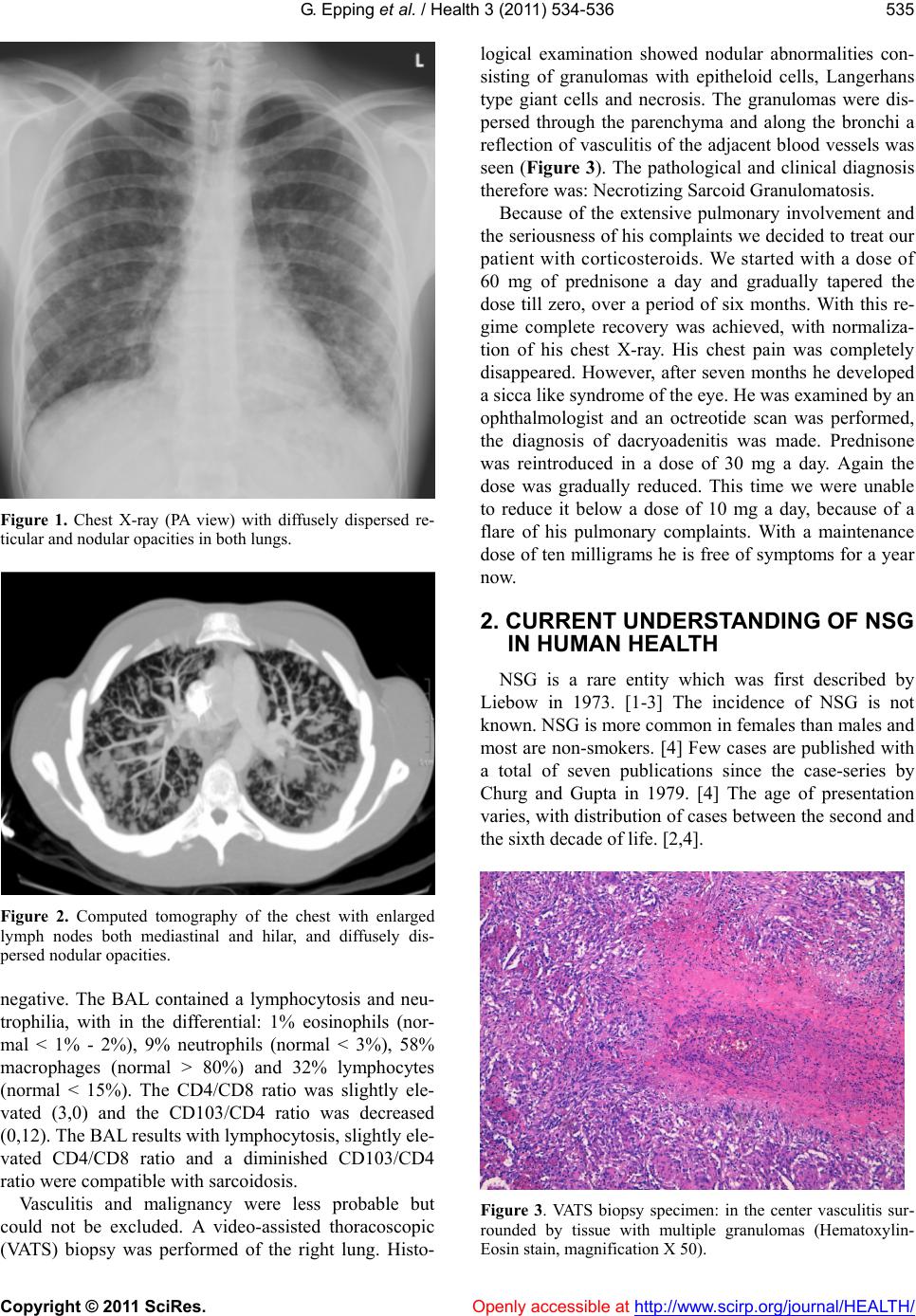 G. Epping et al. / Health 3 (2011) 534-536 Copyright © 2011 SciRes. Openly accessible at http://www.scirp.org/journal/HEALTH/ 535 Figure 1. Chest X-ray (PA view) with diffusely dispersed re- ticular and nodular opacities in both lungs. Figure 2. Computed tomography of the chest with enlarged lymph nodes both mediastinal and hilar, and diffusely dis- persed nodular opacities. negative. The BAL contained a lymphocytosis and neu- trophilia, with in the differential: 1% eosinophils (nor- mal < 1% - 2%), 9% neutrophils (normal < 3%), 58% macrophages (normal > 80%) and 32% lymphocytes (normal < 15%). The CD4/CD8 ratio was slightly ele- vated (3,0) and the CD103/CD4 ratio was decreased (0,12). The BAL results with lymphocytosis, slightly ele- vated CD4/CD8 ratio and a diminished CD103/CD4 ratio were compatible with sarcoidosis. Vasculitis and malignancy were less probable but could not be excluded. A video-assisted thoracoscopic (VATS) biopsy was performed of the right lung. Histo- logical examination showed nodular abnormalities con- sisting of granulomas with epitheloid cells, Langerhans type giant cells and necrosis. The granulomas were dis- persed through the parenchyma and along the bronchi a reflection of vasculitis of the adj acent blood vessels was seen (Figure 3). The pathological and clinical diagnosis therefore was: Necrotizing Sarcoid Granulomatosis. Because of the extensive pulmonary involvement and the seriousness of his complain ts we decided to treat our patient with corticosteroids. We started with a dose of 60 mg of prednisone a day and gradually tapered the dose till zero, over a period of six months. With this re- gime complete recovery was achieved, with normaliza- tion of his chest X-ray. His chest pain was completely disappeared. However, after seven months he developed a sicca like syndrome of the eye. He was examined by an ophthalmologist and an octreotide scan was performed, the diagnosis of dacryoadenitis was made. Prednisone was reintroduced in a dose of 30 mg a day. Again the dose was gradually reduced. This time we were unable to reduce it below a dose of 10 mg a day, because of a flare of his pulmonary complaints. With a maintenance dose of ten milligrams he is free of symptoms for a year now. 2. CURRENT UNDERSTANDING OF NSG IN HUMAN HEALTH NSG is a rare entity which was first described by Liebow in 1973. [1-3] The incidence of NSG is not known. NSG is more common in females than males and most are non-smokers. [4] Few cases are published with a total of seven publications since the case-series by Churg and Gupta in 1979. [4] The age of presentation varies, with distribution of cases between the second and the sixth decade of life. [2,4]. Figure 3. VATS biopsy specimen: in the center vasculitis sur- rounded by tissue with multiple granulomas (Hematoxylin- Eosin stain, magnification X 50). 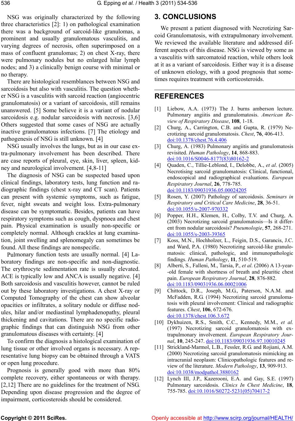 G. Epping et al. / Health 3 (2011) 534-536 Copyright © 2011 SciRes. Openly accessible at http://www.scirp.org/journal/HEALTH/ 536 3. CONCLUSIONS NSG was originally characterized by the following three characteristics [2]: 1) on pathological examination there was a background of sarcoid-like granulomas, a prominent and usually granulomatous vasculitis, and varying degrees of necrosis, often superimposed on a mass of confluent granulomas; 2) on chest X-ray, there were pulmonary nodules but no enlarged hilar lymph nodes; and 3) a clinically benign course with minimal or no therapy. We present a patient diagnosed with Necrotizing Sar- coid Granulomatosis, with extrapulmonary involvement. We reviewed the available literature and addressed dif- ferent aspects of this disease. NSG is viewed by some as a vasculitis with sarcomatoid reaction, while others look at it as a variant of sarcoidosis. Either way it is a disease of unknown etiology, with a good prognosis that some- times requires treatment with corticosteroids. There are histological resemblances between NSG and sarcoidosis but also with vascu litis. The question wheth- er NSG is a vasculitis with sarcoid reaction (angiocentric granulomatosis) or a variant of sarcoidosis, still remains unanswered. [5] Some believe it is a variant of nodular sarcoidosis e.g. nodular sarcoidosis with necrosis. [3,6] Others suggested that some cases of NSG are actually inactive granulomatous infections. [7] The etiology and pathogenesis of NSG is still unknown . [4] REFERENCES [1] Liebow, A.A. (1973) The J. burns amberson lecture. Pulmonary angiitis and granulomatosis. American Re- view of Respiratory Disease, 108, 1-18. [2] Churg, A., Carrington, C.B. and Gupta, R. (1979) Ne- crotizing sarcoid granulomatosis. Chest, 76, 406-413. doi:10.1378/chest.76.4.406 NSG usually involves the lungs, but as in our case ex- tra-pulmonary involvement has been described. There are case reports of pleural, eye, skin, liver, spleen, kid- ney and neurological involvement. [4,8-11] [3] Churg, A. (1983) Pulmonary angiitis and granulomatosis revisited. Human Pathology, 14, 868-883. doi:10.1016/S0046-8177(83)80162-2 [4] Quaden, C., Tillie-Leblond, I., Delobbe, A., et al. (2005) Necrotising sarcoid granulomatosis: Clinical, functional, endoscopical and radiographical evaluations. European Respiratory Journal, 26, 778-785. doi:10.1183/09031936.05.00024205 The diagnosis of NSG can be suspected based upon clinical findings, laboratory tests, lung function and ra- diographic findings (chest x-ray and CT scan). Patients can present with systemic symptoms, such as fatigue, fever, night sweats and weight loss. Extra-pulmonary disease can be symptomatic. Besides, patients can have respiratory symptoms such as cough, dyspnoea and chest pain. Physical examination is usually non-specific or completely normal. Although crackles at lung examina- tion, joint swelling and splenomegaly can sometimes be found. All these findings are nonspecific. [5] Rosen, Y. (2007) Pathology of sarcoidosis. Seminars in Respiratory and Critical Care Medicine, 28, 36-51. doi:10.1055/s-2007-970332 [6] Popper, H.H., Klemen, H., Colby, T.V. and Churg, A. (2003) Necrotizing sarcoid granulomatosis—Is it differ- ent from nodular sarcoidosis? Pneumologie, 57, 268-271. doi:10.1055/s-2003-39365 [7] Koss, M.N., Hochholzer, L., Feigin, D.S., Garancis, J.C. and Ward, P.A. (1980) Necrotizing sarcoid-like granulo- matosis: clinical, pathologic, and immunopathologic findings. Human Pathology, 11, 510-519. Pulmonary function tests are usually normal. [4] La- boratory findings are non-specific and non-diagnostic. The erythrocyte sedimentation rate is usually elevated. ACE is typically low and ANCA is usually negative. [4] Both sarcoidosis an d vasculitis however, cannot be ruled out by these laboratory investigations. A chest X-ray or Computed Tomography of the chest can show alveolar opacities or infiltrates, a solitary nodule or diffuse nod- ules, hilar and/or mediastinal lymphadenopathy, pleural thickening and cavitations. There are no specific radio- graphic findings that can distinguish NSG from other granulomatous diseases with certainty. [4] [8] Alberti, S., Falleni, M., Tarsia, P., et al. (2006) A 13-year- -old female with shortness of breath and pleuritic chest pain. European Respiratory Journal, 28, 876-882. doi:10.1183/09031936.06.00021006 [9] Chittock, D.R., Joseph, M.G., Paterson, N.A.M. and McFadden, R.G. (1994) Necrotizing sarcoid granuloma- tosis with pleural involvement: Clinical and radiographic features. Chest, 106, 672-676. doi:10.1378/chest.106.3.672 [10] Dykhuizen, R.S., Smith, C.C., Kennedy, M.M., et al. (1997) Necrotizing sarcoid granulomatosis with ex- trapulmonary involvement. European Respiratory Jour- nal, 10, 245-247. doi:10.1183/09031936.97.10010245 To confirm the diagnosis a histological examination of lung tissue or other involved organs is necessary. A rep- resentative lung biopsy can be obtained through a VATS or open lung procedure. [11] Strickland-Marmol, L.B., Fessler, R.G. and Rojiani, A.M. (2000) Necrotizing sarcoid granulomatosis mimicking an intracranial neoplasm: Clinicopathologic features and re- view of the literature. Modern Pathology, 13, 909-913. doi:10.1038/modpathol.3880162 Prognosis is generally good with more than 80% complete recovery, either spontaneous or with therapy. [2,12] There are no guidelines for the treatment of NSG. Depending upon disease progression and the degree of impairme nt, cort i cost eroids should be consid ered . [12] Lynch III, J.P., Kazerooni, E.A. and Gay, S.E. (1997) Pulmonary sarcoidosis. Clinics In Chest Medicine, 18, 755-785. doi:10.1016/S0272-5231(05)70417-2 |