

Paper Menu >>
Journal Menu >>
 Materials Sciences and Applicatio n, 2011, 2, 988-992 doi:10.4236/msa.2011.28133 Published Online August 2011 (http://www.SciRP.org/journal/msa) Copyright © 2011 SciRes. MSA A Study of the Optical Properties in ZnWO4 Nanorods Synthesized by Hydrothermal Method# Nguyen Van Minh1*, Nguyen Manh Hung1,2 1Center for Nano Science and Technology, and Department of Physics, Hanoi National University of Education, Hanoi, Vietnam; 1,2 Hanoi University of Mining and Geology, Dong Ngac, Tu Liem, Hanoi, Vietnam. Email: *[email protected]; [email protected] Received May 17th, 2011; revised April 20th, 2011; accepted June 7th, 2011. ABSTRACT We investigate the effect of synthesized time on the structure, as well as optical properties in ZnWO4 nano rod prepared by hydrothermal method. The prepared rods were characterized by X-ray diffraction (XRD), scanning electron micros- copy (SEM), Raman scattering, absorption and photoluminescent (PL) spectra techniques. The size and morphology of ZnWO4 nano-rod can be controlled by adjusting the reaction time. The resultant sample is a pure phase of ZnWO4 without any impurities. The results showed that the optical property of ZnWO4 nanoparticles obviously relied on their rod sizes. Keywords: ZnWO4 Nano Rod, Raman Spectroscopy, Absorption, Photoluminescence 1. Introduction Zinc tungstate (ZnWO4) with a wolframite structure has been of practical interest for a long time because of its attractive luminescence [1]. ZnWO4 has been applied as a possible new material for microwave amplification by stimulated emission of radiation [2], scintillator [3] and optical hole burning lattice material [4], etc. Recently, new applications for this material have emerged, includ- ing large-volume scintillators for high-energy physics [5] In particular, ZnWO4, also known by its mineral name sanmartinite, is a wide-gap semiconductor, with band gap energy close to 4 eV [6], and is a promising material for the new generation of radiation detectors. ZnWO4 has been prepared by different routes such as the Czochralski method [7], sintering of WO3 and ZnO or ZnCO4 powders [8], reaction in aqueous solution fol- lowed by heating of the precipitate [9], heating of ZnO thin films with WO3 vapor [10], sol-gel reaction [11], and hydrothermal reaction over an extensive period [12]. However, ZnWO4 particles prepared by these routes are relatively large in particle size and irregular in morphol- ogy. Furthermore, higher calcining temperature is still needed. It is very significant whether in fundamental or applied field to explore new routes to ZnWO4, especially for ZnWO4 crystallites with nanometer size, which would have unique properties compared to traditional products [13,14]. To obtain nanosized powders, the solid state methods have several problems, because the WO3 has a tendency to vaporize at high temperatures [15], nonho- mogeneous compounds might be easily formed during the solid-state and melting processing and the tempera- ture for the solid state reaction is relatively high [16]. These problems could be solved by applying the hydro- thermal method. However, a few studies on the chemical synthesis of zinc tungstate by the hydrothermal method have been reported. Furthermore, very few papers were concerned with effects of the size and morphology on the optical properties of ZnWO4 nanoparticles. In this work, we report the synthesis of ZnWO4 nano rod by hydrothermal method at a low temperature of 180˚C and investigate their structure, Raman scattering, absorption and photoluminescence. 2. Experiment Zinc tungstate (ZnWO4) nanoparticles were prepared by the hydrothermal reaction of Zn(NO3)2·6H2O and Na2WO4·2H2O at temperature of 180˚C, and various re- action times (2, 4, 6 and 8 h). In a typical procedure for the preparation of sample, Zn(NO3)2·6H2O (1 mmol) in water (10 ml) was added Na2WO4·2H2O (1 mmol) in water (20 ml) with vigorous stirring. H2O was added to make 40 ml of the solution, and pH of the solution was #This work has been supported by The Vietnam’s National Foundation for Science and Technology Development (NAFOSTED). 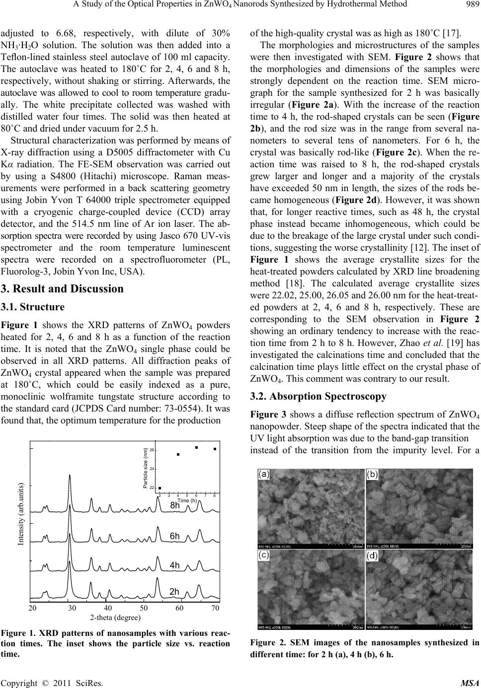 A Study of the Optical Properties in ZnWONanorods Synthesized by Hydrothermal Method989 4 adjusted to 6.68, respectively, with dilute of 30% NH3·H2O solution. The solution was then added into a Teflon-lined stainless steel autoclave of 100 ml capacity. The autoclave was heated to 180˚C for 2, 4, 6 and 8 h, respectively, without shaking or stirring. Afterwards, the autoclave was allowed to cool to room temperature gradu- ally. The white precipitate collected was washed with distilled water four times. The solid was then heated at 80˚C and dried under vacuum for 2.5 h. Structural characterization was performed by means of X-ray diffraction using a D5005 diffractometer with Cu Kα radiation. The FE-SEM observation was carried out by using a S4800 (Hitachi) microscope. Raman meas- urements were performed in a back scattering geometry using Jobin Yvon T 64000 triple spectrometer equipped with a cryogenic charge-coupled device (CCD) array detector, and the 514.5 nm line of Ar ion laser. The ab- sorption spectra were recorded by using Jasco 670 UV-vis spectrometer and the room temperature luminescent spectra were recorded on a spectrofluorometer (PL, Fluorolog-3, Jobin Yvon Inc, USA). 3. Result and Discussion 3.1. Structure Figure 1 shows the XRD patterns of ZnWO4 powders heated for 2, 4, 6 and 8 h as a function of the reaction time. It is noted that the ZnWO4 single phase could be observed in all XRD patterns. All diffraction peaks of ZnWO4 crystal appeared when the sample was prepared at 180˚C, which could be easily indexed as a pure, monoclinic wolframite tungstate structure according to the standard card (JCPDS Card number: 73-0554). It was found that, the optimum temperature for the production 2345678 22 24 26 Particle size (nm) Time (h) y( ) 2h 4h 6h 8h 20 30 40 50 60 70 2-theta (degree) Figure 1. XRD patterns of nanosamples with various reac- tion times. The inset shows the particle size vs. reaction time. of the high-quality crystal was as high as 180˚C [17]. The morphologies and microstructures of the samples were then investigated with SEM. Figure 2 shows that the morphologies and dimensions of the samples were strongly dependent on the reaction time. SEM micro- graph for the sample synthesized for 2 h was basically irregular (Figure 2a). With the increase of the reaction time to 4 h, the rod-shaped crystals can be seen (Figure 2b), and the rod size was in the range from several na- nometers to several tens of nanometers. For 6 h, the crystal was basically rod-like (Figure 2c). When the re- action time was raised to 8 h, the rod-shaped crystals grew larger and longer and a majority of the crystals have exceeded 50 nm in length, the sizes of the rods be- came homogeneous (Figure 2d). However, it was shown that, for longer reactive times, such as 48 h, the crystal phase instead became inhomogeneous, which could be due to the breakage of the large crystal under such condi- tions, suggesting the worse crystallinity [12]. The inset of Figure 1 shows the average crystallite sizes for the heat-treated powders calculated by XRD line broadening method [18]. The calculated average crystallite sizes were 22.02, 25.00, 26.05 and 26.00 nm for the heat-treat- ed powders at 2, 4, 6 and 8 h, respectively. These are corresponding to the SEM observation in Figure 2 showing an ordinary tendency to increase with the reac- tion time from 2 h to 8 h. However, Zhao et al. [19] has investigated the calcinations time and concluded that the calcination time plays little effect on the crystal phase of ZnWO4. This comment was contrary to our result. 3.2. Absorption Spectroscopy Figure 3 shows a diffuse reflection spectrum of ZnWO4 nanopowder. Steep shape of the spectra indicated that the UV light absorption was due to the band-gap transition instead of the transition from the impurity level. For a Intensity (arb.units) Figure 2. SEM images of the nanosamples synthesized in different time: for 2 h (a), 4 h (b), 6 h. Copyright © 2011 SciRes. MSA 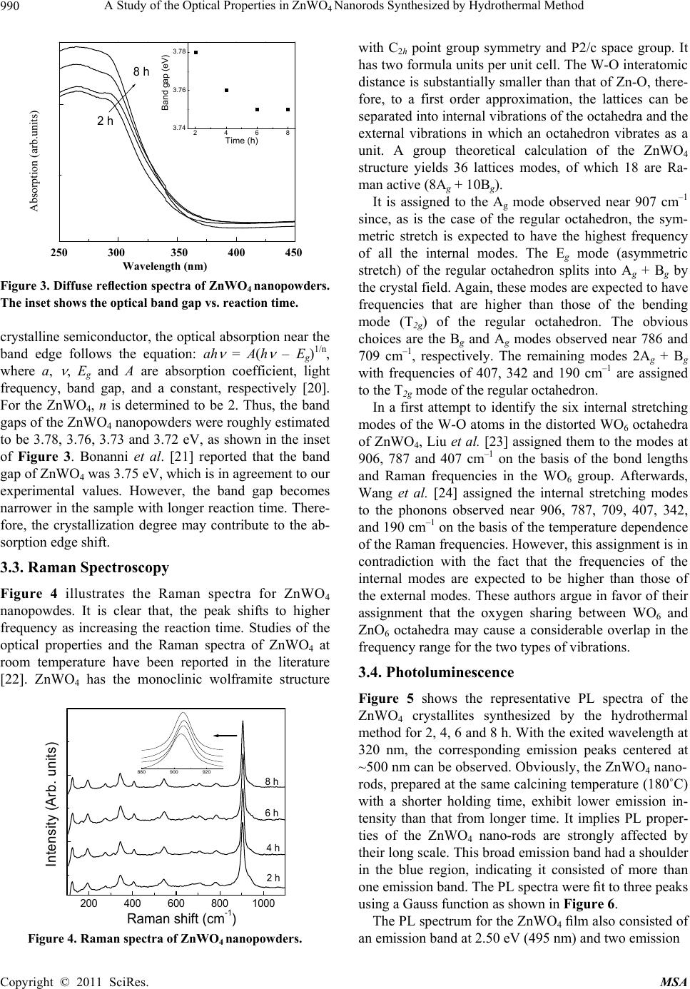 A Study of the Optical Properties in ZnWONanorods Synthesized by Hydrothermal Method 990 4 8 h 2 h Ab so rp tio n (a rb . u n its) 2468 3.74 3.76 3.78 Band gap (eV) Time (h) 250 300 350 400 450 Wavelength (nm) Figure 3. Diffuse reflection spectra of ZnWO4 nanopowders. The inset shows the optic al band gap vs. reaction time. crystalline semiconductor, the optical absorption near the band edge follows the equation: ah = A(h – Eg)1/n, where a, , Eg and A are absorption coefficient, light frequency, band gap, and a constant, respectively [20]. For the ZnWO4, n is determined to be 2. Thus, the band gaps of the ZnWO4 nanopowders were roughly estimated to be 3.78, 3.76, 3.73 and 3.72 eV, as shown in the inset of Figure 3. Bonanni et al. [21] reported that the band gap of ZnWO4 was 3.75 eV, which is in agreement to our experimental values. However, the band gap becomes narrower in the sample with longer reaction time. There- fore, the crystallization degree may contribute to the ab- sorption edge shift. 3.3. Raman Spectroscopy Figure 4 illustrates the Raman spectra for ZnWO4 nanopowdes. It is clear that, the peak shifts to higher frequency as increasing the reaction time. Studies of the optical properties and the Raman spectra of ZnWO4 at room temperature have been reported in the literature [22]. ZnWO4 has the monoclinic wolframite structure 880 900 920 200 400 600 8001000 8 h 6 h 4 h 2 h Intensity (Arb. units) Raman shift (cm -1 ) Figure 4. Raman spectra of ZnWO4 nanopowders. with C2h point group symmetry and P2/c space group. It has two formula units per unit cell. The W-O interatomic distance is substantially smaller than that of Zn-O, there- fore, to a first order approximation, the lattices can be separated into internal vibrations of the octahedra and the external vibrations in which an octahedron vibrates as a unit. A group theoretical calculation of the ZnWO4 structure yields 36 lattices modes, of which 18 are Ra- man active (8Ag + 10Bg). its) Absorption (arb.un It is assigned to the Ag mode observed near 907 cm–1 since, as is the case of the regular octahedron, the sym- metric stretch is expected to have the highest frequency of all the internal modes. The Eg mode (asymmetric stretch) of the regular octahedron splits into Ag + Bg by the crystal field. Again, these modes are expected to have frequencies that are higher than those of the bending mode (T2g) of the regular octahedron. The obvious choices are the Bg and Ag modes observed near 786 and 709 cm–1, respectively. The remaining modes 2Ag + Bg with frequencies of 407, 342 and 190 cm–1 are assigned to the T2g mode of the regular octahedron. In a first attempt to identify the six internal stretching modes of the W-O atoms in the distorted WO6 octahedra of ZnWO4, Liu et al. [23] assigned them to the modes at 906, 787 and 407 cm–1 on the basis of the bond lengths and Raman frequencies in the WO6 group. Afterwards, Wang et al. [24] assigned the internal stretching modes to the phonons observed near 906, 787, 709, 407, 342, and 190 cm–1 on the basis of the temperature dependence of the Raman frequencies. However, this assignment is in contradiction with the fact that the frequencies of the internal modes are expected to be higher than those of the external modes. These authors argue in favor of their assignment that the oxygen sharing between WO6 and ZnO6 octahedra may cause a considerable overlap in the frequency range for the two types of vibrations. 3.4. Photoluminescence Figure 5 shows the representative PL spectra of the ZnWO4 crystallites synthesized by the hydrothermal method for 2, 4, 6 and 8 h. With the exited wavelength at 320 nm, the corresponding emission peaks centered at ~500 nm can be observed. Obviously, the ZnWO4 nano- rods, prepared at the same calcining temperature (180˚C) with a shorter holding time, exhibit lower emission in- tensity than that from longer time. It implies PL proper- ties of the ZnWO4 nano-rods are strongly affected by their long scale. This broad emission band had a shoulder in the blue region, indicating it consisted of more than one emission band. The PL spectra were fit to three peaks using a Gauss function as shown in Figure 6. The PL spectrum for the ZnWO4 film also consisted of an emission band at 2.50 eV (495 nm) and two emission Copyright © 2011 SciRes. MSA 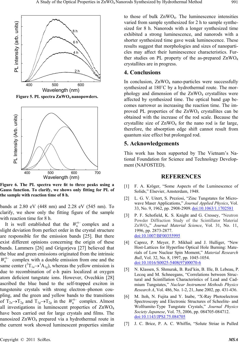 A Study of the Optical Properties in ZnWONanorods Synthesized by Hydrothermal Method991 4 400 500 600 8 h 6 h 4 h 2 h PL intensity (arb. units) Wavelength (nm) Figure 5. PL spectra ZnWO4 nanopowders. 400 500 600 700 PL Intensity (Arb. units) W avelength (nm) Figure 6. The PL spectra were fit to three peaks using a Gauss function. To clarify, we shows only fitting for PL of the sample with reaction time of 8 h. bands at 2.80 eV (448 nm) and 2.28 eV (545 nm). To clarify, we show only the fitting figure of the sample with reaction time for 8 h. It is well established that the complex and a slight deviation from perfect order in the crystal structure are responsible for the emission bands [25]. But there exist different opinions concerning the origin of these bands. Lammers [26] and Grigorjeva [27] believed that the blue and green emissions originated from the intrinsic complex with a double emission from one and the same center (3T1u1A1g), whereas the yellow emission is due to recombination of e-h pairs localized at oxygen atom deficient tungstate ions. However, Ovechkin [28] ascribed the blue band to the self-trapped exciton in tungstenite crystals with strong electron–phonon cou- pling, and the green and yellow bands to the transitions of T2uT2g and T1gT2g in the complex. Almost all investigations in luminescent properties of ZnWO4 have been carried out for large crystals and films. The nanosized ZnWO4 prepared via a hydrothermal route in the current work showed luminescent properties similar to those of bulk ZnWO4. The luminescence intensities varied from sample synthesized for 2 h to sample synthe- sized for 8 h. Nanorods with a longer synthesized time exhibited a strong luminescence, and nanorods with a shorter synthesized time gave weak luminescence. These results suggest that morphologies and sizes of nanoparti- cles may affect their luminescence characteristics. Fur- ther studies on PL property of the as-prepared ZnWO4 crystallites are in progress. 6 6 W 6 6 W 6 6 W 4. Conclusions In conclusion, ZnWO4 nano-particles were successfully synthesized at 180˚C by a hydrothermal route. The mor- phology and dimension of the ZnWO4 crystallites were affected by synthesized time. The optical band gap be- comes narrower as increasing the reaction time. The im- proved PL properties of the ZnWO4 crystallites can be obtained with the increase of the rod scale. Because the crystallite size of ZnWO4 for the nano rod is far large, therefore, the absorption edge shift cannot result from quantum size effect but prolonged rod. 5. Acknowledgements This work has been supported by The Vietnam’s Na- tional Foundation for Science and Technology Develop- ment (NAFOSTED). REFERENCES [1] F. A. Kröger, “Some Aspects of the Luminescence of Solids,” Elsevier, Amsterdam, 1948. [2] L. G. V. Uitert, S. Preziosi, “Zinc Tungstates for Micro- wave Maser Applications,” Journal Applied Physics, Vol. 33, No. 9, 1962, pp. 2908-2909. doi:10.1063/1.1702581 [3] P. F. Schofield, K. S. Knight and G. Cressey, “Neutron Powder Diffraction Study of the Scintillator Material ZnWO4,” Journal Material Science, Vol. 31, No. 11, 1996, pp. 2873-2877. doi:10.1007/BF00355995 [4] Caprez, P. Meyer, P. Mikhail and J. Hulliger, “New Host-Lattices for Hyperfine Optical Hole Burning: Mate- rials of Low Nuclear Spin Moment,” Material Research Bull, Vol. 32, No. 8, 1997, pp. 1045-1054. doi:10.1016/S0025-5408(97)00070-6 [5] N. Klassen, S. Shmurak, B. Red’kin, B. Ille, B. Lebeau, P. Lecoq and M. Schneegans, “Correlations between Struc- tural and Scintillation Characteristics of Lead and Cad- mium Tungstates,” Nuclear Instrument Methods Physics Research A, Vol. 486, No. 1-2, 21, June 2002, pp. 431-436. [6] M. Itoh, N. Fujita and Y. Inabe, “X-Ray Photoelectron Spectroscopy and Electronic Structures of Scheelite- and Wolframite-Type Tungstate Crystals,” Journal Physics Society Japanese, Vol. 75, 2006, pp. 084705-084712. doi:10.1143/JPSJ.75.084705 [7] J. C. Brice, P. A. C. Whiffin, “Solute Striae in Pulled Copyright © 2011 SciRes. MSA  A Study of the Optical Properties in ZnWO4 Nanorods Synthesized by Hydrothermal Method Copyright © 2011 SciRes. MSA 992 Crystals of Zinc Tungstate,” British Journal Applied Physics, Vol. 18, No. 5, 1967, pp. 581-586. doi:10.1088/0508-3443/18/5/304 [8] A. R. Phani, M. Passacantando, L. Lozzi and S. Santucci, “Structural Characterization of Bulk ZnWO4 Prepared by Solid State Method,” Journal Material Science, Vol. 35, No. 19, 2000, pp. 4879-4883. doi:10.1023/A:1004809804206 [9] L. Honeycutt and A. Kuzmin, J. Purans “Communication and Design Course; Local atomic and electronic structure of tungsten ions in AWO4 Crystals of Scheelite and Wolframite Types,” Radiat Measurement, Vol. 33, No. 5, 2001, pp. 583-586. [10] A. Henglein, “Estimated Distributions of Electronic Redox Levels in aq/eaq −, Haq+/Haq and Some Other Systems,” General Introductory Chemistry, Vol. 78, No. 10, 1974, pp. 1078-1084. doi:10.1002/bbpc.19740781016 [11] M. Bonanni, L. Spanhel, M. Lerch, E. Fuglein and G. Muller, “Conversion of Colloidal ZnO-WO3 Heteroag- gregates into Strongly Blue Luminescing ZnWO4 Xe- rogels and Films,” Chemistry Material, Vol. 10, No. 1, 1998, pp. 304-310. doi:10.1021/cm9704591 [12] F.-S. Wen, X. Zhao, H. Huo, J.-S. Chen, E. Shu-Lin and J.-H. Zhang, “Hydrothermal Synthesis and Photolumi- nescent Properties of ZnWO4 and Eu3+-Doped ZnWO4,” Materials Letters, Vol. 55, No. 3, 2002, pp. 152-157. doi:10.1016/S0167-577X(01)00638-3 [13] M. H. Huang, S. Mao, H. Feick, H. Yan, Y. Wu, H. Kind, E. Weber, R. Russo and P. Yang, “Room-Temperature Ultraviolet Nanowire Nanolasers,” Science, Vol. 292, 2001, pp. 1897-1899. doi:10.1126/science.1060367 [14] Y. Cui and C. M. Lieber, “Functional Nanoscale Electronic Devices Assembled Using Silicon Nanowire Building Blocks,” Science, Vol. 2, 2001, pp. 851-853. doi:10.1126/science.291.5505.851 [15] K. Kuribayashi, M. Yoshimura, T. Ohta and T. Sata, “Processes in the Reaction of Yttrium Oxide with. Tung- sten Trioxide,” Bull Chemistry Sciences Japanese, Vol. 50, No. 11, 1977, pp. 2932-2934. doi:10.1246/bcsj.50.2932 [16] R. C. Pullar, S. Farrah and N. M. Alford, “MgWO4, ZnWO4, NiWO4 and CoWO4 Microwave Dielectric Ce- ramics,” Journal of the European Ceramic Society, Vol. 27, No. 2-3, 2007, pp. 1059-1063 [17] H. Fu, J. Lin, L. Zhang and Y. Zhu, “Photocatalytic Ac- tivities of a Novel ZnWO4 Catalyst Prepared by a Hydro- thermal Process,” Applied Catalysis A: General, Vol. 306, No. 7, 2006, pp. 58-67. doi:10.1016/j.apcata.2006.03.040 [18] K. N. P. Kumar, K. Keizer and A. J. Burggraaf, “Textural Evolution and Phase Transformation in Titania Membran- es: Part 1 Unsupported Membranes,” Journal Material Chemistry, Vol. 3, No. 11, 1993, pp. 1141-1149. doi:10.1039/jm9930301141 [19] X. Zhao, W. Yao, Y. Wu, S. Zhang, H. Yang and Y. Zhu, “Fabrication and Photoelectrochemical Properties of Po- rous ZnWO4 Film,” Journal of Solid State Chemistry, Vol. 179, No. 8, 2006, pp. 2562-2570. [20] M. A. Butler, “Photoelectrolysis and Physical Properties of the Semiconducting Electrode WO2,” Applied Physics, Vol. 48, No. 5, 1977, pp. 1914-1920. [21] M. Bonanni, L. Spanhel, M. Lerch, E. Fuglein and G. Muller, “Conversion of Colloidal ZnO-WO3 Heteroag- gregates into Strongly Blue Luminescing ZnWO4 Xe- rogels and Films,” Chemics Material, Vol. 10, No. 1, 1998, pp. 304-310. doi:10.1021/cm9704591 [22] A. Kalinko and A. Kuzmin, “Raman and Photolumines- cence Spectroscopy of Zinc Tungstate Powders”, Journal of Luminescence, Vol. 129, No. 10, 2009, pp. 1144-1147. doi:10.1016/j.jlumin.2009.05.010 [23] Y. Liu, H. Wang, G. Chen, Y. D. Zhou, B. Y. Gu and B. Q. Hu, “Analysis of Raman spectra of ZnWO4 single crystals,” Journal Applied Physics, Vol. 64, No. 9, 1988, pp. 4651-4653. doi:10.1063/1.341245 [24] H. Wang, F. D. Medina, Y. D. Zhou and Q. N. Zhang, “Temperature Dependence of the Polarized Raman Spectra of ZnWO4 Single Crystals,” Physics Re- views B, Vol. 45, No. 18, 1992, pp. 10356-10362. doi:10.1103/PhysRev B.45.103 56 [25] G. Blasse, M. J. J. Lammers and D.S. Robertson, “Structure and Bonding, the Luminescence of Cad- mium Tungstate (CdWO4),” Physics Status Solidi A, Vol. 63, 1981, pp. 569-572. [26] L. Grigorjeva, R. Deych, D. Millers and S. Chernov, “Time-Resolved Luminescence and Absorption in Cd- WO4,” Radiation Measurement, Vol. 29, No. 3-4, 1998, pp. 267-271. [27] A. E. Ovechkin, V. D. Ryzhikov, G. Tamulaitis and A. Žukauskas, “Luminescence of ZnWO4 and CdWO4 Cry- stals,” Physics Status Solidity A, Vol. 103, No. 1, 1987, 285-290. doi:10.1002/pssa.2211030133 |