

Paper Menu >>
Journal Menu >>
 Surgical Science, 2011, 2, 228-231 doi:10.4236/ss.2011.25051 Published Online July 2011 (http://www.SciRP.org/journal/ss) Copyright © 2011 SciRes. SS The surgical Treatment and Outcome of Pathological Fracture in Patients with Giant Cell Tumor of Bone Tomoaki Torigoe1, Atsuhiko Terakado1, Yoshiyuki Suehara1, Taketo Okubo1, Tatsuya Takagi1, Kazuo Kaneko1, Yasuo Yazawa2 1Department of Orthopaedic Surgery, Juntendo University School of Medicine, Tokyo, Japan 2Department of Orthopaedic Oncology, Saitama International Medical Center, Saitama, Japan E-mail: [email protected] Received April 12, 2011; revised June 13, 2011; accepted June 24, 2011 Abstract Between 1992 and 2008, we treated 35 patients with giant cell tumor (GCT) of bone, seven of the 35 pre- sented with a pathological fracture. The fractures were located in the femur in five, and in the humerus and radius in one patient each. The surgical treatments were curettage in six cases and wide resection in the distal radius case. Two of the seven patients developed local recurrence, giving a local recurrence rate of 29%. The local recurrence rate in GCT patients without a pathological fracture was 21%. There was a tendency for there to be a higher recurrence rate associated with fractured GCT, but no statistically significant difference was observed between the two groups. Therefore, it was considered that a pathological fracture was not a risk factor for local recurrence in GCT. Keywords: Giant Cell Tumor of Bone, Pathological Fracture 1. Introduction There have been several reports that a pathological frac- ture in subjects with a pr imary malignant bone tu mor is a risk factor for local recurrence and survival [1]. Scully reported that pathological fractu re was a statistically risk factor for local recurrence in osteosarcoma [2 ]. Although a giant cell tumor (GCT) of bone is considered to have the potential for local recurrence [3], it is unclear whether a pathological fracture is a risk factor for local recurrence in GCT cases. We studied the relationship between pathological fractures and local recurrence in GCT cases. 2. Patients and Methods We retrospectively reviewed 35 patients with GCT in the extremities treated at Juntendo University Hospital and Juntendo Orthopedic Oncology Collaboration (JOOC) affiliated hospitals between 1992 and 2008. The clinical and radiological records of all 35 patients were reviewed. Clinical and radiological evidence of a fracture had been seen either at diagnosis or during the course of preopera- tive treatment. A histopathological diagnosis was ob- tained in all of the cases. Statistical comparisons were made using a Chi-squared analysis and the Fisher exact test. 3. Results In total, seven of 35 patients had a pathological fracture before the initial surgery, four of whom were male, and 3 were female, and they had a median age of 25 years (18 to 36) (Table 1). The median follow-up was 35 months (5 to 73). The tumor was located in the femur in five, and in the humerus and radius in one patient each. In the fe- mur fractures, four were distal, and one was proximal. In the humerus, the fracture was in the proximal humerus, and in the radius, the fracture was in the distal portion. The fractures had been present at diagnosis in six pa- tients, and developed after biopsy in one patient. Six fractures occurred after trauma and one occurred sponta- neously. According to the grading system of Campanacci, two lesions were grade I, two were grade II, and three were grade III. The surgical treatment was curettage of the tumor with high speed burring in six, and wide resec- tion with a vascularized fibula graft in one distal radius ase. Chemical adjuvant treatment was indicated in five c 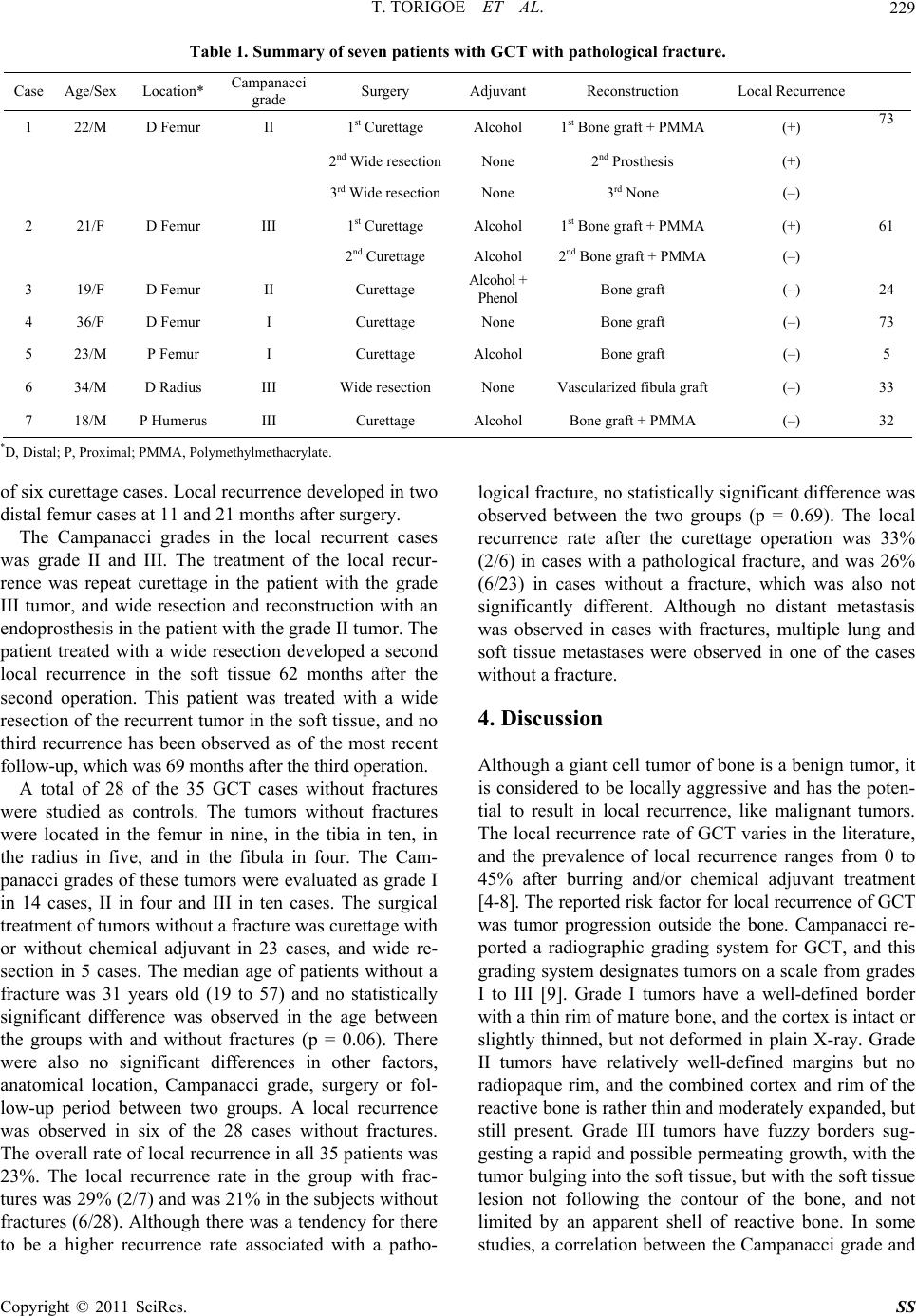 T. TORIGOE ET AL. Copyright © 2011 SciRes. SS 229 Table 1. Summary of seven patients with GCT with pathological fracture. Case Age/SexLocation* Campanacci grade Surgery Adjuvant Reconstruction Local Recurrence 1 22/M D Femur II 1st Curettage Alcohol 1st Bone graft + PMMA (+) 73 2 nd Wide resectionNone 2nd Prosthesis (+) 3 rd Wide resectionNone 3rd None (–) 2 21/F D Femur III 1st Curettage Alcohol 1st Bone graft + PMMA (+) 61 2 nd Curettage Alcohol 2nd Bone graft + PMMA (–) 3 19/F D Femur II Curettage Alcohol + Phenol Bone graft (–) 24 4 36/F D Femur I Curettage None Bone graft (–) 73 5 23/M P Femur I Curettage Alcohol Bone graft (–) 5 6 34/M D Radius III Wide resection None Vascularized fibula graft (–) 33 7 18/M P Humerus III Curettage Alcohol Bone graft + PMMA (–) 32 *D, Distal; P, Proximal; PMMA, Pol ymethylmethacrylate. of six curettage cases. Local recurrence developed in two distal femur cases at 11 and 21 months after surgery. The Campanacci grades in the local recurrent cases was grade II and III. The treatment of the local recur- rence was repeat curettage in the patient with the grade III tumor, and wide resection and reconstruction with an endoprosthesis in the patient with the grade II tumor. The patient treated with a wide resection developed a second local recurrence in the soft tissue 62 months after the second operation. This patient was treated with a wide resection of the recurrent tumor in the soft tissue, and no third recurrence has been observed as of the most recent follow-up, which was 69 m onths after the third operati on. A total of 28 of the 35 GCT cases without fractures were studied as controls. The tumors without fractures were located in the femur in nine, in the tibia in ten, in the radius in five, and in the fibula in four. The Cam- panacci grades of these tumors were evaluated as grade I in 14 cases, II in four and III in ten cases. The surgical treatment of tumors without a fracture was curettage with or without chemical adjuvant in 23 cases, and wide re- section in 5 cases. The median age of patients without a fracture was 31 years old (19 to 57) and no statistically significant difference was observed in the age between the groups with and without fractures (p = 0.06). There were also no significant differences in other factors, anatomical location, Campanacci grade, surgery or fol- low-up period between two groups. A local recurrence was observed in six of the 28 cases without fractures. The overall rate of local recurren ce in all 35 patients was 23%. The local recurrence rate in the group with frac- tures was 29 % (2/7 ) and was 2 1% in the su bject s withou t fractures (6/28). Although ther e was a tendency for there to be a higher recurrence rate associated with a patho- logical fracture, no statistically significant difference was observed between the two groups (p = 0.69). The local recurrence rate after the curettage operation was 33% (2/6) in cases with a pathological fracture, and was 26% (6/23) in cases without a fracture, which was also not significantly different. Although no distant metastasis was observed in cases with fractures, multiple lung and soft tissue metastases were observed in one of the cases without a fracture. 4. Discussion Although a giant cell tu mor of bone is a benign tumor, it is considered to be locally aggressive and has the poten- tial to result in local recurrence, like malignant tumors. The local recurrence rate of GCT varies in the literature, and the prevalence of local recurrence ranges from 0 to 45% after burring and/or chemical adjuvant treatment [4-8]. The reported risk factor for local recurrence of GCT was tumor progression outside the bone. Campanacci re- ported a radiographic grading system for GCT, and this grading system designates tumors on a scale from grades I to III [9]. Grade I tumors have a well-defined border with a thin rim of mature bone, and the cortex is intact or slightly thinned, but not deformed in plain X-ray. Grade II tumors have relatively well-defined margins but no radiopaque rim, and the combined cortex and rim of the reactive bone is rather thin and moderately expanded, bu t still present. Grade III tumors have fuzzy borders sug- gesting a rapid and possible permeating growth, with the tumor bulging into the soft tissue, but with the soft tissue lesion not following the contour of the bone, and not limited by an apparent shell of reactive bone. In some studies, a correlation between the Campanacci grade and  T. TORIGOE ET AL. 230 recurrence rate was reported [10,11]. Prosser reported that the overall local recurrence rate of GCT was 19%, and the recurrence rate of the tumors with extraskeletal extension (Campanacci Grade III) was 29% [12]. In osteosarcoma, a primary bone malignancy, Sculley reported that a pathological raised concerns about tumor dissemination by the hematoma, and should be consid- ered to be a contraindication to limb salvage surgery [13]. Abudu reported that limb sparing surgery with adequate margins of excision could be achieved in many patients with pathological fractures due to primary osteosarcoma without compromising survival, but the risk of local re- currence was significant [14]. It is possible that a patho- logical fracture in GCT may release tumor cells into the surrounding soft tissues like in osteosarcoma, and Coo- per reported that GCT tumor cells were observed in the synovium of the ankle joint after a distal tibia p athologi- cal fracture and in the soft tissues around a fracture site in the distal femur [15]. Campanacci reported a possibil- ity that a pathological fracture was the risk factor in GCT, but the relationship was not clear in his report [9]. O’donnell reported that the overall local recurrence rate of GCT was 25%, and that the local recurrence rate of GCT in patients with a pathological fracture was 50%, thus indicating that a pathological fracture was associ- ated with an increased recurrence rate [16]. Conversely, some reports have suggested that there is a lower local recurrence rate in GCT patients with a fracture than in those without a fracture, but the surgeons in this report were more inclined to perform extensive surgery for le- sions with pathological fractures, because they considered there was a risk of tumor cell contamination into the sur- rounding tissues [8, 17]. Dreinhöfer reported that four of 10 patients with fractured GCT at diagnosis had local recurrence after the curettage surgery, and that the local recurrence rate in patients with fractured GCT was similar to that in patients with non-fractured GCT [18]. He sug- gested that extensive surgery, such as an en-bloc resection and reconstruction with tumor prosthesis, was not neces- sary for GCT with pathological fracture as the first opera- tion, and extensive treatment should be considered only when a local recurrence occurs after a curettage surgery. In our present study, no significant difference was ob- served in the local recurrence rate between GCT patients with and without fractures. We theref ore do not consider a pathological fracture to be a definite risk factor for lo- cal recurrence in GCT, and a curettage operation with adjuvant treatment should therefore be advocated for treating such tumors. 5. References [1] J. A. Bramer, A. A. Abudu, R. J. Grimer, S. R. Carter, and R. M. Tillman, “Do Pathological Fractures Influence Survival and Local Recurrence Rate in Bony Sarcomas?” European Journal of Cancer, Vol. 43, No. 13, September 2007, pp. 1944-1951. doi:10.1016/j.ejca.2007.07.004 [2] S. P. Scully, H. T. Temple, R. J. O’Keefe, H. J. Mankin and M. Gebhardt, “The Surgical Treatment of Patients with Osteosarcoma Who Sustain a Pathologic Fracture,” Clinical Orthopaedics and Related Research, No. 324, March 1996, pp. 227-32. doi:10.1097/00003086-199603000-00028 [3] H. R. Blackley, J. S. Wunder, A. M. Davis, L. M. White, R. Kandel and R. S. Bell, “Treatment of Giant-Cell Tu- mors of Long Bones with Curettage and Bone-Grafting,” Journal of Bone and Joint Surgery American Volume, Vol. 81, No. 6, June 1999, pp. 811-820. [4] D. C. Dahlin, R. E. Cupps and E. W. Johnson Jr., “Gi- ant-Cell Tumor: A Study of 195 Cases,” Cancer, Vol. 25, No. 5, May 1970, pp. 1061-1070. doi:10.1002/1097-0142(197005)25:5<1061::AID-CNCR 2820250509>3.0.CO;2-E [5] F. M. Klenke, D. E. Wenger, C. Y. Inwards, P. S. Rose and F. H. Sim, “Recurrent Giant Cell Tumor of Long Bones: Analysis of Surgical Management,” Clinical Or- thopaedics and Related Research, No. 469, April 2011, pp. 1181-1187. doi:10.1007/s11999-010-1560-9 [6] F. Vult von Steyern, H. C. Bauer, C. Trovik, A. Kivioja, P. Bergh, P. Holmberg Jörgensen, G. Follerås and A. Rydholm, “Treatment of Local Recurrences of Giant Cell Tumour in Long Bones after Curettage and Cementing. A Scandinavian Sarcoma Group Study,” Journal of Bone and Joint Surgery British Volume, Vol. 88, No. 4, April 2006, pp. 531-535. doi:10.1302/0301-620X.88B4.17407 [7] P. J. McGrath, “Giant-Cell Tumour of Bone: An Analysis of Fifty-Two Cases,” Journal of Bone and Joint Surgery British Volume, Vol. 54, No. 2, May 1972, pp. 216-229. [8] S. E. Larsson, R. Lorentzon and L. Boquist, “Giant-Cell Tumor of Bone. A Demographic, Clinical, and Histopa- thological Study of All Cases Recorded in the Swedish Cancer Registry for the Years 1958 through 1968,” Jour- nal of Bone and Joint Surgery American Volume, Vol. 57, No. 2, March 1975, pp. 167-73. [9] M. Campanacci, N. Baldini, S. Boriani and A. Sudanese, “Giant-Cell Tumor of Bone,” Journal of Bone and Joint Surgery American Volume, Vol. 69, No. 1, January 1987, pp. 106-114. [10] F. M. Klenke, D. E. Wenger, C. Y. Inwards, P. S. Rose and F. H. Sim, “Giant Cell Tumor of bone: Risk Factors for Recurrence,” Clinical Orthopaedics and Related Re- search, Vol. 469, No. 2, February 2011, pp. 591-599. doi:10.1007/s11999-010-1501-7 [11] S. D. Pals and R. M. Wilkins, “Giant Cell Tumor of Bone Treated by Curettage, Cementation, and Bone Grafting,” Orthopedics, Vol. 15, No. 6, June 1992, pp. 703-708. [12] G. H. Prosser, K. G. Baloch, R. M. Tillman, S. R. Carter and R. J. Grimer, “Does Curettage without Adjuvant Therapy Provide Low Recurrence Rates in giant-Cell Tumors of Bone?” Clinical Orthopaedics and Related Research, No. 435, June 2005, pp. 211-218. doi:10.1097/01.blo.0000160024.06739.ff Copyright © 2011 SciRes. SS  T. TORIGOE ET AL. Copyright © 2011 SciRes. SS 231 [13] S. P. Scully, M. A. Ghert, D. Zurakowski, R. C. Thomp- son and M. C. Gebhardt, “Pathologic Fracture in Os- teosarcoma: Prognostic Importance and Treatment Im- plications”, Journal of Bone and Joint Surgery American Volume, Vol. 84, No. 1, January 2002, pp. 49-57. [14] A. Abudu, N. K. Sferopoulos, R. M. Tillman, S. R. Carter and R. J. Grimer, “The Surgical Treatment and Outcome of Pathological Fractures in Localised Osteosarcoma,” Journal of Bone and Joint Surgery British Volume, Vol. 78, No. 5, September 1996, pp. 694-698. [15] K. L. Cooper, J. W. Beabout and D. C. Dahlin, “Giant Cell Tumor: Ossification in Soft-Tissue Implants,” Radi- ology, Vol. 153, No. 3, December 1984, pp. 597-602. [16] R. J. O’Donnell, D. S. Springfield, H. K. Motwani, J. E. Ready, M. C. Gebhardt and H. J. Mankin, “Recurrence of Giant-Cell Tumors of the Long Bones after Curettage and Packing with Cement,” Journal of Bone and Joint Sur- gery American Volume, Vol. 76, No. 12, December 1994, pp. 1827-1833. [17] D. J. McDonald, F. H. Sim, R. A. McLeod and D. C. Dahlin, “Giant-Cell Tumor of Bone,” Journal of Bone and Joint Surgery American Volume, Vol. 68, No. 2, February 1986, pp. 235-242. [18] K. E. Dreinhöfer, A. Rydholm, H. C. Bauer and A. Kreic- bergs, “Giant-Cell Tumours with Fracture at Diagnosis. Curettage and Acrylic Cementing in Ten Cases,” Journal of Bone and Joint Surgery Britihs Volume, Vol. 77, No. 2, March 1995, pp. 189-193. |